
Abstract
The expression of the β4 integrin subunit protein in pancreatic cancer was investigated using routine immunohistochemical methods on paraffin-embedded archival material. Forty-eight cases of pancreatic ductal adenocarcinoma were immunostained with a monoclonal antibody to the β4 integrin subunit, and the extent of staining was compared with that seen in non-cancerous pancreatic tissues, including 15 separate cases of chronic pancreatitis and 6 sections from normal pancreas. We found that the β4 integrin subunit protein was overexpressed in the majority of pancreatic carcinoma cases tested, whereas chronic pancreatitis and normal pancreas did not display substantial levels of expression.
P
Studies using global gene expression profiling techniques have identified certain genes differentially expressed in pancreatic carcinoma. Further study of these genes may identify potential markers that could be utilized for early diagnosis and improve our understanding of the biological basis of the disease. Some of these overexpressed genes, particularly those that code for membrane-bound proteins, may also be useful for targeted therapy.
Two recent gene profiling studies of pancreatic carcinoma (Logsdon et al. 2003; Crnogorac-Jurcevic et al. 2003) report overexpression of β4 integrin, a transmembrane protein that heterodimerizes with the α6 integrin to form a laminin receptor. The α6β4 integrin is a hemidesmosome component expressed in the basal aspect of epithelial cells of multiple organ systems (Nievers et al. 1999). In the pancreas, β4 integrin expression is localized to the pancreatic ducts, especially large- and medium-sized ducts, but is not seen in association with acini or islets (Jiang et al. 2002).
Overexpression of α6β4 integrin has been linked to tumor aggressiveness in malignancies of several organ systems including carcinoma of the breast (Mercurio and Rabinovitz 2001). Studies have suggested that this integrin heterodimer may promote invasion of malignant cells by stimulating cell motility and activating intracellular signaling pathways essential for invasion (Mercurio et al. 2001).
In this report, we sought to verify the presence of β4 integrin protein overexpression in pancreatic adenocarcinoma by immunohistochemical analysis in formalin-fixed, paraffin-embedded archival sections to evaluate its potential use as a tumor marker for pancreatic cancer. We analyzed 48 cases of invasive pancreatic ductal adenocarcinoma (4 well differentiated, 35 moderately differentiated, 9 poorly differentiated) and compared the findings to sections of benign pancreas including 15 cases of chronic pancreatitis and 6 cases of morphologically normal pancreas. In 4 of the cancer cases, sufficient areas of chronic pancreatitis were also identified for analysis. In addition, morphologically benign-appearing large- to intermediate-sized ducts were identified in 35 of the cancer cases for use as internal controls.
The sections were stained with a monoclonal antibody against β4 integrin subunit (Novocastra clone ELF1; Novocastra Laboratories Ltd., Newcastle upon Tyne, UK) at a 1:40 dilution using the standard Ventana BenchMark automated staining system and the iView detection kit (Ventana Medical Systems; Tucson, AZ). The automated stainer was set to a standard protocol consisting of a deparaffinization routine followed by cell conditioning/antigen retrieval steps. Medium Cell Conditioner #1 (pH 8.0–9.0) (Ventana Medical Systems) was applied for 8 min at 95C and then for 20 min at 100C. Following repeated rinsing steps, the primary antibody was applied for an incubation time of 32 min (ambient temperature). Amplifiers A and B (Ventana Medical Systems) were applied each for 8 min followed by consecutive 8 min applications of biotin immunoglobulin, streptavidin horseradish peroxidase, and DAB.
For evaluating levels of β4 integrin subunit expression in each tumor section, we used as a reference the observed β4 integrin staining levels in normal large pancreatic ducts. For cancer sections, adjacent normal-appearing ducts were used for internal controls. Accordingly, the scoring scheme consisted of four categories: 1. negative—no detectible staining, 2. weak/ normal—staining intensity equivalent to that in normal pancreatic ducts, 3. moderately increased—staining intensity moderately stronger than in normal pancreatic ducts, and 4. strongly increased—markedly increased staining compared with normal pancreatic ducts. We also noted the staining distribution pattern for β4 integrin subunit in each section analyzed.
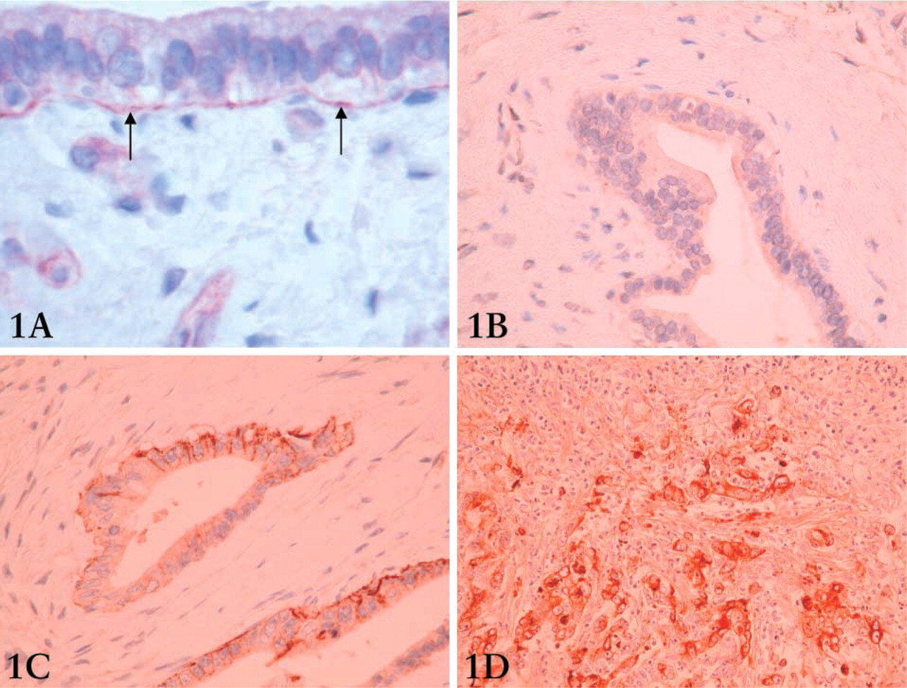
Sections of benign pancreas all demonstrated relatively weak linear staining along the basement membrane of large ducts (Figure 1A). Staining in these ducts was linear in distribution and occasional areas of discontinuous staining were seen. Smaller ducts almost invariably displayed no staining (only rare focal staining seen). The majority of pancreatic adenocarcinoma cases (44/48 or 92%) showed moderately increased to strongly increased staining for β4 integrin subunit. Only one case of chronic pancreatitis demonstrated moderately increased staining, while the additional fourteen cases possessed weak β4 integrin subunit expression similar to that seen in normal pancreas (Figure 1B). One of the four areas of chronic pancreatitis associated with pancreatic adenocarcinoma also displayed moderately increased β4 integrin expression.
(
Staining patterns in the cases of pancreatic adenocarcinoma were somewhat heterogeneous, and a patchy distribution of β4 integrin subunit expression was observed in several of the cases. Primarily, however, β4 integrin subunit was observed in association with the invasive front of the pancreatic tumors. The staining seen at the edge of the tumor-stromal interface displayed abundant staining thickness of varying intensity (Figure 1C). Cytoplasmic and/or membranous staining patterns (Figure 1D) were occasionally identified (five cases).
This study confirms that overexpression of β4 integrin subunit protein in pancreatic adenocarcinoma does occur in the majority of cases and supports the recent findings of gene microarray studies. Our study suggests that β4 integrin subunit protein may be a useful biomarker in pancreatic cancer. Further studies are required to investigate the value of β4 integrin staining in the differentiation of pancreatic carcinoma from chronic pancreatitis, a problem that may arise in the diagnostic arena, particularly with small biopsy specimens. It is interesting to note that we have also observed β4 integrin overexpression in cases of ampullary carcinoma (data not shown). High throughput technologies, such as tissue microarray, would be useful to validate these preliminary findings.
Our results are consistent with those from reports describing upregulation or neoexpression of β4 integrin in various carcinomas. The α6β4 integrin heterodimer is thought to be involved in the generation of traction forces required for cell migration (Rabinovitz et al. 2001). In addition to its function as a laminin receptor in normal epithelial cells (in which expression is restricted to the basal surface), the α6β4 integrin appears to activate signaling pathways that promote invasion (Shaw et al. 1997). Further study of these pathways and their downstream effectors may provide new therapeutic targets for pancreatic carcinoma as well as other cancers known to overexpress β4 integrin.
In summary, our results demonstrate that β4 integrin subunit protein is commonly overexpressed in pancreatic adenocarcinoma and that its relative expression is markedly elevated compared with chronic pancreatitis. This study provides an incentive for further investigation as to the role of β4 integrin in the pathogenesis of pancreatic cancer and in cancer biology in general.