
Abstract
We are developing a reference device to be used in the validation of immunohistochemical imaging of biomarkers by microscopy. The prototype device consists of p53 protein immobilized at various concentrations on a glass slide. The device is designed as a reference control to be used with assays that incorporate commercially available anti-p53 antibodies. p53 protein was characterized by mass spectrometry and covalently immobilized through amide linkage to the (3-aminopropyl)trietoxysilane-modified glass surface. This procedure is reproducible and provides a chemically stable product in high yield. The surface-bound protein was shown to be immunoreactive by its specific interaction with anti-p53 antibody (Ab) and detection by absorbance and fluorescence spectroscopy. Also, comparison was made with microscopic images of Ab-stained tissue samples, known to stain positive for p53. Further development will be required to establish accurate surface protein concentrations in the range required for specific clinical applications.
Keywords
Immunohistochemistry by microscopy is used to localize antigens in tissue sections that are visualized by interaction with specific labeled antibodies. This can be an advantage over traditional histochemical staining techniques that identify only a limited number of proteins and tissue structures; however, antigen quantification by IHC is highly dependent on methods, reagents, and materials. The different protocols used to visualize immunohistochemical reactions and the concentrations and specificities of the antibody (Ab) reagents are important sources of variability (Taylor 1992, 2000, 2007). Improved methods and standards for quantifying these biomarkers are needed for a wide range of applications in clinical diagnostics (Taylor 1992, 2000, 2007; Barker 2003; Michaud 2005; Goldstein et al. 2007). For example, improved measurements of cancer biomarkers, such as p53 and telomerase, are needed for accurate assessment of their ability to determine the presence of cancer and its extent of progression. In this regard, we have developed improved methods to measure telomerase activity and have produced a telomerase lysate material as a candidate reference material for these assays (Atha et al. 2003; Hess et al. 2004; Jakupciak et al. 2004, 2005). We have also developed an IHC method for telomerase quantification that can be used in a wide range of histological and Ab staining applications (Jakupciak et al. 2009).
Although protein biomarkers play critical roles in clinical research and diagnostics, they are often in low abundance and difficult to purify and accurately measure. Some biomarkers, such as human telomerase, require cofactors or associated proteins for activity and stability, which are usually removed in purification (Martin-Rivera and Basco 2001). Human p53, on the other hand, is more easily purified and in a concentration range that is measurable in serum (~16 pg/ml) by ELISA (Chow et al. 2001). Because of its relative stability, availability, and widespread use in research and diagnostics, we chose the p53 protein as a model in our development of a reference device for immunohisto-chemical imaging.
p53 is a tumor suppressor protein that interacts with normal DNA to play a key role in monitoring genetic changes that are harmful to the cell. Studies have shown that p53 is linked to the development and progression of cancer (Hollstein et al. 1991; Vogelstein and Kinzler 1992). It is considered the most frequently altered gene in human cancer with more than 50% of tumors expressing abnormal p53. Inherited mutation in p53 causes Li-Fraumeni syndrome in which loss of one copy of p53 gene results in tumor formation. Previously, we developed improved methods to detect p53 single-point mutations and a reference panel to use in detecting certain mutations in clinical tissue samples (Atha et al. 1998; O'Connell et al. 1998, 2003, 2005; Wenz et al. 1998; Sunar-Reeder et al. 2004). Recent studies indicate that nuclear accumulation of p53 is a potential marker for the development of squamous cell lung cancer in smokers (Piyathilake et al. 2003). Also, it was reported that nuclear accumulation of p53 is a potential prognostic marker of colorectal cancer (Manne et al. 1997a, 1998; Manne 2007).
In this report, we describe a candidate reference device that could be used for quality control to assess the reproducibility and accuracy of IHC measurements. The device consists of p53 protein, immobilized on a glass slide. Previous studies have reported the immobilization of purified proteins or peptides on glass slides or other matrices for immunohistochemical analyses of different markers (Millar and Williams 1982; Nibbering and van Furth 1987; Larsson and Hougaard 1994; Sompuram et al. 2002; Bogen et al. 2009). However, we are specifically assessing the use of this method in the immunohistochemical analysis of p53, for which many commercial antibodies are available. Cell preparations or cultured cell lines can also be used for quality control in IHC assay systems, but the advantages of a protein immobilization approach are that it is less cumbersome and more reproducible, as it is not susceptible to changes in protein expression, especially if paraffin-embedded tissues are used as controls (Otali et al. 2009). The use of the entire p53 protein would also result in immunoreactivity of a wider range of p53 antibodies than if a selected peptide sequence was used as the antigen.
In our measurements, the reactivity of the immobilized p53 was tested spectroscopically using commercially available immunohistochemical antibodies and probes, commonly used for microscope imaging. Although further analysis will be required to establish accurate surface protein concentrations and reproducibility, in our view, this method would allow users in various research and diagnostic labs to compare results using a reference device that is known to contain a specified amount of the correct target antigen, p53.
Materials and Methods
The prototype reference device contains a covalently immobilized biomarker protein on a glass slide. Protein concentration and activity were characterized by previously established biochemical and physical methods as described below.
Characterization of p53 Preparation
A commercial preparation of recombinant wild-type human p53 protein, listed as 95% pure, was obtained from ProteinOne (Bethesda, MD). We used two-dimensional (2D) gel chromatography and matrix-assisted laser desorption ionization time of flight (MALDI-TOF) mass spectrometry to verify that the preparation contained the designated p53 protein (Ligner et al. 1997; Merrick et al. 2001) as follows:
2D Gel Chromatography. For the first dimension, 200 ng of the p53 preparation was applied to an immobilized pH gradient strip (pH 3-10). The second dimension was run on 10% PAGE. After silver staining, the predominant protein spots were excised from the gel, extracted in buffer, and individually analyzed as described below.
Mass Spectrometric Analysis—Trypsin Digest. For mass spectrometric analysis, 1 μg or 10 pmol protein was used in a volume of 15 μl. Digestion buffer [5 mmol/liter dithiothreotol, 1% (w/v) Rapigest in NH4HCO3] was added. The sample was heated in a water bath at 100C for 5 min, then heated at 60C for 30 min and cooled to room temperature. To alkylate the reduced cysteine thiols so that the disulfide bonds cannot re-form, 3 μl of 0.5 M iodoacetamide was added to a final concentration of 15 mmol/liter and the sample was placed in the dark for 60 min. Trypsin (0.5 μl of 1 μg/μl stock solution in 50 mmol/liter acetic acid) was added to the sample, for a 2:1 substrate/trypsin mass ratio. The sample was incubated at 37C, with shaking, for at least 12 hr. Trifluoroacetic acid (TFA; 0.5 μl) was added to the sample so that the final TFA concentration was 0.5% (v/v). The sample was incubated at 37C for 45 min, centrifuged at 13,000 rpm for 10 min, and transferred to new vial. The sample was evaporated to near dryness (~7 μl) so that as much sample as possible could be loaded on to the ZipTip C18 (Millipore; Billerica, MA) to remove any salts or remaining surfactants that could suppress the MALDI process. The peptides were eluted off the ZipTip bed using 5 μl [60% v/v acetonitrile (ACN), 40% v/v H2O, 0.3% v/v TFA] into a microcentrifuge tube, evaporated to dryness, and reconstituted in 5 μl matrix solution [saturated α-cyano-4-hydroxycinnamic acid (CHCA) in 30% v/v ACN, 70% v/v H2O, 0.1% v/v TFA]. Final peptide concentration was ~2 pmol/μl (assuming 10 pmol protein, 1 mol protein = 1 mol peptide, and no loss). The peptides were then spotted on the MALDI target plate.
Mass Spectrometric Analysis. An Applied Biosystems (Foster City, CA) 4700 MALDI-TOF/TOF Spectrometer was used to collect the spectra of peptides and fragmentation spectra of individual proteins obtained after 2D gel separation of the commercial p53 preparation. The peptides/proteins were identified by comparing experimentally determined masses with expected peptide masses, based on trypsin cleavage (at Arg and Lys residues) of the known amino acid sequence. p53 (55 kDa, pI = 6.5) was identified in one of the six spots removed from the 2D gel. As shown in Figure 1, 14 peptides (30% sequence) were identified in this protein. In three of the remaining 2D gel spots, protein contaminants were identified as cytokeratins 9 and 10 (59-62 kDa, pI = 5.09-5.1) using 12-15 peptides (28-39% sequence; data not shown).
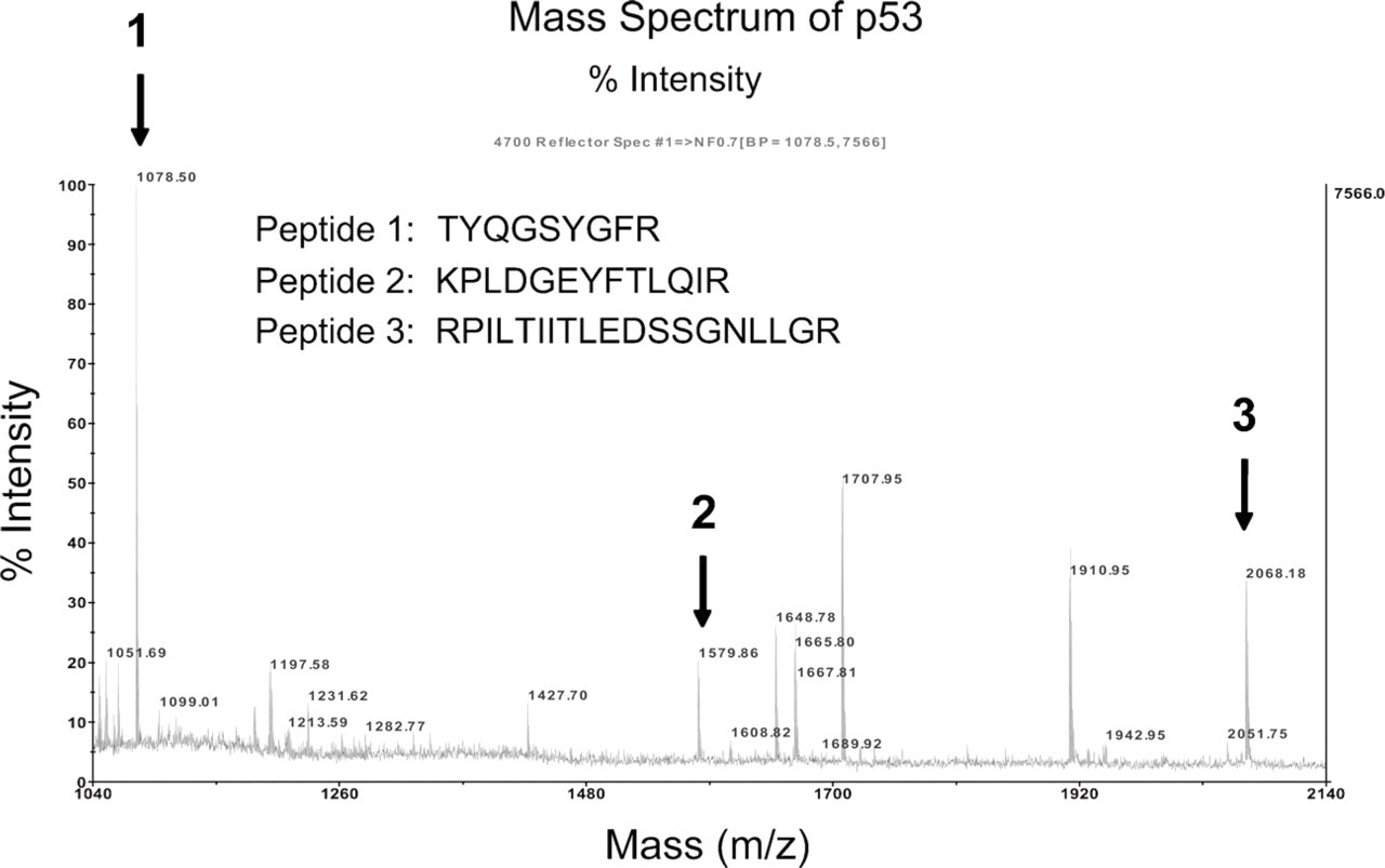
Mass spectrum of p53 used for immobilization on a glass slide. Three primary sequences, indicated by arrows, are shown from trypsin-digested fragments that uniquely identify p53. G, glycine; L, leucine; I, isoleucine; F, phenylalanine; P, proline; S, serine; T, threonine; Y, tyrosine; N, asparagine; Q, glutamine; D, aspartic acid; E, glutamic acid; K, lysine; R, arginine; H, histidine.
Immobilization of p53
The original p53 preparation was immobilized on glass microscope slides (UltraGaps coated slides; Corning Microarray Products, Corning, NY), containing a γ-aminopropylsilane layer. As shown in Figure 2, the protein was immobilized through amide linkage to the (3-aminopropyl)triethoxysilane-modified glass surface using previously described methods (Weetall 1993). Briefly, the slides were dried overnight at 100C, followed by activation in 2.5% (w/v) solution of glutaraldehyde in PBS (pH = 7.4) for 1 hr. Residual glutaraldehyde was removed by a thorough slide wash in pure PBS and drying in N2 gas flow. Protein solution (10 μl of 0.05 mg/ml) was spotted on marked slide areas, and slides were kept overnight at 4C in a 100% humid environment. The slides were then rinsed several times in PBS and placed in 1 M emanolamine blocking solution for 30 min. Following a PBS rinse, slides were incubated with Ab solutions. This method has been shown to be robust while maintaining the activity of the original protein (Willamson et al. 1989; Wang et al. 2004; Choi et al. 2008).
Characterization of Immobilized p53
The immobilized protein was shown to be immunoactive by its specific interaction with anti-p53 Ab as verified by comparison with immobilized BSA controls. The immobilized protein was incubated 1 hr at room temperature with mouse monoclonal Ab (clone: BP53.12; Zymed-Invitrogen, Carlsbad, CA) diluted 1:50 (v/v) in PBS 1% BSA. The immunoreactivity of the immobilized p53 was characterized using a combination of IHC, spectroscopy, and microscopy, described as follows.
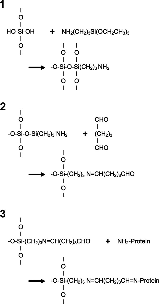
Diagram of protein immobilization chemistry. The p53 protein was immobilized through amide linkage to the (3-aminopropyl) trietoxysilane-modified glass surface using previously described methods (Weetall 1993). Diagram modified from Weetal with permission. Appl Biochem Biotechnol 41:157-188, 1993.
Chromogenically Labeled Ab Assay. After the immobilized p53 protein was incubated with the mouse monoclonal Ab, the slide was gently rinsed in PBS, followed by a 30-min incubation with biotin-conjugated rabbit anti-mouse secondary Ab and 15-min incubation with streptavidin-conjugated horse radish peroxidase (HRP), and was again gently rinsed in PBS, followed by a 10-min incubation with DAB and hydrogen peroxide (I-view chromogenic system kit; Ventana, Tucson, AZ). Ultraviolet (UV)/visible absorbance of selected slide areas was performed using a Chem2000 fiber optic spectrophotometer (Ocean Optics; Dunedin, FL). The slide was positioned between two 100-μm-diameter optical fiber ends separated by 3 mm. This arrangement provided about 1-mm-diameter light spot on the glass slide. Before the absorbance measurement in the center of the protein immobilization spot, the reference spectrum was recorded by positioning the light beam outside the immobilized protein spot.
Fluorescent Probes. In place of the streptavidin-conjugated HRP, streptavidin conjugated with fluorescent cyanine dye (Cy3; Sigma-Aldrich, St. Louis, MO), 3-nm silicon nanocrystals (Choi et al. 2008), or commercial quantum dots (Qdot 605; Invitrogen, Carlsbad, CA) were used and compared by fluorescence emission. An SLM model LM800 spectrofluorimeter (Horiba Scientific; Edison, NJ) with sample compartment modified to accommodate glass microscope slides at 30-degree incidence to the excitation light was used for measurements of fluorescence. Visual images of the surface-immobilized fluorophores were obtained using a 514-nm laser excitation, with emission filtered by a laser-line notch filter (Kaiser Scientific Systems; Ann Arbor, MI).
Microscopic Imaging. Paraffin-embedded sections (4 μm) of colon tissue, obtained at the University of Alabama at Birmingham (UAB) under Internal Review Board regulations, were prepared using a Ventana Benchmark XT and the I-view chromogenic system as described above. Microscopic images were taken using a Zeiss (Thornwood, NY) Axiovert S-100 microscope and a Sony (Fort Meyers, FL) DSC-S75 camera.
Results
We analyzed the commercial preparation of recombinant wild-type human p53 protein by 2D gel electrophoresis and MALDI-TOF mass spectrometry. Although the preparation used for immobilization contained a few contaminating proteins (cytokeratins 9 and 10), it was determined by mass spectrometry to contain the target p53. This method of characterization by 2D gel electrophoresis and mass spectrometry would be useful in interpreting any crossreactivity that may occur in other Ab systems.
We immobilized the p53 protein on silanized glass slides, which were tested for immunoreactivity by treatment with an anti-p53 primary and a secondary Ab labeled with biotin and detected using chromogenic and fluorescent probes. These IHC methods are commonly used to detect p53 in biological samples (Manne et al. 1997a; Chow et al. 2001). Analysis by absorption spectroscopy indicated successful binding of the anti-p53 Ab and chromogenic staining in slides containing p53 compared with immobilized BSA controls (Figure 3). Figure 3 shows about a 10-fold increase in UV absorbance (300 nm) of the immobilized p53, treated with anti-p53 Ab, compared with the control BSA, treated with the same Ab. A comparable increase was observed at the visible wavelengths (400-600 nm) where the DAB stain is expected to absorb. Control p53 and BSA without Ab produced low absorbance in both the UV and visible wavelengths. This showed that the immobilized p53 was immunoreactive and retained its specific interaction with the anti-p53 Ab. These results are summarized in Table 1.
The amount of absorbed protein was estimated using the measured UV absorbance of the immobilized protein film (given in Table 1), the protein extinction coefficient, and the molecular size to estimate the light path, assuming a monolayer coating. The p53 with extinction coefficient at 280-nm wavelength (∊280) = 17,130 cm−1·M−1 (Bullock et al. 1997) and diameter = 6.82 nm (Tidow et al. 2007) yielded ~0.86 nmol/cm2. The BSA alone with ∊280 = 43,824 cm−1·M−1 (Johnstone and Thorpe 1982) and diameter = 14.1 nm × 4.2 nm × 4.2 nm (Wright and Thompson 1975) yielded ~0.42 nmol/cm2. The estimated diameters and extinction coefficients were combined in estimates of the p53 and BSA treated with anti-p53 Ab. The contribution to the UV absorbance by the tetravalent streptavidin-HRP was considered to be negligible compared with the biotin-labeled IgG. As a result, the p53 + IgG with ∊280 = 210,000 cm−1·M−1 (Johnstone and Thorpe 1982) and diameter = 28.0 nm (Roberts et al. 1995) yielded ~5 nmol/cm2. The BSA + IgG (control) yielded ~0.08 nmol/cm2. These calculations are only to be considered rough estimates of the protein surface concentration as absorbance measurement in such thin films are dependent on several assumptions. The actual concentration of p53 would also require additional measurements (see Discussion).
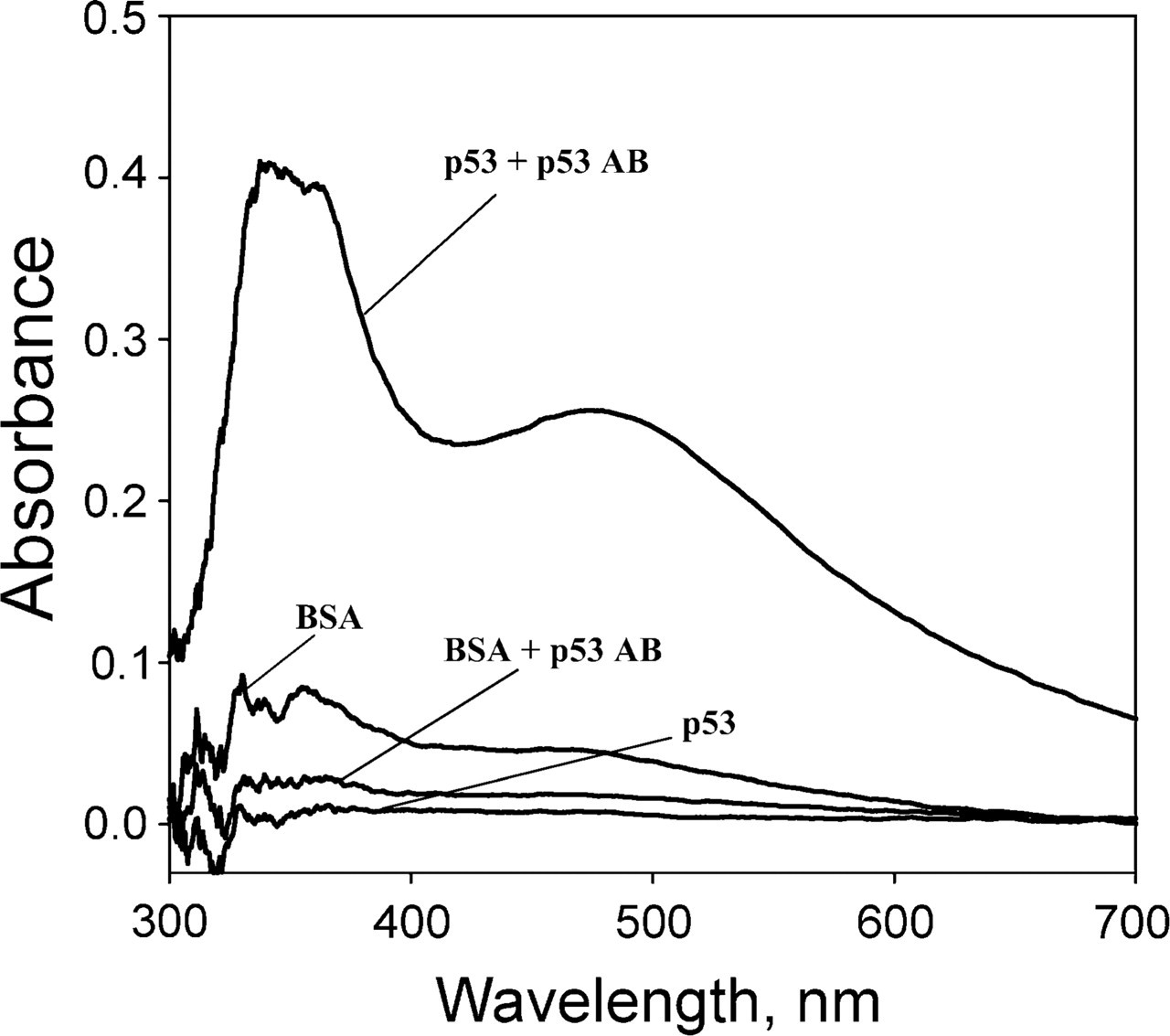
Absorbance spectra of local slide areas using the chromogenic DAB system. Effect of different treatments: p53 + p53 antibody (Ab)—covalently immobilized p53, incubated with anti-p53 Ab; BSA + p53 Ab—covalently immobilized BSA, incubated with anti-p53 Ab; p53—covalently immobilized p53, no Ab; BSA—covalently immobilized BSA, no Ab.
Characterization of immobilized p53 by ultraviolet (UV) and visible absorbance
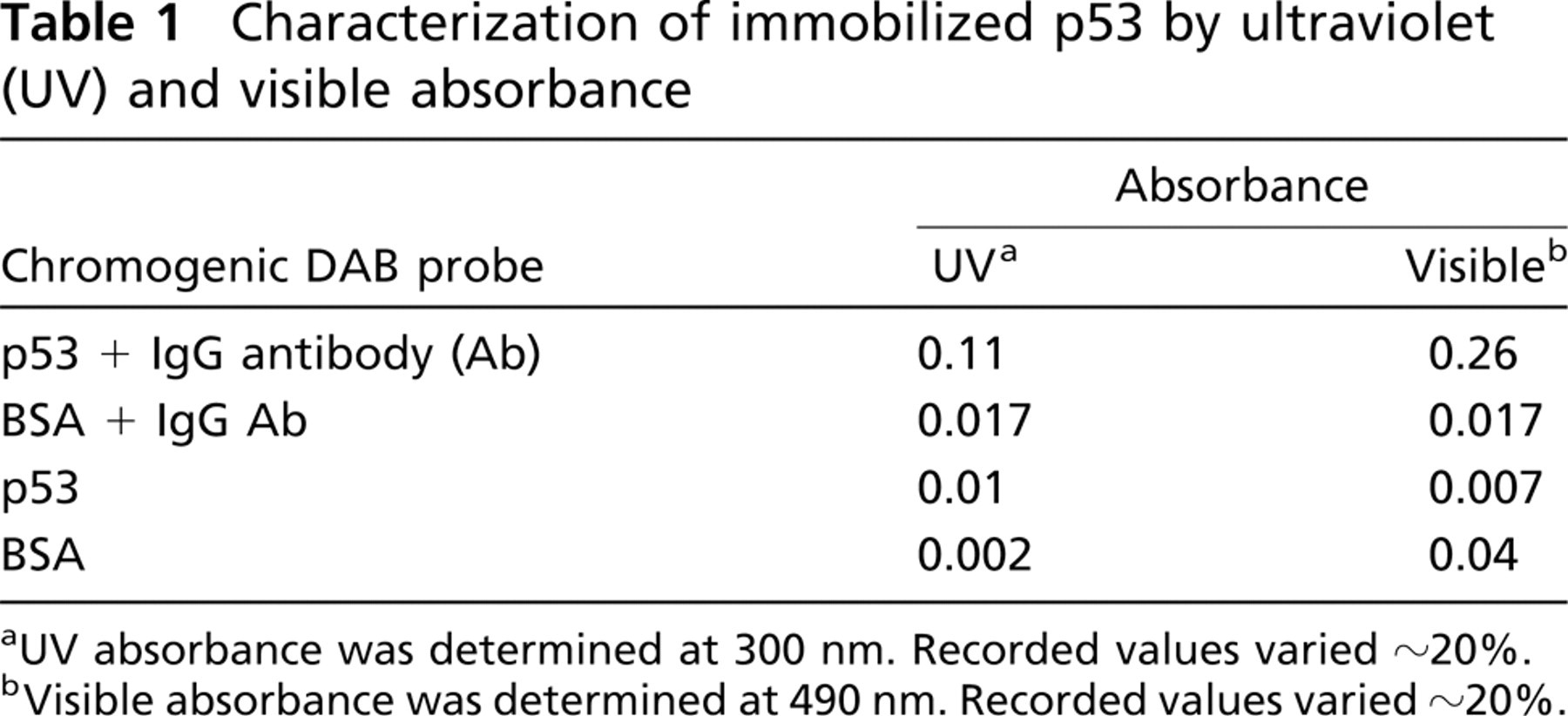
UV absorbance was determined at 300 nm. Recorded values varied ~20%.
Visible absorbance was determined at 490 nm. Recorded values varied ~20%.
Figure 4 presents the fluorescence spectra recorded on four glass slides, containing different fluorescent probe-streptavidin conjugates Cy3, quantum dot 605, and silicon nanocrystal to label the secondary Ab. Absolute recorded emission intensities varied about 20% from sample to sample, but consistently demonstrated preferential Ab binding to the immobilized p53 sites (middle areas of the slides). Comparison of the relative fluorescence intensities, compared with measurements of the blank in the absence of anti-p53 Ab, is shown in Table 2. Cy3-labeled streptavidin showed the highest emission, which permitted visual observation of the p53-labeled area using direct laser illumination at 514 nm.
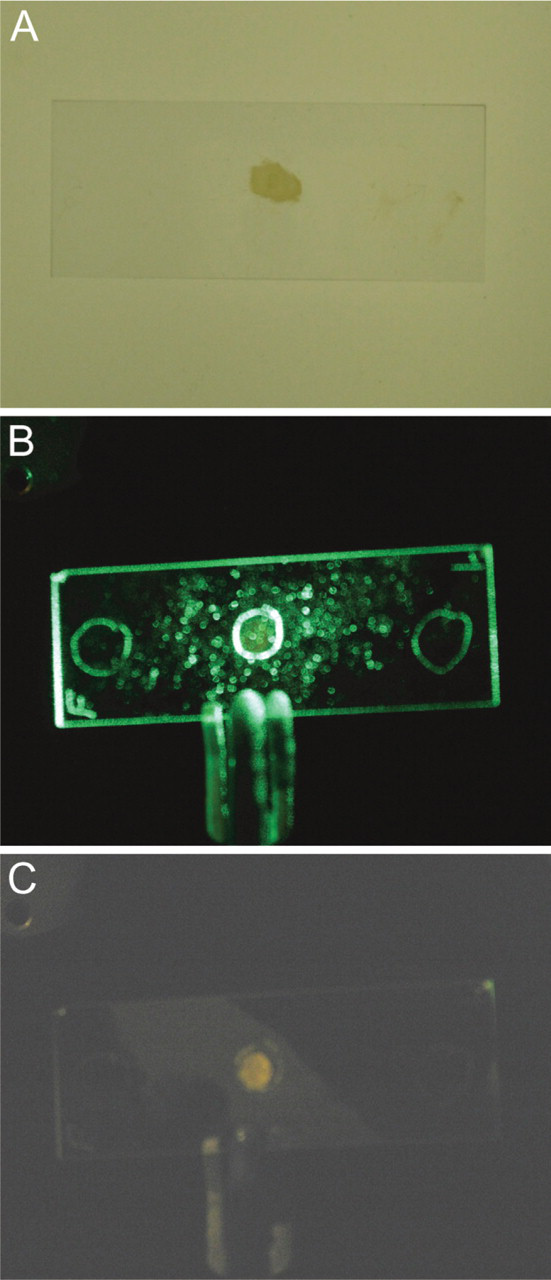
Images of microscope slides containing immobilized p53 are shown in Figure 5. Images were obtained using the chromogenic DAB probe with transillumination (Figure 5A), the fluorescent Cy3 probe with 514-nm laser illumination (Figure 5B), and the fluorescent Cy3 probe with laser-line notch filter illumination (Figure 5C). Immobilized p53 is located in center of slides; it appears as a brown spot under transillumination and as a reddish spot under laser illumination with the notch filter. Immobilized control BSA is located at the ends of the slides and is not visible under either light source.
Figure 6 shows a comparison of microscopic images of chromogenically stained colon tissue sections and chromogenically stained slides containing immobilized p53. The tissue sections show the expected dark brown staining of cell nuclei (Figure 6A), compared with the control tissue without primary anti-p53 Ab (Figure 6B). The images from immobilized p53 result in a dark brown background and particulate matter resulting from the reacted DAB stain. The control slide, in the absence of primary anti-p53 Ab, shows no indication of the brown DAB stain. These results, using the immobilized p53 as a reference, demonstrate that the reagents used to detect the p53 Ab in tissues are functioning properly.
Discussion
The detection of a protein biomarker by IHC depends on several factors. One is the amount of active/immunogenic protein that is accessible in the assay. This of course depends on the particular biomarker and its half-life in the cell and probably other unidentified factors in paraffin-embedded material (Otali et al. 2009). For example, when tissue with no mutation in p53 is stained for p53, no staining for p53 can be detected because of the very short biological half-life of native p53 in cells. In contrast, when there is a point mutation in p53, the half-life of p53 in cells is increased and the nuclear presence of p53 in cellular nuclei can be detected. We selected p53 for this study because of this complex pattern of staining. The staining of immobilized p53 on the reference slides demonstrates that p53 is stable outside the cell and that the reference is designed to be compared with cells that have a mutation in p53, as in Figure 6A.
Characterization of immobilized p53: effect of fluorescent probes
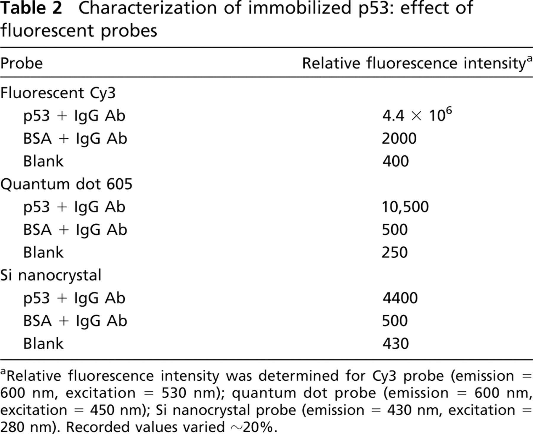
Relative fluorescence intensity was determined for Cy3 probe (emission = 600 nm, excitation = 530 nm); quantum dot probe (emission = 600 nm, excitation = 450 nm); Si nanocrystal probe (emission = 430 nm, excitation = 280 nm). Recorded values varied ~20%.
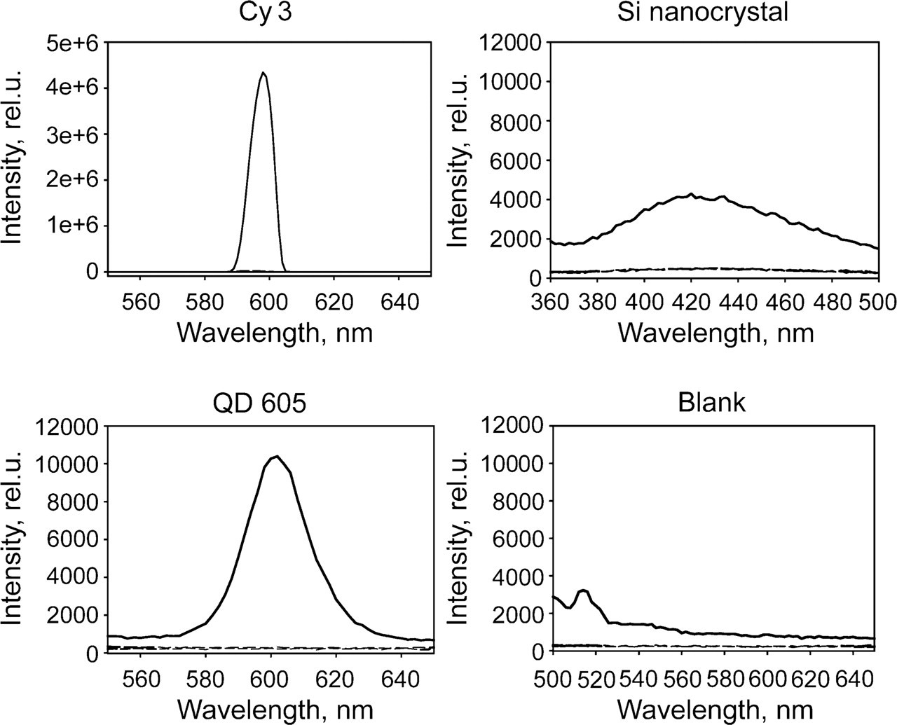
Fluorescent spectra of immobilized p53/p53 Ab/IgG-biotin using different streptavidin-conjugated fluorescent probes (fluorescent Cy3; quantum dot 605; Si nanocrystal; and Blank, no added probe). Dashed lines show BSA controls. Excitation wavelengths for fluorescent Cy3, quantum dot 605, and Si nanocrystal probes were 530, 450, and 280 nm, respectively.
The amount of active/immunogenic protein is also affected by environmental conditions (i.e., specimen preparation) that can influence the conformation and accessibility of the protein. Immobilizing the entire p53 protein rather than a peptide fragment may make the protein more susceptible to conformational effects, such as those that could occur with preparative methods used in antigen retrieval. However, we chose to immobilize the intact protein rather than a peptide fragment as the intact protein should react with a wider range of antibodies and be compatible with a wider range of staining protocols. We realize that this approach is not feasible in the case of other biomarkers, such as HER-2, for which the purified forms are not commercially available.
Another factor is the choice of Ab. The Ab used in this study was produced using a peptide region of the p53 molecule that is outside of commonly mutated regions. In addition to the amount of bound Ab, the resulting signal intensity also depends on the choice of label and its extent of coupling, stability, and optical efficiency. Environmental conditions can affect all of these aspects. Careful calibration of instruments and comparison to a known reference device could serve as a quality control to help reduce uncertainties in these measurements.
Images of microscope slides containing immobilized p53: (
In this report, we describe the first stage in developing a candidate reference device to improve the quality (reproducibility and accuracy) of biomarker imaging using IHC. In this prototype, we have shown that we can immobilize on glass slides a representative protein biomarker and preserve its immunogenicity. This shows that p53, immobilized by previously described methods, can be used in the design of a reference device. The immobilized p53 protein produced a measurable signal when used in a conventional IHC tissue staining protocol.
In our experience, the immobilized p53 is stable when stored covered at 4C in 100% humidity. Measurements were within a 20% variation after storage for several days. However, when stored at room temperature (23C) and humidity (~60%), we found, after a period of about 1 year, that the immobilized p53 loses about one half of its immunoactivity when reacted with fresh reagents. The stability of the probe is another matter. We found that processed slides stored in the dark for a year at room temperature (23C) and humidity (~60%) vary in stability, depending on the probe used. Specifically, the silicon nano-particle probe retained about 25% of its intensity, the quantum dot probe retained about 60% of its intensity, and the Cy3 fluorescent probe lost essentially all of its intensity. Such loss of immunoreactivity in cut paraffin-embedded tissues has been reported previously (Prioleau and Schnitt 1995; Jacobs et al. 1996; Manne et al. 1997b).
The completed reference device would contain the biomarker protein immobilized at various concentrations on a glass microscope slide. Because of variation in the efficiency of immobilization or aggregation, this may result in a non-linear calibration curve. Mixtures of the target p53 and BSA protein may be necessary. The completed device would be specifically designed with the appropriate p53 concentration for use with chromogenically or fluorescently labeled antibodies to provide a calibration vehicle for microscopy. This would include accurate determination of the protein surface concentration by dedicated measurement methods such as quartz crystal microbalance (Tothill 2009). This measurement would provide a basis to adjust the p53 concentration used for immobilization such that the final protein density will match the range observed in the histological sample. The device as a kit could also include the specific reagents needed for the immunostaining steps involved in the detection of the biomarker, e.g., labeled antibodies, buffers.
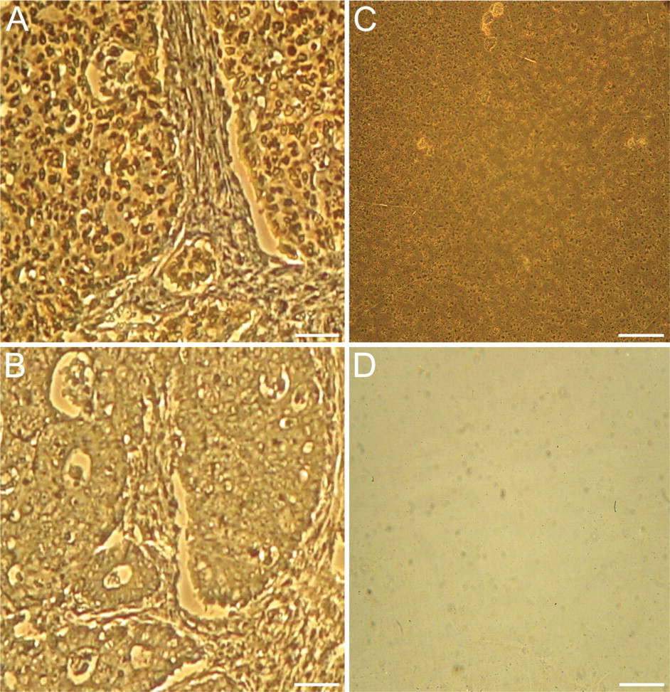
Microscopic images of p53 visualized using chromogenic probe: comparison of colon tissue sections and an immobilized p53 reference. Sequential slide sections taken from a biopsied colon tissue sample were prepared and stained using the Ventana chromogenic DAB system and compared with immobilized p53 reference slides using the same chromogenic system. (
The reference device would provide a tool to verify that IHC reagents and protocols are correct when tested using the reference device in which the biomarker protein is known to be present. This is particularly important with respect to the Ab that is used to detect the biomarker. Compared with immobilized control proteins, the device could be used to estimate nonspecific binding of the Ab. It could also provide a way to estimate the concentration of the biomarker in tissues when the sample is compared with a reference device in which the concentration of the biomarker has accurately been determined. With further development, such a device could impact numerous areas related to cancer diagnostics and be used in a wide range of academic and industrial research.
Certain commercial equipment, instruments, and materials are identified in this article to specify an experimental procedure as completely as possible. In no case does the identification of particular equipment or materials imply a recommendation or endorsement by the National Institute of Standards and Technology nor does it imply that the materials, instruments, or equipment are necessarily the best available for the purpose.
Footnotes
Acknowledgements
This work was funded in part by National Cancer Institute—Early Detection Research Network (EDRN) and National Institute of Standards and Technology (NIST)—Biochemical Science Division jointly under interagency agreement no. YI-CN5001 and by the EDRN Reference Laboratory at UAB (5U24CA086359-10).
The authors thank Dr. Ilarion Turko, NIST, for the mass spectrometry measurements to characterize the p53 used in these experiments; Dr. Chandrakumar Shanmugam, UAB, for help in preparing the tissue slides; and Dr. John P. Jakupciak, Cosmosid, for helpful discussions during the preliminary phase of these experiments.