
Abstract
The mRNA expression pattern of dipeptidyl peptidase (DPP) 8 and DPP9, two DPP4 homologs, was studied previously and showed a broad tissue distribution. In this study, protein expression and activity of DPP8 and DPP9 were investigated in male reproductive tissues of different mammals. Based on specific DPP activities and inhibition profiles, the proline-selective DPP activity in the bovine and rat testis could predominantly be attributed to DPP8/9 and not to DPP4. This is in contrast to the epididymis, where most of the activity was caused by DPP4. Bovine sperm preparations had very low or undetectable DPP8/9 activity. After characterization of polyclonal antibodies specific for DPP8 or DPP9, we could localize both enzymes in seminiferous tubules of the testis. A specific staining for DPP9 was found associated with spermatozoids embedded in the epithelium, just before their release into the lumen, and in spermatids. DPP8 was localized in spermatozoids in an earlier stage of maturation. These findings help to provide insight into the physiological role of DPP4-like enzymes in the male reproductive system. This manuscript contains online supplemental material at http://www.jhc.org. Please visit this article online to view these materials.
Keywords
T
The mRNA expression pattern of DPP8 and DPP9 was studied and showed a broad distribution among human tissues. The highest DPP8 mRNA levels are found in testis and placenta. The enzyme is upregulated in activated T cells and expressed in all B- and T-cell lines examined (Abbott et al. 2000; Qi et al. 2003). The DPP9 mRNA expression levels are high in skeletal muscle, heart, liver, and peripheral blood leukocytes (Olsen and Wagtmann 2002; Qi et al. 2003; Ajami et al. 2004). The DPP8 and DPP9 mRNA expression profiles showed a ubiquitous distribution in different skin cell types (Thielitz et al. 2008a,b). In kidney, small intestine, lung, and pancreas of pig and dog, semiquantitative RT-PCR analysis showed different relative abundance of DPP4-like enzymes, with DPP4 and DPP9 having the highest expression, followed by DPP2 and DPP8 (Wagner et al. 2006). In mice, a higher number of DPP8 and DPP9 transcripts compared with DPP4 were present in colon, brain, skin, and thymus (Helmuth et al. 2008).
Only very recently, the expression of DPP8 and DPP9 started to be studied at the protein and/or activity level. High expression levels of DPP8/9 were reported in rat and human brain (Frerker et al. 2007; Stremenova et al. 2007; Busek et al. 2008). Also, human leukocytes contained DPP8/9 activity (Maes et al. 2007a). DPP8 and DPP9 were upregulated in the bronchi after induction of experimental asthma in the rat (Schade et al. 2008). The precise functions of these enzymes in vivo are still unknown. There is some evidence that, in leukocytes, they are involved in immunoregulation. Inhibition of DPP8 and DPP9 suppresses mitogen-stimulated T-cell responses, whereas selective inhibition of DPP4 and DPP2 does not (Reinhold et al. 2008).
Despite the similar substrate specificity on X-Pro—containing chromogenic and fluorogenic substrates at neutral pH, the DPP4-like enzymes can be discriminated using selective inhibitors. The selective DPP2 inhibitor N-(4-chlorobenzyl)-4-oxo-4-(1-piperidinyl)-1,3-(S)-butane-diamine dihydrochloride (UAMC00039) (Senten et al. 2004), the selective DPP4 inhibitor (3R)-3-amino-1-[3-(trifluoromethyl)-5,6-dihydro-[1,2,4]triazolo[4,3-α] pyrazin-7(8H)-yl]-4-(2,4,5-trifluorophenyl)butan-1-one (sitagliptin) (Kim et al. 2005), and the DPP8/9 inhibitor (2S,3R)-2-amino-1-(isoindolin-2-yl)-3-methylpentan-1-one (allo-Ile-isoindoline) (Lankas et al. 2005) were used in our study (Figure 1).
Recently, we purified proline-selective DPPs clearly different from DPP2 and DPP4 from bovine testes (Dubois et al. 2008). One of them was identified as DPP9. Here, we studied the distribution of the activity and cell-specific expression of DPP8 and DPP9 in the male reproductive system. Because no antibody preparations against these enzymes have been studied thoroughly before, we included a comparison and characterization here. Based on activity and inhibition profiles and immuno-blotting, we showed the expression of DPP8 and DPP9 in the testis, epididymis, and sperm. IHC stains were used to localize DPP8 and DPP9 in these tissues.
Materials and Methods
Materials
The DPP2 inhibitor UAMC00039 and the DPP8/9 inhibitor allo-Ile-isoindoline were synthesized as described (Senten et al. 2004; Lankas et al. 2005). The DPP4 inhibitor sitagliptin was extracted from Januvia tablets (Merck; Vienna, Austria). Bovine tissues were kindly provided by a local slaughterhouse. Tissues of Wistar rats were obtained from the animalarium of the University of Antwerp. After the animals were sacrificed, tissues were excised, either immediately frozen in liquid nitrogen and stored at −80C until use or fixed in 4% paraformaldehyde in PBS for 24 hr. Bovine sperm was obtained from the “Vlaamse Rundveeverbetering” (Oosterzele, Belgium). DPP2 and DPP4 were purified from human seminal plasma (De Meester et al. 1996; Maes et al. 2005). Recombinant human DPP8 (rDPP8) was expressed and purified as described (Chen et al. 2004). DPP9 was purified from bovine testes (Dubois et al. 2008). The rabbit polyclonal anti-DPP9 antibody was generated by Eurogentec as described in Dubois et al. (2008). Rabbit polyclonal antibodies against the spacer region of DPP4 (ab28340), the aminoterminal end (ab42075), the spacer region (ab42076), and the catalytic domain (ab42077) of DPP8; the aminoterminal end (ab42078), the spacer region (ab42079), and the catalytic domain (ab42080) of DPP9; and the chicken polyclonal antibody against DPP9 (ab26177) were obtained from Abcam (Cambridge, UK) (Table 1). Ala-Pro-p-nitroanilide (-pNA) and Gly-Pro-pNA were purchased from Bachem (Budendorf, Switzerland).
Tissue Homogenate and Sperm Preparations
Tissues were homogenized in lysis buffer (LB; 1% octylglucoside, 10 mM EDTA, and 70 μg/ml aprotinin in 0.05 M HEPES buffer, pH 7.0; 5 ml/g) (Maes et al. 2007a). After incubation for 1 hr, the homogenized tissue was centrifuged for 10 min at 3000 X g, and the resulting supernatant was subsequently centrifuged for 1 hr at 20,000 X g at 4C. The final supernatant was filtered.
Bull sperm samples were pooled. Differential centrifugation of bovine sperm was performed according to Vanhoof et al. (1992). In brief, bovine sperm was centrifuged at 900 X g for 10 min at 4C. Spermatozoa were obtained from the pellet. The supernatant was centrifuged at 105,000 X g for 2 hr to precipitate the prostasomes. The final supernatant contained the seminal fluid. For clarification, 1% octylglucoside was added to the seminal fluid. The seminal fluid was centrifuged at 13,000 X g at 4C, and the supernatant was used. The spermatozoa and prostasomes were lysed as described in Maes et al. (2007a). Subcellular fractionation of spermatozoa was performed according to Cooper (2000). Cells were ruptured by a single freeze-thaw cycle, followed by 3 × 15-sec sonication on ice.
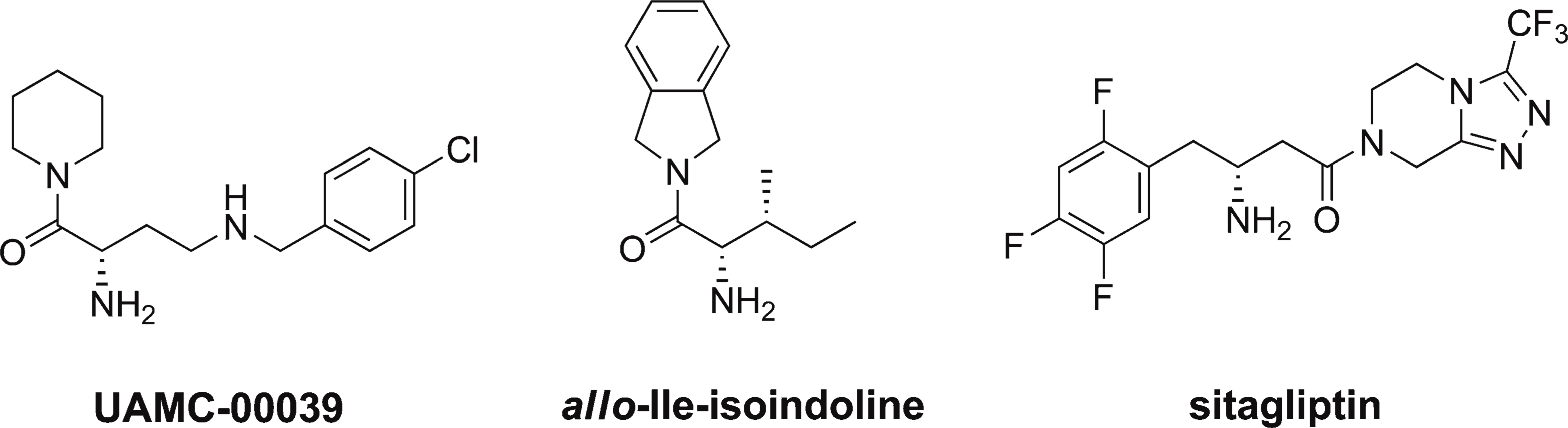
Structures of the dipeptidyl peptidase (DPP) inhibitors used: the DPP2 inhibitor N-(4-chlorobenzyl)-4-oxo-4-(1-piperidinyl)-1,3-(S)-butanediamine dihydrochloride (UAMC00039), the DPP8/9 inhibitor (2S,3R)-2-amino-1-(isoindolin-2-yl)-3-methylpentan-1-one (allo-Ile-isoindoline), and the DPP4 inhibitor (3R)-3-amino-1-[3-(trifluoromethyl)-5,6-dihydro-[1,2,4]triazolo[4,3-α]pyrazin-7(8H)-yl]-4-(2,4,5-trifluorophenyl)butan-1-one (sitagliptin).
Characteristics of the polyclonal affinity purified antibodies used for DPP4, DPP8, and DPP9
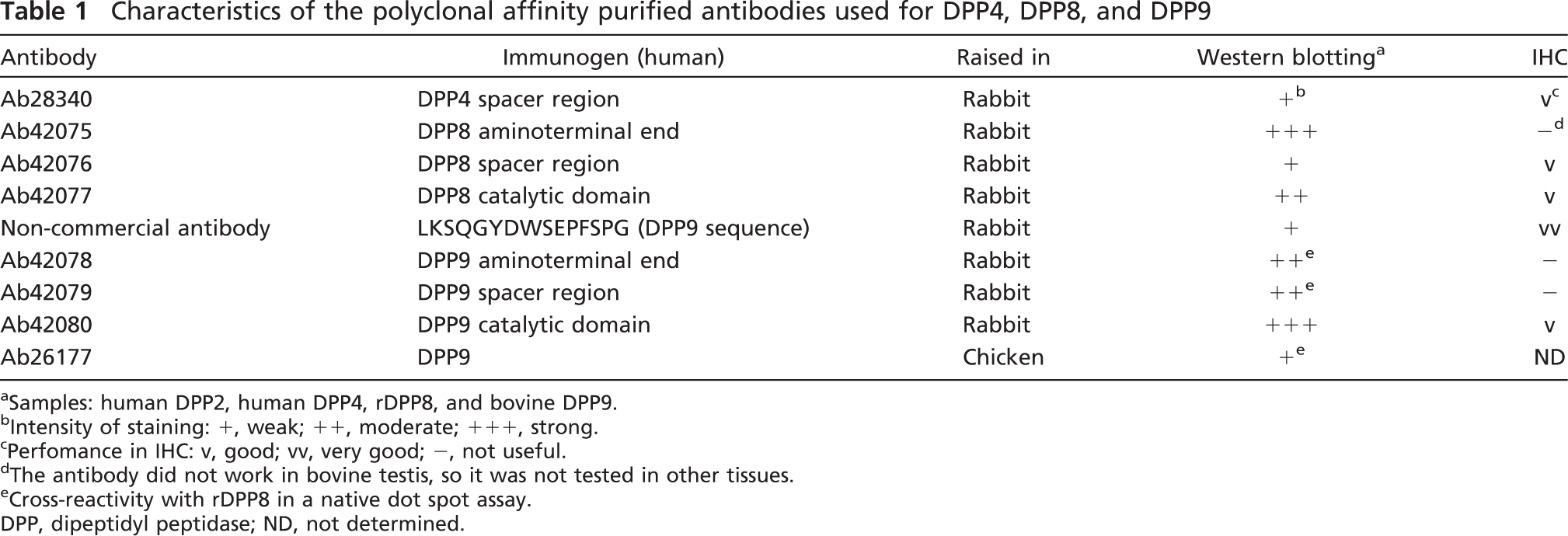
aSamples: human DPP2, human DPP4, rDPP8, and bovine DPP9.
bIntensity of staining: +, weak; ++, moderate; +++, strong.
cPerfomance in IHC: v, good; vv, very good; -, not useful.
dThe antibody did not work in bovine testis, so it was not tested in other tissues.
eCross-reactivity with rDPP8 in a native dot spot assay.
DPP, dipeptidyl peptidase; ND, not determined.
The resulting samples were used to measure enzyme activities and protein concentration.
For the immunodetection of DPPs in homogenates, tissues were homogenized in 0.02 M HEPES buffer, pH 7.0, containing 1% octylglucoside and a complete protease inhibitor cocktail tablet (Roche; Brussels, Belgium). Apart from this, the procedures were as described above.
Enzyme Assays
Enzymatic activities were determined kinetically in a final volume of 200 μl for 10 min at 37C by measuring the initial velocities of pNA release (405 nm) from the substrate using a Spectramax plus microtiter plate reader (Molecular Devices; Sunnyvale, CA). One unit of enzymatic activity is the amount of enzyme that catalyzes the release of 1 μmol pNA from the substrate per minute under assay conditions. The substrates Ala-Pro-pNA (1 mM in 0.05 M HEPES buffer, pH 7.0, containing 10 mM EDTA, 14 μg/ml aprotinin, and 0.1% Tween 20) and Gly-Pro-pNA (0.5 mM in 0.05 M Tris buffer, pH 8.3, containing 10 mM EDTA and 14 μg/ml aprotinin) were used to probe DPP2, DPP4, DPP8/9, and/or FAP activity (Maes et al. 2007a).
Protein concentration was determined according to Bradford (1976) with BSA (Sigma; Bornem, Belgium) as the standard.
Inhibition Assays
Tissue homogenates, cell lysates, and pure enzyme samples diluted in LB supplemented with ∼10 mg/ml BSA were preincubated for 15 min at 37C with a wide range of inhibitor concentrations. DPP activities were determined as described above. The specific contribution of DPP2 activity to the Ala-Pro-pNA cleavage was determined using the DPP2 inhibitor UAMC00039 (100 nM). The dual DPP8/9 inhibitor allo-Ile-isoindoline (5–5000 nM) served to determine the contribution of both enzymes to the total DPP activity. DPP4 activity was inhibited by its selective inhibitor sitagliptin (50–50,000 nM).
Immunoaffinity Purification of the Rabbit Polyclonal Anti-DPP9 Antibody
The DPP9 peptide (LKSQGYDWSEPFSPG), synthesized by Eurogentec, was immobilized onto cyanogen bromide (CNBr)-activated Sepharose 4B (GE Healthcare; Diegem, Belgium) according to the manufacturer's instructions (Supplementary Materials, Dataset SD1). Rabbit serum (5 ml) diluted 1:2 with PBS was incubated with the DPP9 peptide-Sepharose 4B (1 ml) for 1 hr at room temperature, and the gel was washed successively with PBS, 0.05 M Tris, pH 8.0 and 9.0, and 0.05 M sodium phosphate, pH 6.3, all containing 0.1% Triton X-100 and 0.5 M NaCl. Bound antibodies were eluted with 0.1 M citric acid, pH 3.0, and 0.1 M citric acid, pH 2.5. The fractions (1 ml) were neutralized in 250 μl 1 M Tris, pH 9.0, and visualized by dot spots using horseradish peroxidase (HRP)-conjugated goat anti-rabbit IgG (Bio-source; Merelbeke, Belgium) for 1 hr at 37C. Visualization of the blots occurred by DAB (Roche) diluted in peroxide buffer.
Western Blot Analysis
Western blot was performed as described by Dubois et al. (2008) with slight modifications. For the detection of DPPs in tissue homogenates, blots were blocked with 0.05 M Tris buffer containing 0.15 M NaCl, 0.1% Tween 20, and 5% BSA for 1 hr at room temperature. Blots were incubated with 10% goat serum (Dako; Heverlee, Belgium) for 1 hr at room temperature and with the primary antibodies for 1 hr at room temperature and overnight at 4C. HRP-conjugated goat anti-rabbit IgG (1:10,000) was used as a secondary antibody in a 2-hr incubation at room temperature. After each incubation step the blots were washed with 0.05 M Tris buffer containing 0.15 M NaCl and 0.1% Tween 20. Visualization of the blots occurred by OptiGo (IsogenLife Science; Sint-Pieters-Leeuw, Belgium) using the supersignal west femto maximum sensitivity substrate (Pierce; Erembodegem, Belgium).
Histochemical Activity Assay
The substrate H-Gly-L-Pro-1-hydroxy-4-naphtylamide hydrochloride was synthesized in-house, and the histochemical activity assay was performed according to Schade et al. (2008) with slight modifications. Sections were preincubated for 5 min at 37C with the inhibitors UAMC00039 (100 nM), sitagliptin (10 μM), and/or allo-Ile-isoindoline (5 μM) in 0.1 M phosphate buffer, pH 7.8. Subsequently, the sections were incubated for 20 hr at 37C in 0.1 M phosphate buffer, pH 7.8, containing 0.25 mM H-Gly-L-Pro-1-hydroxy-4-naphtylamide hydrochloride dissolved in DMSO (final: 0.1% DMSO in solution; Acros Organics, Geel, Belgium), 0.25 mM nitro blue tetrazolium (NBT; Sigma), and the inhibitors at concentrations mentioned above. The sections were washed (three times) with 0.1 M phosphate buffer, pH 7.8, and fixed in 4% paraformaldehyde. Sections were counterstained with 0.5% methyl green and mounted in 75% glycerol. Controls consisted of sections incubated without substrate.
IHC
After fixation, tissues were washed (three times) with PBS for 30 min, dehydrated, and embedded in paraffin, and 4-μm sections were cut. After deparaffinization and rehydration, antigen retrieval was performed for 15 min in a microwave in citrate buffer (pH 6.0; DPP8) or Tris/EDTA buffer (pH 9.0; DPP4 and DPP9; Dako). The sections were treated for 10 min with 3% H2O2 in TBS to block endogenous peroxidase activity and incubated subsequently with 10% non-immune goat serum to minimize nonspecific antibody binding. Sections were incubated with the primary polyclonal antibody diluted in TBS containing 0.3% Triton X-100 and 1% BSA for 1 hr at room temperature, followed by incubating overnight at 4C. Incubation with biotinylated polyclonal goat anti-rabbit IgG (1:200; Dako) and streptavidin HRP (1:200; Dako) were each carried out for 30 min at room temperature. Immunoreactivity was visualized by applying aminoethylcarbazole (Dako). Sections were counter-stained with hematoxylin and mounted in 75% glycerol. Controls consisted of (a) sections incubated with TBS containing 0.3% Triton X-100 and 1% BSA instead of the specific primary antibody, and (b) sections incubated with normal rabbit serum at the same dilution as of the specific primary antibody. Control slides were processed simultaneously in the same session to eliminate interexperimental variations and yielded non-detectable staining, showing specificity of the staining results.
Results
Activity of DPPs in Male Reproductive Tissues and Sperm Preparations
The distribution of the different proline-selective DPP activities in male bovine and rat reproductive tissues and in bovine sperm preparations was determined using Ala-Pro-pNA at pH 7.0 and Gly-Pro-pNA at pH 8.3 (Maes et al. 2007a). Ala-Pro-pNA is cleaved by DPP2, DPP4, DPP8, DPP9, and FAP at neutral pH, whereas the cleavage of Gly-Pro-pNA at pH 8.3 is catalyzed by the same enzymes with the exception of DPP2, which is not active at this alkaline pH. The choice of the selective inhibitors (Figure 1) allowed us to determine the contribution of DPP2, DPP4, and DPP8/9 to the total Ala-Pro-pNA cleaving activity. FAP has been reported not to be present in normal tissues (Wang et al. 2008); therefore, we did not anticipate interference by FAP in our assays. At concentrations used to inhibit DPP2, DPP8/9, and DPP4 completely, none of the inhibitors used (Figure 1) affected rFAP activity (Kim et al. 2005; Van der Veken et al. 2008). In the presence of all three inhibitors, hardly any Ala-Pro-pNa-cleaving activity (≤12%) was measured in the male reproductive tissues (Supplementary Materials, Table ST1). Therefore, we concluded that there was no significant interference from FAP in our experimental setup.
The specific activities of individual proline-selective DPPs outlined in Figures 2 and 3 were calculated based on the inhibition profiles using 5 μM allo-Ile-isoindoline (Supplementary Materials, Figures SF1 and SF2).
In the bovine testis and both reproductive tissues of the rat, the contribution of DPP2 was two to three times higher than in the bovine epididymis (Figure 2A). A high DPP4 activity was found in the epididymis of both species compared with the testis (Figure 2). The bovine epididymis exhibited the highest DPP4 vs DPP2 and DPP8/9 activity. In the bovine and rat testis, DPP8/9 exceeded DPP4 activity (Figure 2).
In the cytosol of bovine spermatozoa, almost all of the Ala-Pro-pNA-cleaving activity was attributed to DPP2 (Figure 3A). In the spermatozoa lysate and seminal fluid, the contribution of DPP2 was ∼70–75%. The prostasomes contained the highest DPP4 vs DPP2 and DPP8/9 activity (Figure 3). All the bovine sperm preparations had very low or undetectable DPP8/9 activity.
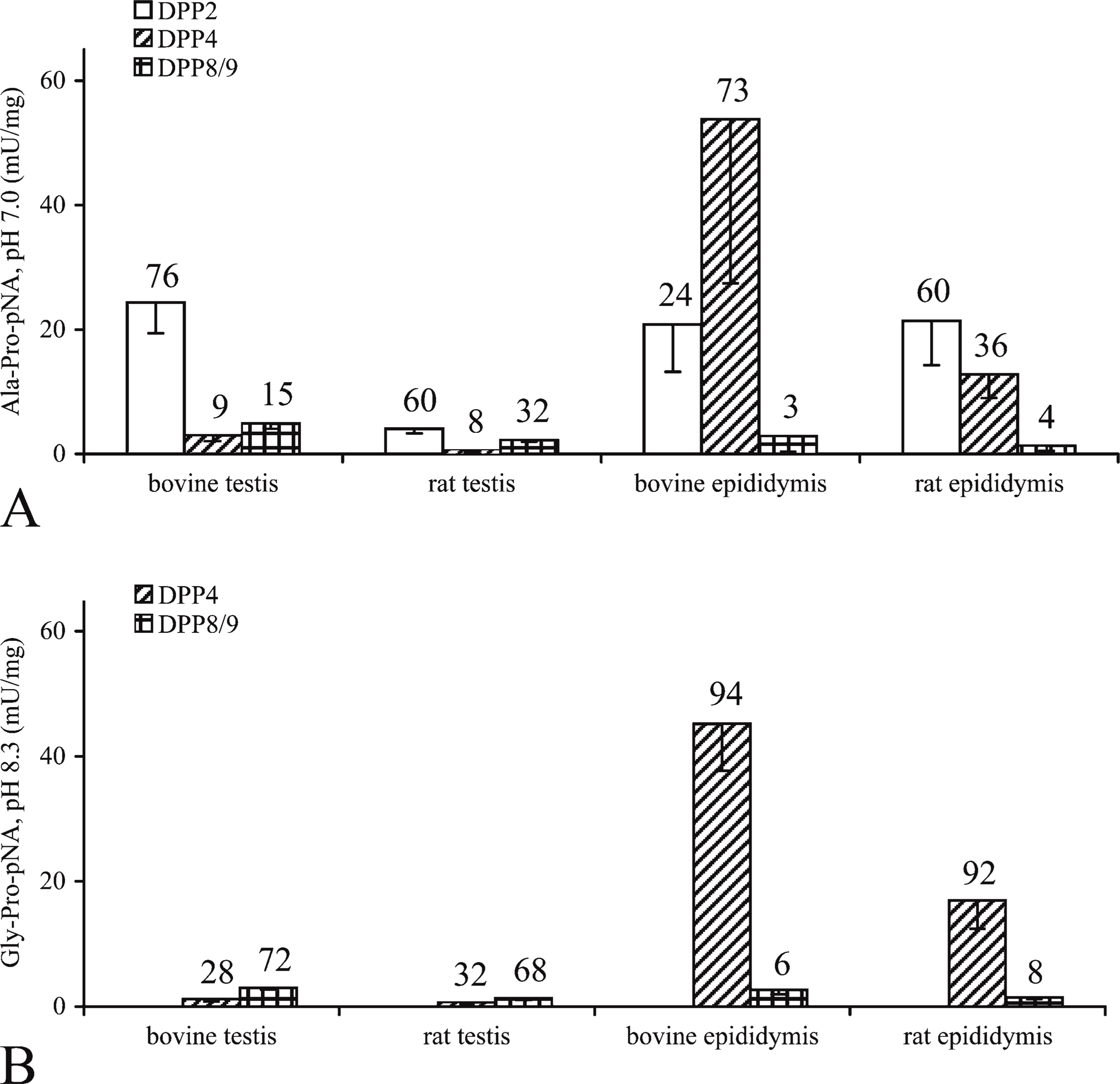
DPP-specific activity in bovine and rat male reproductive tissues. DPP activity in the tissue homogenates was calculated based on inhibition profiles using UAMC00039 for DPP2, allo-Ile-isoindoline for DPP8/9, and residual activity as DPP4. Specific activities (mU/mg) are represented as the mean ± SD of six separate experiments. Above each bar, the percentages of specific DPP activity toward the total DPP activity are represented. (
Detection of DPPs in Bovine Male Reproductive Tissue Homogenates by Immunoblotting
First, we characterized the polyclonal anti-DPP antibodies because this has not been done thoroughly before. All the anti-DPP9 antibodies recognized the native purified bovine DPP9 and DPP9 in bovine testes homogenate in a native dot spot assay. Ab42078, ab42079, and ab26177 showed some cross-reactivity with rDPP8. After SDS gel electrophoresis, the anti-DPP9 antibody preparations reacted with denatured DPP9 purified from bovine testes. All anti-DPP9 antibodies were raised against human DPP9 but cross-reacted with the bovine DPP9 enzyme. All anti-DPP8 antibody preparations reacted with rDPP8, and human DPP4 was recognized by ab28340 after SDS gel electrophoresis (Table 1).
To analyze the presence of DPPs in bovine male reproductive tissues, immunoblotting was performed. DPP9 (ab42080) could be detected in the bovine testis homogenate, whereas the expression in the bovine epididymis was too low to be observed unambiguously on Western blot (Figure 4A). DPP8 (ab42075) showed a similar expression pattern as DPP9 (Figure 4B). A faint band for the expression level of DPP4 (ab28340) was only seen in the bovine epididymis homogenate (Figure 4C).
Histochemical Localization of DPPs in Male Reproductive Tissues
The histochemical activity assay was performed on the male bovine reproductive tissues. Bovine epididymis sections incubated with UAMC00039 (Figures 5A and 5B) and with UAMC00039 and allo-Ile-isoindoline (Figures 5C and 5D) showed a strong DPP activity, pointing to a high contribution of DPP4. This was reflected in an intensely blue color. DPP4 was active in the epithelium of the epididymis, with a stronger reaction at the basal zone. On the other hand, neither the sections incubated with UAMC00039 and sitagliptin (Figures 5E and 5F) nor the sections treated with all three inhibitors (Figures 5G and 5H) showed specific staining. Therefore, DPP8/9 activity was undetectable in the bovine epididymis. In the bovine testis, no DPP activity was visible. No staining was detectable on control sections incubated in the presence of NBT while omitting the substrate (data not shown).
Most of the antibodies were tested for their applicability in IHC studies of proline-selective DPP expression in bovine and rat testis and epididymis (Table 1). For the names of the different germ cells in the seminiferous tubules, we refer to Wheater et al. (1979). Figures 6A, 6I, 6E, and 6M show an overview of the bovine testis. In the bovine testis, a specific staining for DPP9 (non-commercial antibody and ab42080) was found in spermatozoids embedded in the epithelium, just before their release into the lumen (Figures 6A–6C, arrows). In the rat testis, we saw a DPP9 specific staining (non-commercial antibody) in spermatids (Figure 7A, right arrow) and in spermatozoids (Figure 7A, left arrow) in seminiferous tubules. In the bovine testis, a specific staining for DPP8 (ab42076) was observed in spermatozoids, which were situated more deeply in the epithelium than the spermatozoids that expressed DPP9 (Figure 6H, arrows). The DPP8 staining in these “early” spermatozoids was less pronounced than the staining for DPP9 in the later stage spermatozoids. The tubule in Figures 6B–6D was in a different stage of development compared with the one in Figures 6F–6H. These images show that the expression of DPP8 and DPP9 differs in tubules at different stages of development.
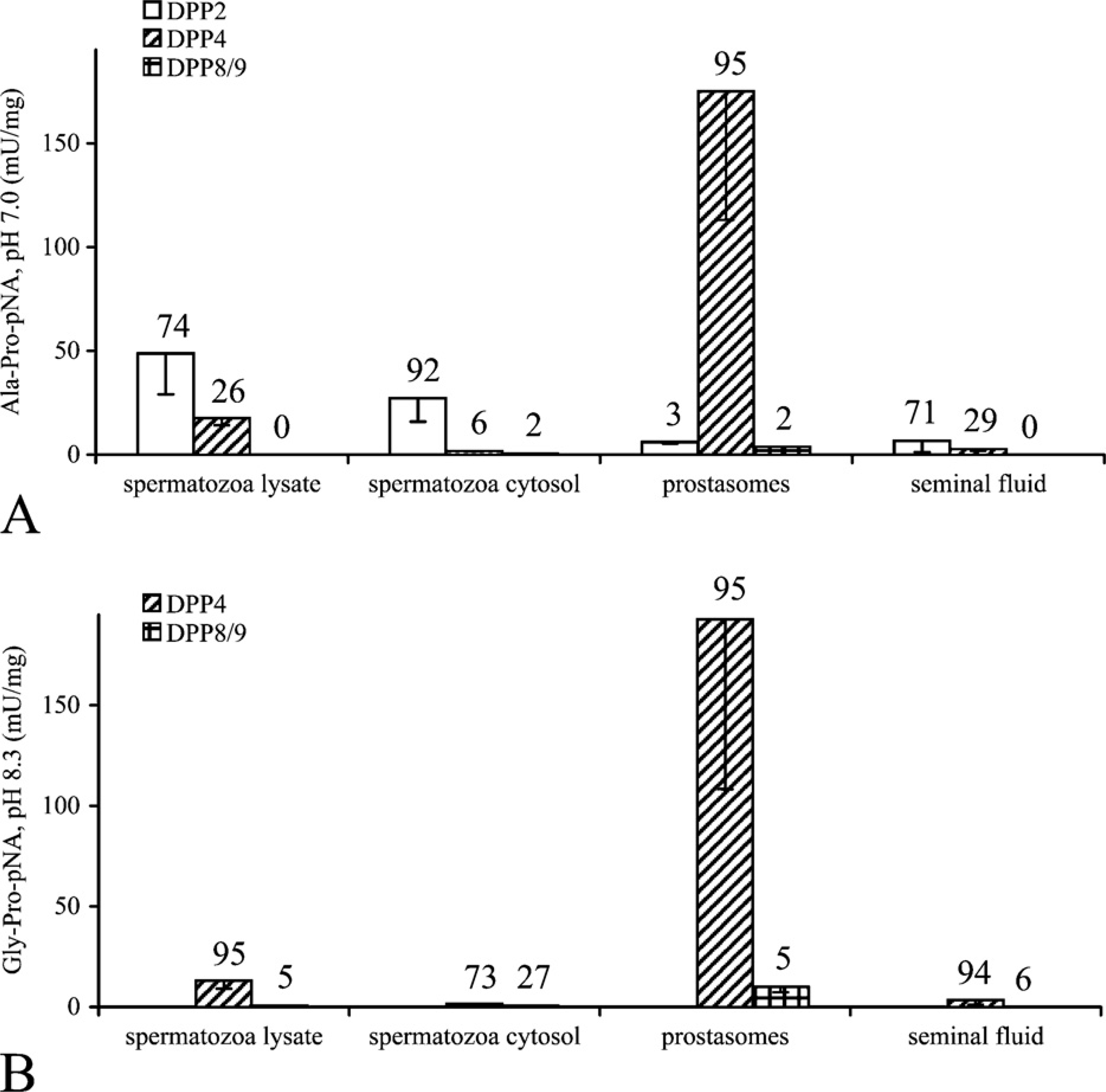
DPP-specific activity in bovine sperm. DPP activity in pooled bovine sperm preparations was calculated based on inhibition profiles using UAMC00039 for DPP2, allo-Ile-isoindoline for DPP8/9, and residual activity as DPP4. Specific activities (mU/mg) are represented as the mean ± SD of two separate experiments. Above each bar, the percentages of specific DPP activity toward the total DPP activity are represented. (
In the bovine epididymis, a DPP9-specific staining (non-commercial antibody) was found associated with cytoplasmic droplets in the lumen (Figure 7B, left arrow). They can be derived from the testis or from the epithelium of the epididymis itself. In addition, DPP9 was found associated with secreted vesicles from the epithelium (Figure 7B, right arrow). In the rat epididymis, a less pronounced staining for DPP8 (ab42077) and DPP9 was seen in the secreted vesicles (data not shown). A DPP4-specific staining (ab28340) was shown in the sperm cells and cytoplasmic droplets from the testis or from the epithelium of the epididymis in the lumen (Figure 7C, arrows). Figures 6I–6P and 7D–7F are control sections.
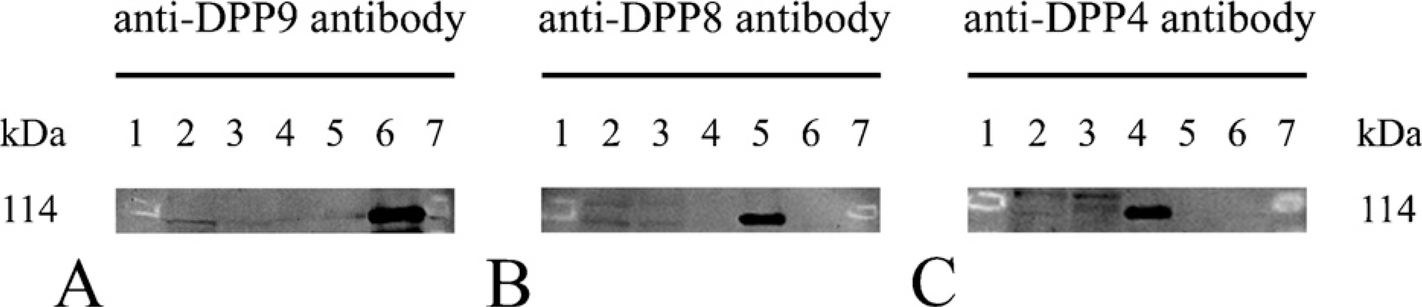
Detection of DPPs in male bovine reproductive tissues by Western blot after SDS gel electrophoresis. Samples were resolved by SDS-7.5% PAGE under reducing conditions: bovine testis homogenate (Lane 2; 40 μg), bovine epididymis homogenate (Lane 3; 40 μg), human DPP4 (Lane 4), rDPP8 (Lane 5), and bovine DPP9 (Lane 6). Molecular mass standards are shown in Lanes 1 and 7. Analysis occurred by immunoblotting with the anti-DPP9 antibody ab42080 (
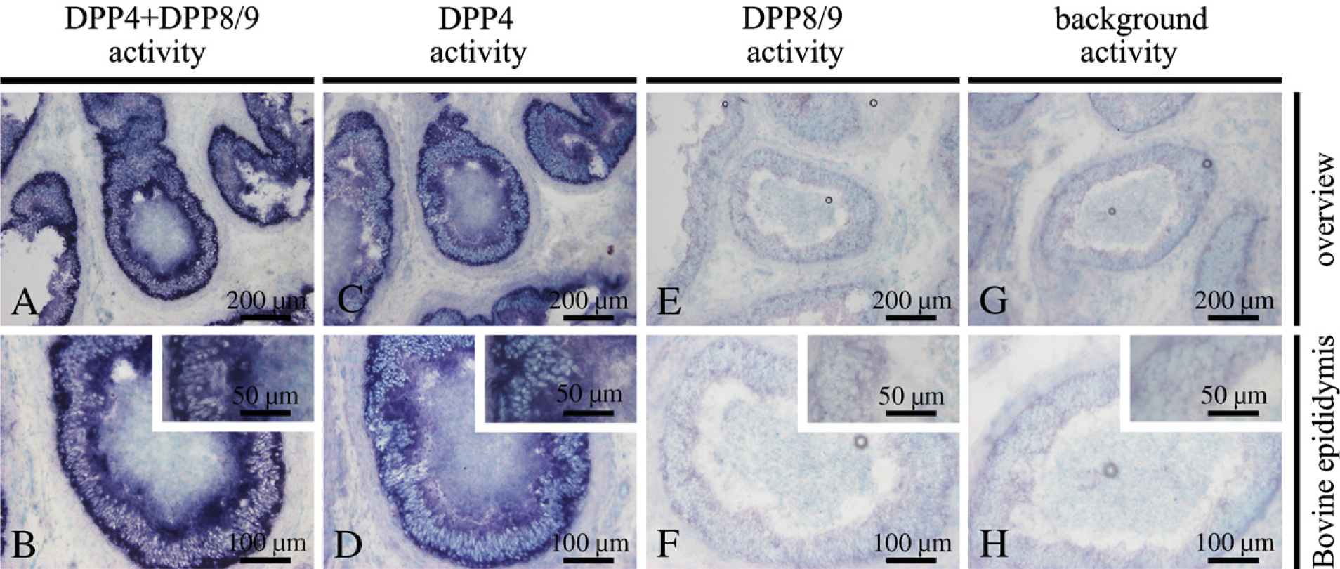
Detection of DPP activities with a histochemical activity assay in bovine epididymis. Cryosections of bovine epididymis were incubated with the substrate H-Gly-L-Pro-1-hydroxy-4-naphtylamide hydrochloride and nitro blue tetrazolium in the presence of different combinations of DPP inhibitors: UAMC00039 to detect DPP4 and DPP8/9 activity (
Discussion
In this study, using several selective DPP inhibitors and polyclonal anti-DPP antibodies, the distribution of DPP2, DPP4, DPP8, and DPP9 was documented in the testis, epididymis, and sperm of bulls and rats. For the first time, the expression and localization of DPP8 and DPP9 were studied in the male reproductive system.
Based on proline-selective DPP activities and inhibition profiles, we showed that the contribution of DPP8/9 activity was high in the bovine and rat testis in contrast to the epididymis. The testis of both species contained more DPP8/9 than DPP4 activity, but the DPP2 activity was still higher. In the bovine epididymis, a lower DPP2 activity than DPP4 activity was shown. An opposite result was seen in the rat epididymis. Vanha-Perttula (1973a,b) mentioned the presence of DPP2 and DPP4 activity in the rat testis. Agrawal and Vanha-Perttula (1986) reported a high DPP2 activity in the bovine testis and epididymis. In the bovine testis, the total DPP activity measured using Gly-Pro-β-naphtylamide was low. Considering the results of this study, most of the activity measured by Agrawal and Vanha-Perttula (1986) in the bovine testis was probably DPP8/9. In fact, all the “DPP4 activity” measurements reported in literature before 2000 might be mixtures of DPP4/8/9. In the bovine cauda epididymis, DPP4 activity was higher than DPP2 activity (Agrawal and Vanha-Perttula 1986). In the case of the epididymis, DPP4-like activity is indeed DPP4. DPP8/9 showed a very low or undetectable activity in bovine seminal plasma preparations. DPP2 activity in bovine seminal fluid and spermatozoa was higher than in prostasomes. Vanha-Perttula (1984) reported that human seminal fluid and prostasomes contained high DPP2 activity levels compared with spermatozoa. We found extremely high DPP4 activities in bovine prostasomes, as was observed in men (Vanhoof et al. 1992). In bulls, the seminal vesicles are the predominant source of DPP4 (Vanha-Perttula et al. 1990), whereas in men, the prostate gland is the predominant source (Wilson et al. 1998). This suggests a species specificity in sex accessory gland source of secretory DPP4. Taken together, these results suggest that the distribution of the DPPs varies between species.
The findings from the experiments with the selective DPP inhibitors and polyclonal anti-DPP antibodies are complementary. Native dot spots and Western blot results showed that most of the polyclonal antibodies had a high specificity. However, ab42078, ab42079, and ab26177 showed some cross-reactivity with native rDPP8. We found that DPP8 and DPP9 were detectable in the testis and DPP4 in the epididymis of bulls on Western blot, which is in agreement with the results of activity measurements mentioned previously. The reactivity of the anti-human DPP antibodies with the bovine enzymes can be explained by the high degree of sequence conservation among different species. More importantly, several preparations (the non-commercial anti-DPP9 antibody, ab42080, ab42076, ab42077, and ab28340) were suited for the analysis of DPP distribution in the male reproductive system by IHC. In the bovine testis, DPP9 was localized in spermatozoids embedded in the epithelium. In the rat testis, the spermatids also showed DPP9 staining. The small difference between both species could be explained by the fact that various stages, the number of stages, and the overall duration of the stages of the spermatid development (spermatogenesis) in the testis are species dependent. In the bull, for example, 14 stages that include round, elongating, and elongated spermatids have been identified (Berndston and Desjardins 1974). DPP8 was localized in spermatozoids in an earlier stage of spermatogenesis than the ones positive for DPP9. Although it is difficult to make a distinction between the cytoplasm and nucleus of the spermatozoids, both enzymes were most probably present in the cytoplasm. In Figures 6, 7A, and 7D, we clearly see that not all the seminiferous tubules are in the same stage of development, a phenomenon called the spermatogenic wave. Therefore, not all tubules show similar enzyme staining. In the epididymis, DPP9 was localized in cytoplasmic droplets derived from the testis or epididymis itself and in secreted vesicles originating from the epithelium. These vesicles also contained DPP8. Both enzymes were not found in the spermatozoa in the lumen of the epididymis, which is corroborated by the low DPP8/9 activity in the bovine sperm preparations. In contrast, these spermatozoa showed specific DPP4 staining. The epididymal sections incubated with the anti-DPP antibodies (non-commercial anti-DPP9 antibody, ab42077, and ab28340) showed a slightly more intense red coloring in the epithelium compared with the control. However, we could not conclude unambiguously that this staining was specific. As reported by Agrawal and Vanha-Perttula (1986), the histochemical activity assay showed DPP4 activity in the epithelium of the bovine epididymis. A strong reaction was seen in the basal part of the epithelium and a weaker reaction at the apical zone. Unfortunately, we could not detect any DPP8/9 activity in the bovine epididymis, and no staining was visible in the bovine testis. This is possibly caused by (a) insufficient sensitivity of the method for detection of DPP8/9, (b) in situ inhibition of enzyme activity, (c) inhibition of enzyme activity during the conventional histochemical procedure, or (d) the low specific activity in these tissues (<5 mU/mg; Figure 2). In a dot spot analysis, the substrate H-Gly-L-Pro-1-hydroxy-4-naphtylamide hydrochloride was more sensitive to DPP4 than to DPP8 and DPP9 (data not shown). Given that the specific DPP8/9 activity in the testis and epididymis of the rat is lower than the specific DPP8/9 activity in the corresponding bovine tissues (<3 mU/mg), the histochemical activity assay was not performed on these rat tissues. Dikov et al. (1999) mentioned DPP4-like activity in the epithelium of the rat epididymis. Our biochemical data support the conclusion that DPP4 is responsible for this activity. In general, we showed that the antibodies ab42080 and ab42075 are suitable for immunoblotting experiments by their intense staining and no cross-reactivity. The non-commercial anti-DPP9 antibody is the best antibody for IHC because it showed a specific staining in all the tissues used in this study.
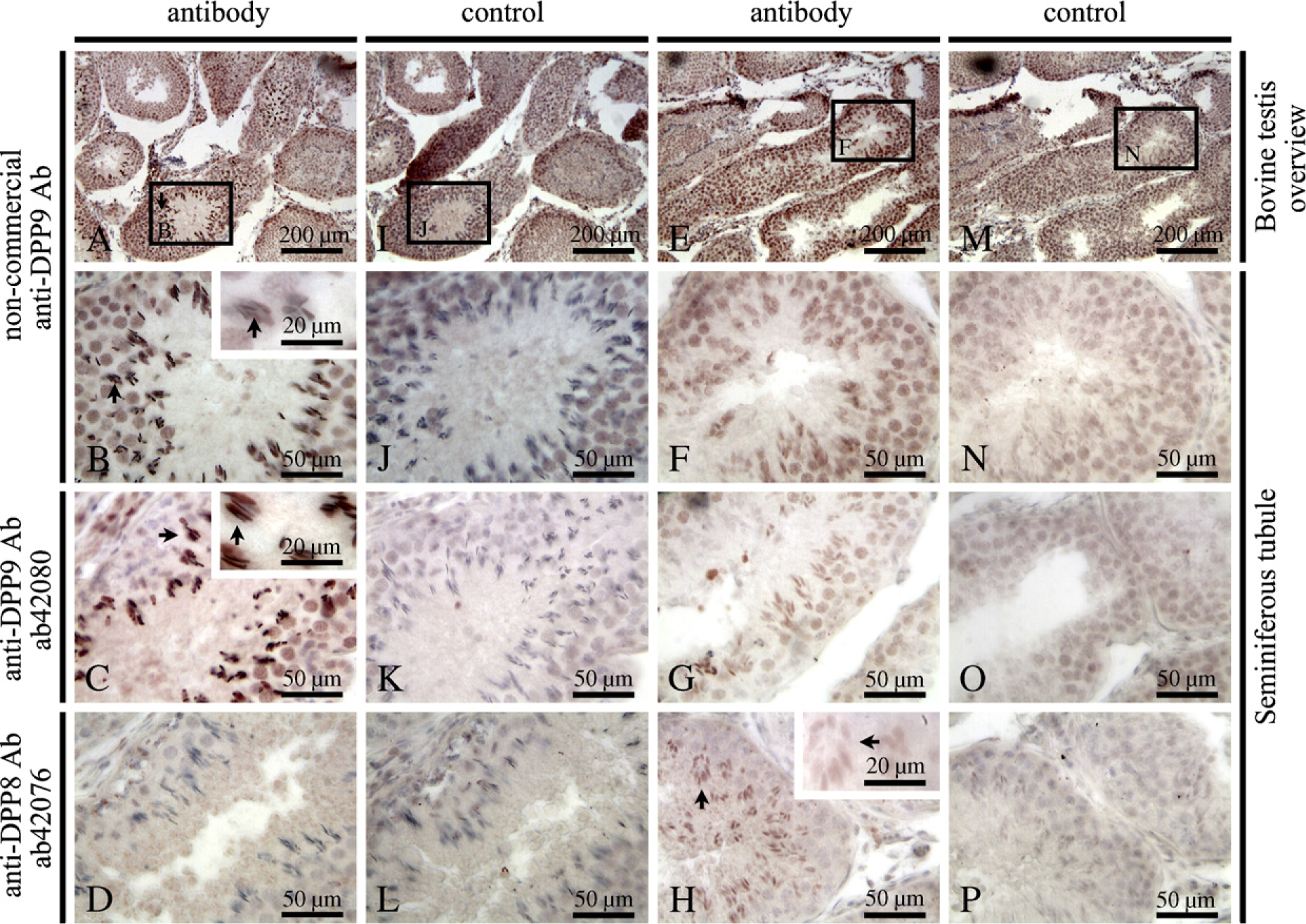
IHC localization of DPPs in bovine testis. Paraffin sections of the bovine testis were incubated with the non-commercial anti-DPP9 antibody (
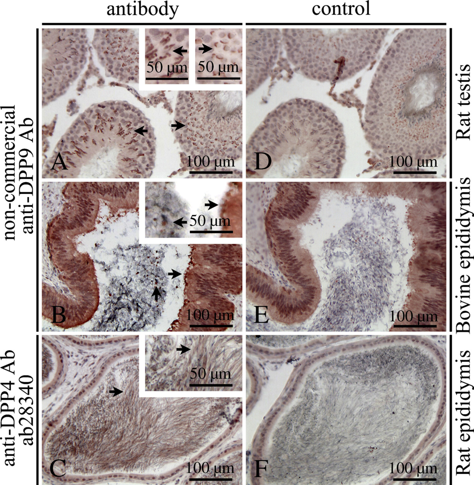
IHC localization of DPPs in male bovine and rat reproductive tissues. Paraffin sections of the rat testis, bovine epididymis, and rat epididymis are shown. Staining of rat testis (
Proteases and protease inhibitors are supposed to be involved in the events pertinent to germ cell movement in the seminiferous epithelium during spermatogenesis (Fritz et al. 1993; Le Magueresse-Battistoni 2007). Epididymal epithelial cells are engaged in absorption of material derived from the maturing spermatozoa. DPP8 and/or DPP9 may be involved in such processes. The site-specific activity and expression pattern of DPP4, DPP8, and DPP9 in the male reproductive system suggest differential functional roles during late stages of spermatogenesis. The in vivo substrates and corresponding physiological roles of the individual proline-selective peptidases are currently unknown. Apart from this, the tissue distribution pattern of DPP8 and DPP9 proteins needs to be studied further. In the near future, the adaptation of existing methods may allow visualization of DPP8/9 activity in living cells (Boonacker and Van Noorden 2001). Moreover, the ongoing development of specific inhibitors for DPP8 or DPP9 (Van der Veken et al. 2007b,2008; Van Goethem et al. 2008) will facilitate further physiological studies.
In conclusion, we showed a high expression of DPP8 and DPP9 in the bovine and rat testis at both the protein and activity levels. Both enzymes are localized in specific sperm cells of late stages of spermatogenesis. In contrast, the epididymis and sperm preparations contained low levels of DPP8 and DPP9. Our findings suggest that these proteases may contribute to male gamete development.
Footnotes
Acknowledgements
This study was supported by the Fund for Scientific Research-Flanders (Belgium; F.W.O.-Vlaanderen) and the University of Antwerp (B.O.F.). We thank Prof. P. Bols for providing the bovine sperm.