
Abstract
A carcinoid tumor was found as a solitary soft mass in the wall of the rectum adjacent to the anorectal junction in an adult Holstein cow. Microscopically, the tumor involved the submucosa and partly invaded the muscular layer. It consisted of a compact arrangement of a great number of large polygonal cells and a small number of small dark cells, some of which showed argyrophilia (Grimelius positive). Immunohistochemically, both types of tumor cells were positive for vimentin, keratin, and S-100 protein and weakly positive for neuron-specific enolase (NSE), whereas they were negative for some endocrine markers such as chromogranin A, insulin, glucagon, somatostatin, serotonin, adrenocorticotropic hormone, and calcitonin. Electron microscopy revealed membrane-bound secretory granules in the cytoplasm of some small dark cells. In such a poorly differentiated carcinoid, the morphologic characteristics of the small dark cells were strong evidence for the diagnosis. This is the first description of a poorly differentiated carcinoid developing in the rectum of a cow.
Carcinoids are rare neoplasms derived from neuroendocrine cells that are widely distributed in the gastrointestinal tract, tracheobronchial tree, genitourinary tracts, biliary tract, and pancreatic ducts of humans and animals. Gastrointestinal (GI) carcinoids in humans have been ontogenetically classified as foregut (duodenum, small intestine, appendix, and cecum), midgut (right colon) and hindgut (left colon and rectum) tumors.7 In humans, GI carcinoids compose 74% of all carcinoid tumors; the most common site of GI carcinoids is the small bowel (29%), followed by the appendix (19%), the rectum (13%), and the other sites (13%).7 GI carcinoid tumor has been reported in a cow,4 dogs,1,11,17 cats,3,12 and a horse.10 As rectal tumors in cattle, adenoma, adenocarcinoma, fibroma, and fibrosarcoma have been reported,2,16 but not carcinoid tumors. This paper is the first description of a poorly differentiated carcinoid arising in the rectum of a cow.
A Holstein cow of more than 6 years of age presented with a 2-week history of episodic bloody stool, mild anemia (erythrocyte count 4.78 × 10⁶/μl; hemoglobin 7.95 g/dl; packed cell volume, 21%) and mild leucopenia (leukocyte count 3.55 × 10³/μl). Rectal examination revealed the presence of a soft mass measuring approximately 5 cm in diameter in the mucosa of the rectum. The cow was euthanized by exsanguination after administration of xylazine hydrochloride at the owner's request.
At necropsy, a dark red, polypoid soft mass measuring 8 × 5 × 4 cm was located in the posterior rectal mucosa adjacent to the anorectal junction. Its protuberant mucosal surface was ulcerated and hemorrhagic, and the cut surface of the mass was gray-white with dark red spots and lobulated by fibrous tissue. There were no gross lesions in the other organs.
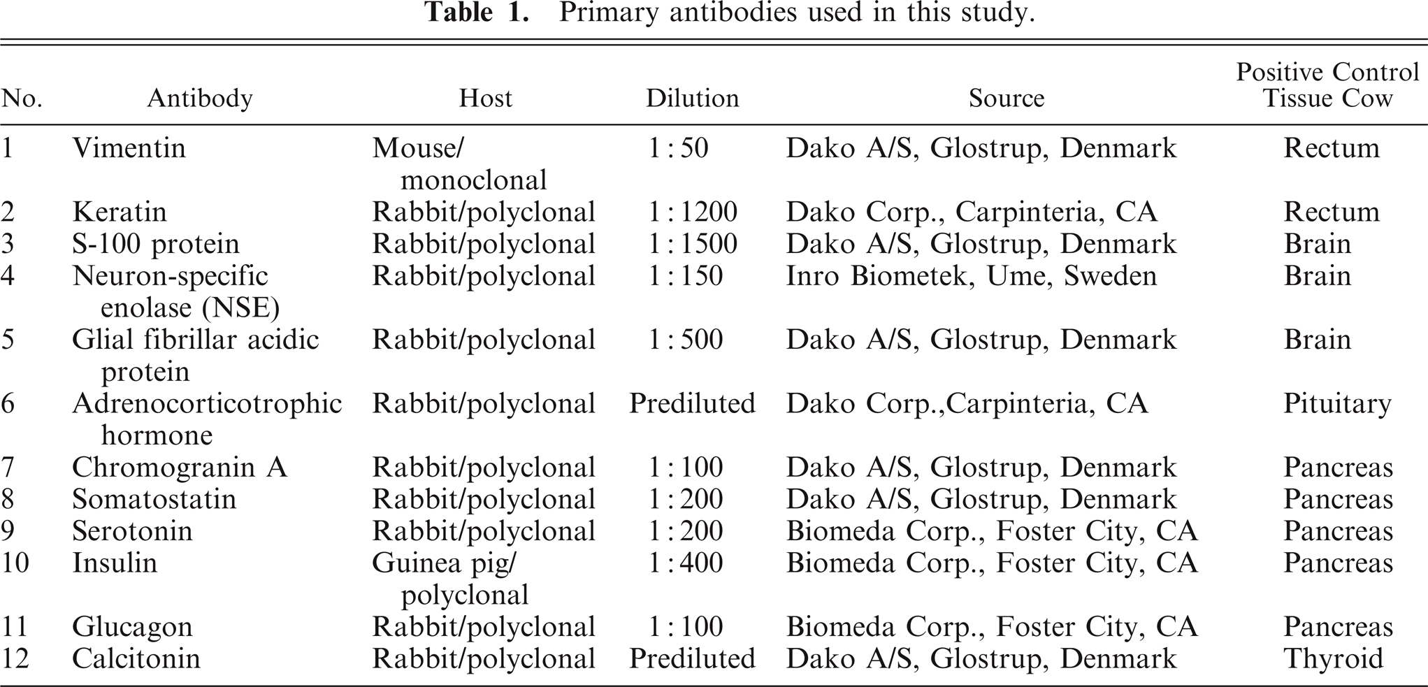
The mass was fixed in 10% neutral-buffered formalin and embedded in paraffin wax. Paraffin sections (3 μm) were stained with hematoxylin and eosin (HE), Masson's trichrome, toluidine blue, alcian blue and periodic acid-Schiff, Watanabe's silver, Fontana-Masson's argentaffin, and Grimelius' argyrophil stains. Additional serial sections were immunostained with a streptavidin–biotin complex method.18 The 12 primary antibodies used are shown in Table 1. After being reacted with primary antibodies, sections were incubated with biotinylated goat anti-rabbit IgG (Dako A/S, Glostrup, Denmark), anti-mouse IgG (Dako A/S), or anti-guinea pig IgG (Vector Lab., Burlingame, CA) antibody, followed by peroxidase-conjugated streptavidin (Dako A/S). Finally, the reaction to each antigen was visualized by addition of diaminobenzidine tetrahydrochloride chromogen and counterstained with hematoxylin. The validation of these 12 antibodies in cattle was confirmed by a positive reaction with the normal tissues listed in Table 1 or by a negative reaction on replacement with normal serum of mouse, rabbit, or guinea pig. For electron microscopic examination, small pieces of the fresh tumor tissues were fixed in 2.5% glutaraldehyde, postfixed in 1% osmium tetraoxide, and embedded in epoxy resin. Ultrathin sections were stained with uranyl acetate and lead citrate.
Primary antibodies used in this study.
Histologically, the tumor involved the submucosal layer and partly invaded the muscular layer. The mucosal surface covering the tumor was severely ulcerated. The tumor consisted of solid nests of a large number of large polygonal cells and a small number of small dark cells with thin fibrovascular stroma (Figs. 1, 2). The large polygonal cells had a hypochromatic nucleus with apparent nucleoli and abundant cytoplasm of faintly eosinophilic tint (Fig. 2). They frequently showed mitotic figures (Fig. 2). The small dark cells had an oval hyperchromatic nucleus, and some were positive with Grimelius' stain for argyrophilic granules (Figs. 2, 3). The ratio of large polygonal cells to small dark cells was approximately 9:1. Both types of tumor cell were negative with Fontana-Masson's stain for argentaffin granules. No metastatic lesions were observed in any other organs.
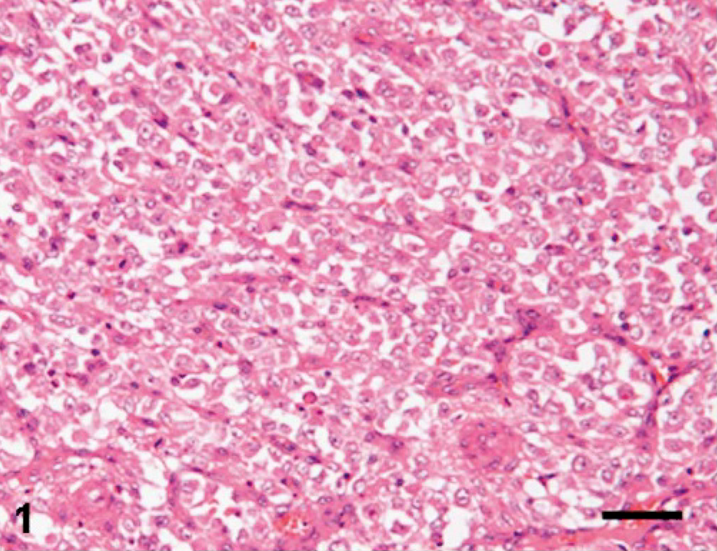
Rectum; carcinoid, cow. Compactly arranged tumor cells are divided into solid nests by delicate fibrovascular septa. HE. Bar = 200 μm.
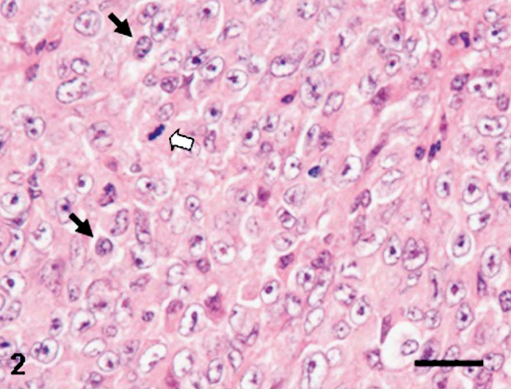
Rectum; carcinoid, cow. Higher magnification of Fig. 1. Each nest is composed of large polygonal cells admixed with small dark cells (black arrows). White arrow indicates mitotic figure. HE. Bar = 50 μm.
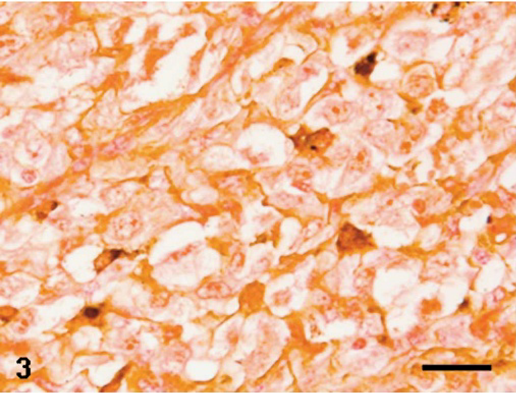
Rectum; carcinoid, cow. Small tumor cells show argyrophilia, whereas large polygonal cells are nonreactive. Grimelius' stain. Bar = 50 μm.
Immunohistochemically, both types of tumor cell were strongly positive for vimentin (Fig. 4). With keratin stain, positive reactivity was seen in almost all of the small dark cells and most of the large polygonal cells (Fig. 5). Strong staining for S-100 protein was found consistently in the small dark cells and sometimes in small clusters of the large polygonal cells (Fig. 6). Some large polygonal cells and small dark cells were weakly positive for neuron-specific enolase (NSE). The tumor cells were consistently negative for chromogranin A, insulin, glucagon, serotonin, adrenocorticotropic hormone, calcitonin, somatostatin, and glial fibrillary acidic protein.
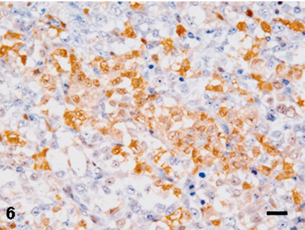
Rectum; carcinoid, cow. S-100–positive reaction is seen in dispersed tumor cells or small clusters of tumor cells. Immunohistochemistry for S-100 protein with hematoxylin counterstain. Bar = 50 μm.
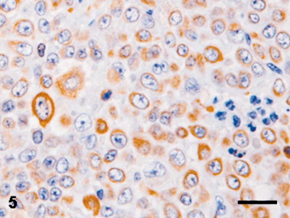
Rectum; carcinoid, cow. Almost all tumor cells are positive for keratin. Immunohistochemistry for keratin with hematoxylin counterstain. Bar = 50 μm.
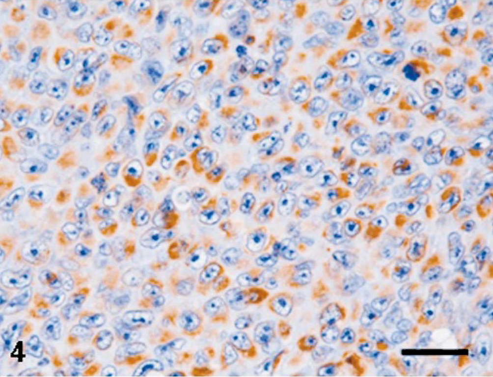
Rectum; carcinoid, cow. Almost all tumor cells are positive for vimentin. Immunohistochemistry for vimentin with hematoxylin counterstain. Bar = 50 μm.
Ultrastructurally, the large polygonal cells showed frequent nuclear irregularity, and the cytoplasm included abundant free ribosomes, some mitochondria, and rough endoplasmic reticulum (Fig. 7). The small dark cells had a round, hyperchromatic nucleolus and narrow cytoplasm rich in well-developed Golgi apparatuses and mitochondria. In addition, some contained varying amounts of round, electron-dense, membrane-bound cytoplasmic granules that measured 80–500 nm in diameter. These secretory granules had a small electron-dense core or mottled content (Fig. 7 inset). Granules were rarely observed in the large polygonal cells.
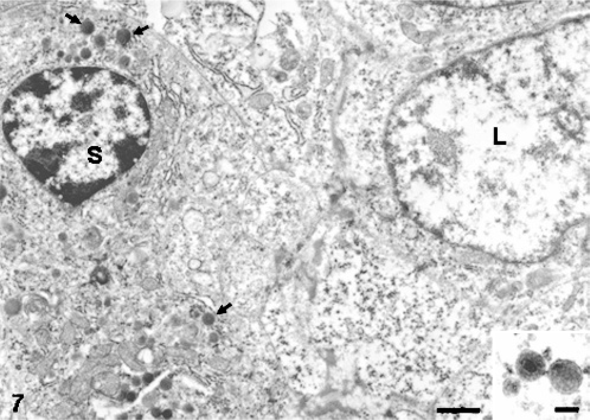
Rectum; carcinoid, cow. Electron microscopy of a small dark cell (S) and a large polygonal cell (L). Small numbers of electron-dense secretory granules (arrows) are seen in the cytoplasm of the former. Bar = 3 μm. Uranyl acetate and lead citrate stain. Inset: higher magnification of secretory granules. They have a small electron-dense core or mottled content. Bar = 500 nm.
This tumor comprised types of cells that differed from each other in their morphology. Immunohistochemistry demonstrated that both were positive for S-100 protein, NSE, vimentin, and keratin, and ultrastructurally, both types of cell had secretory granules to varying degrees, although there were very few in the large polygonal cells. Therefore, it was concluded that the types of cell were of the same cell lineage.
The large polygonal cells, the main cellular constituents of this tumor, were too poorly differentiated to easily identify their origin, although they were considered to be of neurogenic nature because of their positivity for S-100 protein and NSE. The small dark cells, which were the minor population, contained argyrophilic secretory granules characterized by electron-dense, membrane-bound profiles. The definitive diagnosis of carcinoid was made by the characteristics of the small dark cells.
Malignant carcinoid arising in the colon of a cow reported by Cho and Archibald4 was argentaffin positive, whereas in this case, it was argyrophilic. There were some differences in ultrastructural characteristics of secretory granules in these tumors; the argentaffin granules in the other bovine tumor were irregular and variable in shape,4 whereas those in our tumor had a round profile. These differences agree with the different ultrastructural features of argentaffin and argyrophil carcinoids seen in the human appendix vermiformis carcinoid.5 A review of silver impregnation of 62 human carcinoids15 suggested a correlation between its reactive characteristics and distribution: either nonreactive or argyrophil type is predominant in foregut-derived carcinoids, the argentaffin type in midgut-derived carcinoids and the nonreactive type specifically in hindgut-derived carcinoids.
Immunohistochemical results revealed coexpression of the epithelial marker cytokeratin and the mesenchymal marker vimentin in most tumor cells. This could be explained by the occasional expression of both cytokeratin and vimentin in the undifferentiated carcinoma.9,14,19 For example, in canine mammary tumors, the well-differentiated type is positive for cytokeratin but not for vimentin, whereas anaplastic carcinoma expresses not only cytokeratin but also vimentin.9 Vimentin can be considered to be a marker of undifferentiated carcinoma related to a poor clinical outcome.13 Another possible interpretation of vimentin expression could be related to the site of this tumor because the rate of vimentin-positive findings in human rectal carcinoids is approximately 5 times higher than that in other GI carcinoids.6
The biochemical contents of secretory granules in human rectal carcinoids have been reported to be multihormonal peptide and amine.8 We did not attempt to identify the biological nature of the secretory products in this tumor.