
Abstract
This report documents 2 cases of branchioblastomas in koi carp (Cyprinus carpio). Macroscopically, both cases were characterized by well-demarcated, pale red nodular masses located at the left first branchial arch and the right pseudobranch, respectively. Histologically, the neoplasias were composed of blast-like cells that differentiated into cartilage and branchial lamellae embedded in abundant fibrous connective tissue. Based on these findings, a branchioblastoma was diagnosed.
Fish, like mammals, are frequently affected by neoplastic proliferations. These neoplasms are classified according to mammalian tumor classifications. Most commonly, such skin neoplasias as papillomas, fibromas, and fibrosarcomas are diagnosed.7 Other organs are less frequently affected.7 Neoplasms of the gills are very rare, and the few published cases include papillomas, squamous cell carcinomas, fibromas, chondromas, and branchioblastomas.7,10,12,13
The term branchioblastoma refers to a neoplastic proliferation of primitive, blast-like branchial cells with the capacity to differentiate into epithelial and mesenchymal cells. Based on its macroscopic and histologic growth characteristics, this tumor is considered benign. Its locally expansive growth, however, is incompatible with normal gill function and is therefore lethal. This report documents 2 independent cases of spontaneously occurring branchioblastomas in koi carp.
The first animal was a 9-year-old male, 48-cm long, red-black-white European-bred koi. It was kept for several years in a backyard pond of 20 m3 in southern Germany with a water recirculation system including state-of-the art mechanical and biological filtration. The total stock consisted of 15 koi carp. The fish were fed a commercial pellet diet. Previous medications included formalin-malachite green, potassium permanganate, and mebendazole as antiparasitic treatments. Clinically, the koi presented with anorexia, lethargy, and respiratory problems. There were bilateral masses overlying the gills. Additionally, the koi had a mild infestation with external parasites that was not considered to be of clinical relevance. The presumptive diagnosis of a gill neoplasm with guarded prognosis was given, and because the tumor was inoperable, no further treatment was initiated. The animal died 2 months later. At necropsy, there was 1 solid, firm mass, 7 × 4 × 1 cm, originating from the base of the left first branchial arch and extending to the right branchial chamber. The mass was pale red and multilobulated. No other lesions were observed. A sample of the tumor fixed in 10% formalin was sent to the Centre for Fish and Wildlife Health (FIWI) for further analysis.
The second koi was a Japanese-bred Shiro Utsuri (black and white) 8-year-old male with a length of 67 cm. The fish was kept for 7 years in a backyard pond of 25 m3 with 20 other koi. The pond was equipped with a water recirculation and standard filtration system. All fish were fed a commercial pellet diet. Two months prior to examination, the koi in this pond were treated with praziquantel because of heavy infestation with Gyrodactylus sp. At that time, no gill alterations were observed. Prior to this, the fish had not been treated with chemicals for at least 4 years.
Two months later the animal presented with sudden inability to close its mouth. Upon clinical examination, a unilateral mass in the right gill chamber leading to a mechanical obstruction of the mouth was identified. Because of poor prognosis, the fish was euthanized and immediately necropsied. There was a multilobulated, 4.5 × 3 × 1 cm red mass originating from the pseudobranch region at the inner surface of the operculum craniolateral to the first branchial arch (Fig. 1). The entire mass was removed, fixed in 10% formalin, and sent to the FIWI for histologic examination.
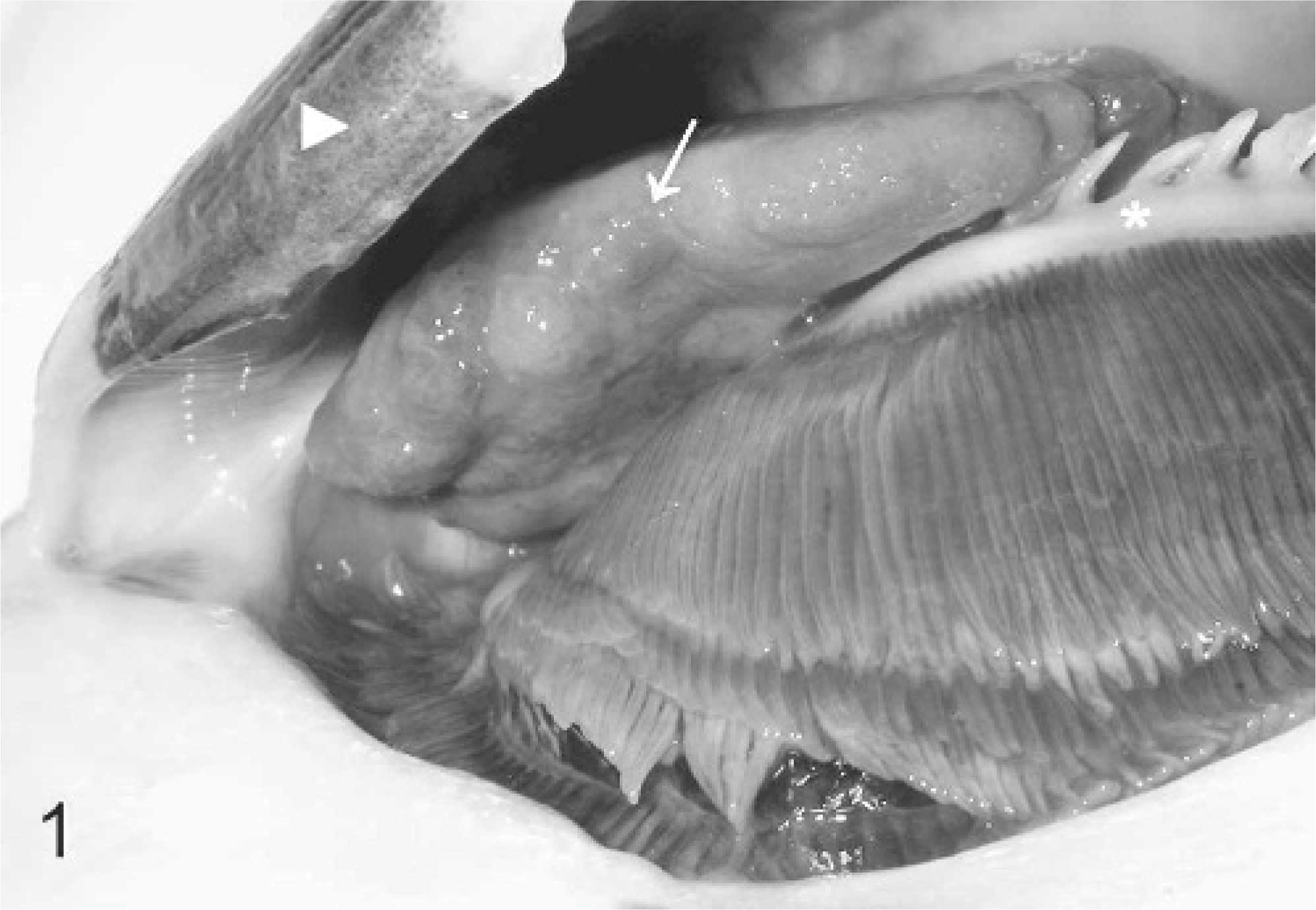
Gills; koi carp No. 2. The operculum (arrowhead) is lifted to provide a view of the inner surface where the pseudobranch is located. A unilateral large, solid, multilobulated neoplasm (arrow) originating from the area of the pseudobranch is present. Note that the first branchial arch (asterisk) is unaffected.
Samples of both masses were routinely processed for histology and stained with HE. Histologically, the neoplasms from both animals appeared similar. They were nonencapsulated but well-demarcated highly cellular neoplasms embedded in abundant fibrovascular stroma. The tumors were composed of a disorganized mixture of 3 cell populations: ill-defined densely cellular aggregates of blastemal cells together with 2 differentiated cellular elements. One part was composed of epithelial cells arranged in branches and folds resembling branchial lamellae. The other part consisted of mesenchymal cells composed of islets of cartilage and pillar cells forming vascular channels filled with erythrocytes in lamella-like structures. These elements were embedded in moderately dense fibrous tissue (Fig. 2). There were a few cystic structures partly lined by goblet cells and filled with mucoid material (Fig. 3). Multifocally, there were small areas of necrosis and a moderate to marked infiltration with lymphocytes, eosinophilic granular cells, and neutrophils within the connective tissue. The mitotic rate was low (<1/40x HPF). No signs of invasive growth were present.
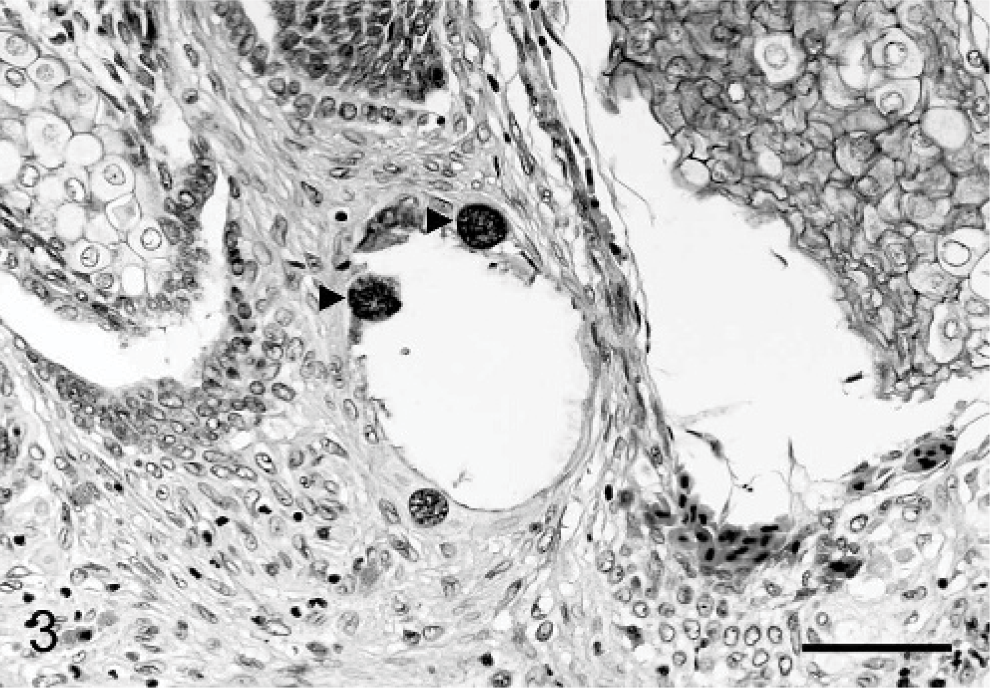
Branchioblastoma; koi carp No. 1. Within the fibrous stroma are a few cystic structures partly lined by goblet cells (arrowheads). Bar = 50 μm. Stain: HE.
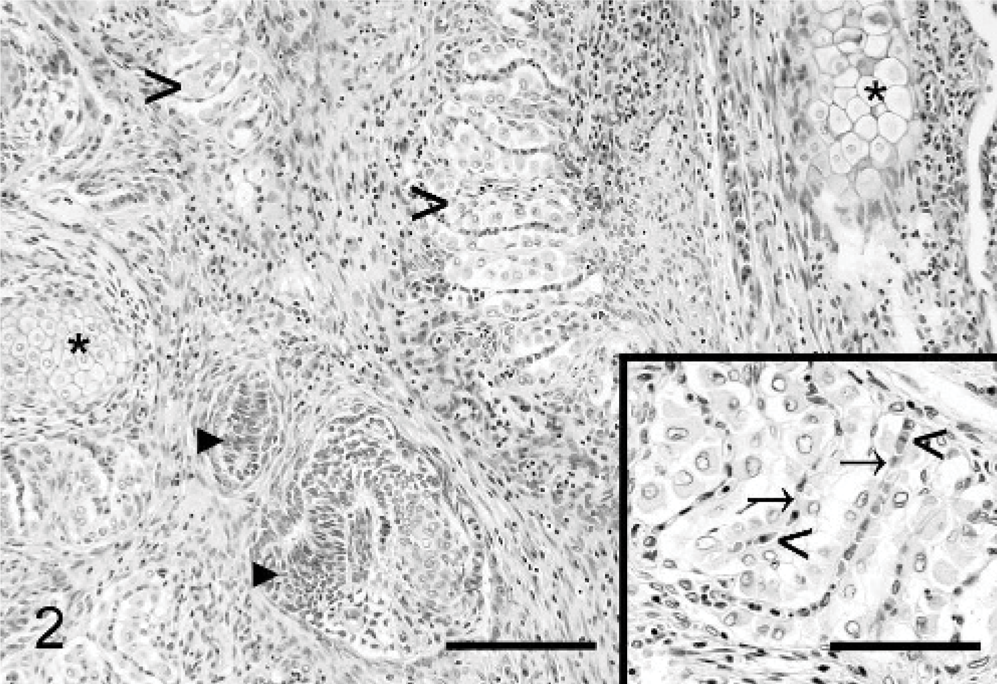
Branchioblastoma; koi carp No. 2. Aggregates of blastemal cells (arrowheads) with differentiation into islets of cartilage (asterisks) and lamella-like structures (open arrowheads). Bar = 100 μm. Inset with higher magnification of the lamella-like structures that are composed of epithelial cells and vascular channels supported by pillar cells (arrows) containing erythrocytes (open arrowheads). Bar = 50 μm. Stain: HE.
Based on these histomorphologic characteristics, both tumors were diagnosed as branchioblastomas.
This neoplasm is rarely reported in fish. Spontaneously occurring branchioblastomas have been described in rainbow trout (Oncorhynchus mykiss) and brown trout (Salmo trutta 8) and one koi carp.13 Wildgoose described a multifocal occurrence of the neoplasm in the gills with expansion and displacement of lamellae.13 In contrast, the cases described here had well-demarcated, noninfiltrating neoplastic masses originating from the branchial arch and from the pseudobranch, respectively. The pseudobranch is a remnant of the mandibular arch.1 No infiltration into the adjacent gill tissue was found, but the expansive tumor growth impaired the normal gill function and led to respiratory impairment.
In fish, neoplasms are frequently reported to be chemically inducible. Branchioblastomas can be experimentally induced by exposure to carcinogens, such as N-methyl-N'-nitro-N-nitrosoguanidine and nifurpirinol, in medaka (Oryzias latipes),3,6 sunshine bass (Morone saxatilis × Morone chrysops),11 Japanese medaka (Oryzias latipes),2,3 channel catfish (Ictalurus punctatus),3 and platyfish cross-swordtail F1 hybrids (Xiphorus maculates cross helleri).6
Formaldehyde and malachite green are known tumor inducers in mammals.4,5,9 There are, however, no reports of tumor induction by these chemicals in fish.
Previous exposure to these chemicals was reported in the first koi stock but not in the second. However, recurrent exposure is likely because they are frequently used for prophylactic antiparasitic and antifungal treatments.
It is not possible to establish a relationship between the administration of these widely used chemicals and the occurrence of tumors in these 2 fish.
Breeding and selection criteria for koi carp are mainly based on the external appearance, with less emphasis on health conditions. Therefore, genetic predispositions for the development of certain tumors are possible. In our cases, there is no indication for such a predisposition because of the different origins and breeds of the 2 fish.
Thus, the single occurrence of tumors in these 2 unrelated and separately maintained populations of koi suggests spontaneous development of the neoplasms.