
Abstract
Venezuelan equine encephalitis (VEE) viruses cause natural outbreaks in humans and horses and represent a significant biothreat agent. The effect of tunicamycin on the course of the disease in mice with VEE was investigated, and the combined effects of these agents was characterized. CD-1 mice given 2.5 μg of tunicamycin had > 1,000-fold more virus in the brain 48 hours after infection with the virulent VEE strain V3000 and >100-fold of the attenuated strain V3034 at all tested times than did untreated mice, indicating enhanced neuroinvasion. Tunicamycin did not alter the viremia profiles of these viruses nor the replication of V3000 in the brain itself. Tunicamycin alone caused ultrastructural blood-brain barrier damage, yet neuroinvasion by V3000 in treated mice appeared to occur via the olfactory system rather than the blood-brain barrier. Tunicamycin-treated, V3000-infected mice also exhibited earlier and more severe weight loss, neurological signs, neuronal infection, neuronal necrosis and apoptosis, and inflammation than untreated, V3000-infected mice. The mean survival time of tunicamycin-treated, V3000-infected mice was 7.3 days versus 9.9 days for untreated, V3000-infected mice. These studies imply that animals that ingest toxins similar to tunicamycin, including the agent of annual ryegrass toxicity in livestock, are conceivably at greater risk from infections by encephalitis viruses and that humans and horses exposed to agents acting similar to tunicamycin may be more susceptible to encephalitis caused by VEE viruses. The exact mechanism of tunicamycin-enhanced neuroinvasion by VEE viruses requires further study.
Venezuelan equine encephalitis (VEE) virus is a mosquito-borne alphavirus that causes major outbreaks of febrile disease and encephalitis among equines and humans in the western hemisphere. Although the virus is found primarily in tropical South and Central America, outbreaks of VEE have extended as far north as Texas in the United States, and nonepidemic strains of VEE virus circulate in portions of southern Florida, producing occasional disease. 15, 29 VEE virus is also a significant concern as a biothreat agent. 23
As with other encephalitis viruses, such as West Nile and St. Louis encephalitis, VEE virus infections typically result in demonstrable invasion of the brain only in a limited number of human cases. 15 Differences in host barriers to neuroinvasion that exist among individuals may help to explain some differences in susceptibility to viral encephalitides. Several studies in recent years have shown that a variety of internal and external factors may alter host barriers to neuroinvasion and facilitate the development of viral encephalitis. For example, lipopolysaccharide can induce structural and functional changes in the blood–brain barrier (BBB) of mice treated experimentally 18, 30, 31 and can enhance viral neuroinvasion and the severity of encephalitis caused by the West Nile virus and Sindbis virus. 18 In addition, tumor necrosis factor α (TNF-α) has been associated with enhanced neuroinvasion by human immunodeficiency virus-1, 32 and neuroinvasion by dengue virus has been attributed to the effects of a virus-induced cytokine called cytotoxic factor. 3 Even chemical agents like pyridostigmine, other acetylcholinesterase inhibitors, and cocaine have been shown to increase viral penetration of the central nervous system. 10, 32
Tunicamycin (TM) is one of a group of antibiotics produced by several species of Streptomyces, 25 and similar toxins are produced by species of Clavibacter. 7 Our laboratory previously showed that TM caused enhanced mortality attributable to Semliki forest virus and encephalomyocarditis virus, which correlated with increased levels of these viruses in the brain. 19 TM is widely used in cell biology for its ability to inhibit N-linked glycosylation of asparagine residues on proteins. Its toxic properties are biologically indistinguishable from those of the corynetoxin responsible for the neurologic condition of livestock in Australia and South Africa known as annual ryegrass toxicity, 6, 13, 21 so TM is now used to study this condition. 7, 9 The underlying impairment in the brain caused by TM appears to be microvascular damage, 7 leading to neuronal injury. TM has also been shown to alter the BBB. 8 In the present study, it is shown that treatment of mice with a low dose of TM enhanced the process of neuroinvasion by virulent and attenuated VEE viruses. Enhanced neuroinvasion caused by TM administration was accompanied by greater clinical and pathologic changes in mice infected with virulent VEE virus and more rapid death from viral encephalitis. These results provide additional evidence that exposure to chemical agents may increase susceptibility to encephalitic viruses. TM could serve as a useful tool in studies aimed at understanding the mechanisms of neuroinvasion by VEE virus and other encephalitic viruses.
Materials and Methods
Mice
Six- to 10-week-old male and female CD-1 mice (Charles River Laboratories, Wilmington, MA) were obtained and used for all in vivo experiments. Mice were housed in cages equipped with microisolators and were provided food and water ad libitum. For the portions of the study involving live VEE virus, mice were housed in a biosafety level 3 (BSL-3) facility accredited by the American Association for Accreditation of Laboratory Animal Care. In conducting research with mice, the investigators adhered to the Guide for the Care and Use of Laboratory Animals (Committee on Care and Use of Laboratory Animals of the Institute of Laboratory Animal Resources, National Research Council, National Institutes of Health Publication No. 86–23, revised 1996).
Viruses and chemical agents
Experiments used the virulent VEE virus strain V3000 and the attenuated strain V3034. 4 This attenuated strain was chosen because it results in limited or no detectable neuroinvasion in mice after peripheral infection, as previously reported. 11 Preparations of VEE virus stocks and virus titrations were also performed as reported. 11 Briefly, virus working solutions were prepared by dilution of virus stocks in 1× phosphate-buffered saline (PBS) containing Ca2+ and Mg2+ supplemented with 0.1% bovine serum. Stock solutions of TM (Calbiochem, San Diego, CA) were prepared by dissolution in dimethylsulfoxide, and working solutions were prepared by further dissolution in sterile PBS.
Mortality study
In the VEE-TM mortality experiment, mice were treated by intraperitoneal injection of 250 μl of TM (5 μg/mouse) 6–12 hours before infection. Mice were then inoculated with 100–1,000 plaque-forming units (pfu; lethal dose) of the virulent VEE virus V3000 by footpad injection of 25 μl of virus stock diluted in sterile PBS. Control mice were treated with PBS before infection. Mice were observed daily, and the numbers of live and dead mice were counted and recorded. The percentage mortality and mean survival time ± standard error of the mean (MST ± SEM) were calculated after an observation period of 14 days.
VEE pathogenesis studies
Multiple experiments were performed to investigate further how TM might alter the pathogenesis of VEE in mice. The first experiment measured the effect of TM administration on key steps in the pathogenesis of lethal VEE. Mice were treated with 2.5 μg TM (TM+) or diluent (TM-). This sublethal dosage was chosen based on a previous experiment that indicated the lethal dose of TM in adult female Swiss mice to be 10 μg per mouse 19 ; in that study, mice treated with TM at 5.0 μg or less exhibited no abnormal physical or pathologic changes. Twelve hours after TM treatment, mice were infected with 25 μl containing 1,000 pfu of V3000 (VEE+) in the footpad. Uninfected control (VEE-) mice were administered virus diluent. All mice were observed daily for clinical signs of disease. Twelve mice were killed at each of the following time points: 6, 12, 24, 48, 72, 96, and 120 hours postinfection (PI). Each group of 12 mice included the following experimental manipulations: VEE+/TM+ (n = 4), VEE+/TM- (n = 4), VEE-/TM+ (n = 2), and VEE-/TM- (n = 2). At each time point, mice were deeply anesthetized with methoxyfluorane and killed by cervical dislocation. Mice were weighed (except the 12-hour group), and necropsies were performed immediately. The axillary vessels were transected, and samples of blood were obtained. Each brain was removed and hemisectioned. One half of each brain was fixed in 10% neutral-buffered formalin, and the remaining half was processed for virus titration. Fixed brains were processed for histologic analysis and immunohistochemistry. Additional mice were treated identically, and the brains were processed for virus titration only.
The second pathogenesis experiment was designed to determine the effect of TM on the replication of virulent VEE virus in the brain. For this experiment, mice were treated with 2.5-μg TM or diluent 24 hours before intracranial infection. This timing was chosen to approximate the effect of TM at the time of peak viremia in the first pathogenesis experiment, i.e., when the virus conceivably had invaded the brain and was beginning to replicate there, based on previous studies. 2 TM-treated and diluent-control mice were infected with 25 μl containing 1,000 pfu of diluted V3000 virus injected into the left cerebral hemisphere. Three VEE+/TM+ and 3 VEE+/TM- mice were killed at 2, 12, 24, 48, and 72 hours PI. Immediately after death, the right half of each brain was removed and processed for virus titration.
The third pathogenesis experiment measured the effect of TM on neuroinvasion by the attenuated strain V3034. Mice were treated with TM or diluent as in the first pathogenesis experiment and infected with 25 μl containing 1,000 pfu of V3034 in the footpad. Four V3034+/TM+ and 4–8 V3034+/TM- mice were killed at 48, 72, and 96 hours PI. Serum and brain samples were harvested at necropsy for virus titration as previously described.
Virus titrations
For determination of VEE virus titers, brain samples were homogenized with sterile pestles in volumes of PBS containing Ca2+/Mg2+ and 0.1% bovine serum to produce 20% (weight/volume) suspensions. Blood samples were allowed to clot on ice. Clotted blood and brain suspensions were centrifuged for 5 minutes using a benchtop centrifuge, and sera and brain supernatants were removed. All samples were stored at -70 to -80°C until virus titration. Titration of virus was performed by standard plaque assay using baby hamster kidney-21 cells as previously described. 11
Histopathology, immunohistochemistry, and electron microscopy
After formalin fixation, tissues were processed routinely and embedded in paraffin blocks. Five-micrometer histologic sections were prepared, mounted on glass slides, and stained with hematoxylin and eosin (HE). Duplicate sections of selected tissues were mounted on silane-coated slides (Sigma Diagnostics, St. Louis, MO) and stained for VEE virus antigen by immunohistochemistry using an immunoperoxidase method (Envision System, DAKO, Carpinteria, CA) according to the manufacturer's recommendations. Briefly, sections were deparaffinized and rehydrated to dH20, then placed in citrate buffer (2.1 g/l, pH 6.0) at 97°C for 30 minutes for antigen retrieval. Sections were then washed in dH20 and blocked for endogenous peroxidase. Sections were incubated with the primary antibody, a rabbit polyclonal antiserum raised against VEE virus, eastern equine encephalitis virus, western equine encephalitis virus, and Sindbis virus (provided by Cindy Rossi and Dr. George Ludwig, US Army Medical Research Institute of Infectious Diseases) diluted 1°5,000, for 30 minutes at room temperature (RT). After washing with 3 changes of PBS, sections were incubated with peroxidase-labeled antibody against rabbit immunoglobulin (DAKO kit) for 30 minutes at RT. Color was developed by incubation in a solution containing 3,3′-diaminobenzidine and H2O2 (DAKO kit) for 7 minutes at RT. Additional sections were treated with nonimmune rabbit serum as negative controls.
For electron microscopy, mice were treated with 2.5 μg of TM as described previously. After 24 hours, the treated and untreated control mice were deeply anesthetized and killed by cervical dislocation. Brains were removed immediately after death and 1-mm cubes of pyriform cortex, neocortex, and thalamus were placed in universal fixative (4% paraformaldehyde, 1% glutaraldehyde) for 2 hours at RT. Following fixation, all samples were washed 3 times in 0.1-M Millonig's phosphate buffer (pH 7.4) and processed for standard transmission electron microscopy. Briefly, tissues were postfixed in 1% osmium tetroxide, dehydrated in ethanol and propylene oxide, and embedded in EMbed-812 resin (Electron Microscopy Sciences, Warrington PA). Ultrathin sections were cut, placed on 200-mesh copper grids, stained with uranyl acetate and lead citrate, and examined with a Phillips CM100 transmission electron microscope. Images were photographed with an AMT side-mounted, Kodak-Megaplus camera.
Statistical analysis
Data were analyzed using the commercially available software program SPSS for Windows, version 10.0 (SPSS Inc., Chicago, IL). In the mortality studies, data for percentage mortality and MST were compared between infected mice treated with TM and infected untreated mice using the t-test for independent samples. In the pathogenesis studies, data obtained for virus titers were compared for TM-treated and untreated mice at equivalent time points, also using the t-test. Brain virus titers were also compared for treated and untreated groups of mice at multiple time points by multiple regression analysis. Mouse body weights were compared for all 4 experimental groups in the V3000 pathogenesis experiment by one-way analysis of variance (ANOVA) using the Tukey post-test. Statistical significance was established at P <.05 a priori. All analyses were 2-tailed tests.
Results
In the initial mortality study, a single dose of TM administered 6–12 hours before infection significantly shortened the survival time of mice infected with virulent VEE virus in the footpad (Table 1), although all mice infected with V3000 died. Subsequent to the mortality study, additional experiments were performed to better characterize the effect of TM administration on VEE in mice.
Effect of TM on the survival time of mice infected with virulent VEE virus.
MST = mean survival time in days; SEM = standard error of the mean.
The reduced mean survival time was statistically significant compared to the untreated (TM−/VEE+) control group (P < 0.05).
First, a serial sacrifice study was performed and several virologic and pathologic parameters were measured. After footpad infection with V3000, mice treated with TM (TM+/VEE+) developed clinical signs sooner than untreated mice (TM-/VEE+). These signs were manifest as progressive weight loss (Fig. 1), lethargy, huddling, and dehydration first observed at 72 hours PI in the TM+/VEE+ mice and at 96 hours PI in the TM-/VEE+ mice. Infected mice in both groups later developed additional clinical signs, including spastic tremors and hindlimb paresis or paralysis. Three of the 4 TM+/VEE+ mice died between 96 and 120 hours PI, before termination of this experiment. The only clinical change evident in uninfected mice treated with TM (TM+/VEE-) was somewhat less rapid weight gain than uninfected, untreated mice (TM-/VEE-). This difference was not statistically significant, possibly because of the low numbers of mice in these 2 experimental groups.
Body weights in TM-treated and untreated mice infected with virulent VEE virus. TM-treated (TM+) or untreated (TM-) CD-1 mice were infected with 1,000 pfu of the virulent V3000 virus (VEE+) in the footpad. Mice were sacrificed (4 per group) and weighed immediately after death at the indicated times PI. TM-treated and untreated uninfected (VEE-) mice (2 per group) were also included. The mean weights for TM+/VEE+ mice were significantly different than those in the corresponding TM-/VEE+ mice at the indicated times (∗). The mean weights of the TM+/VEE- mice were not significantly different than those of the TM-/VEE- mice at all times.
TM-treated and untreated mice had nearly identical levels of VEE virus in the blood following footpad infection with V3000 (Fig. 2). Differences in the virus levels in the brain, however, were apparent between the 2 groups (Fig. 3). The geometric mean virus titer in the brains of TM-treated mice was much higher than in untreated mice at 48 hours PI and at later time points until 120 hours PI. Only 1 of 6 untreated and 2 of 5 TM-treated mice had detectable virus titers at 24 hours PI. No mice had detectable virus before 24 hours (data not shown).
Mean serum titers of TM-treated (solid lines) and untreated (dashed lines) mice infected with 1,000 pfu of virulent VEE virus (V3000) in the footpad and sacrificed at the indicated times PI. In the TM-treated group, only 1 of 4 mice remained alive at 120 hours PI.
Mean brain titers of virulent VEE virus in TM-treated and untreated mice. TM-treated (solid lines) and untreated (dashed lines) mice were infected with 1,000 pfu of V3000 in the footpad and sacrificed at the indicated times PI. The mean brain titer of TM-treated mice was significantly greater than that of untreated mice at 48 hours (∗). Detectable brain titers were not seen before 24 hours PI. In the TM-treated group, only 1 of 4 mice remained alive at 120 hours PI.
By immunohistochemistry, VEE virus antigen was initially detectable in the brains of 3 of 4 TM-treated mice at 24 hours PI. At this time, antigen was present primarily in the olfactory bulbs and olfactory-associated regions of the brain proper, such as olfactory nuclei and the lateral olfactory tracts. By 48 hours PI, at least a few antigen-positive cells were present in all regions of the brain, although the olfactory regions and the thalamus contained the most abundant amounts of antigen. In untreated mice, virus antigen was not detectable in the brain until 48 hours PI. Virus antigen also first appeared in the olfactory structures of these mice and spread to all regions of the brain by 72 hours PI. The amounts of antigen accumulated progressively throughout the time course of the study in both TM-treated and untreated mice. At any given time point, the amount of antigen present in a particular region of the brain was demonstrably greater in TM-treated mice (Fig. 4) than in untreated mice (Fig. 5).
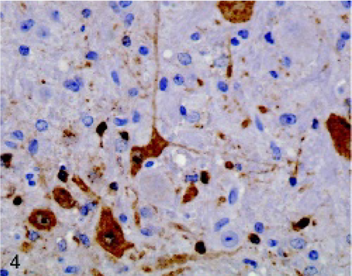
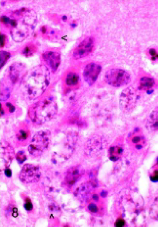
Brain; mouse No. 69. Typical of TM-treated mice 96 hours after infection with virulent VEE virus, virus antigen is abundant in the dorsal medulla oblongata, including within large neurons and small cells, which could be degenerate neurons or glial cells. Immunoperoxidase method using rabbit polyclonal antibody to multiple alphaviruses; hematoxylin counterstain.
Brain; mouse No. 65. Typical of untreated mice 96 hours after infection with virulent VEE virus, virus antigen is limited in the dorsal medulla oblongata (compare to Fig. 4). Immunoperoxidase method using rabbit polyclonal antibody to multiple alphaviruses; hematoxylin counterstain.
There were no differences evident in the pattern of virus spread through the brains of TM-treated versus untreated mice. In effect, the immunohistochemistry results indicated that in both groups of mice, VEE virus invaded the brain via the olfactory apparatus and spread via olfactory circuits to all parts of the brain in a fashion consistent with previous reports. 24, 27 Antigen was not evident within endothelial cells, and there was no perivascular pattern of antigen deposition in either TM-treated or untreated mice to indicate that VEE virus entered the brain directly from the bloodstream (i.e., through the BBB). In both TM-treated and untreated mice, VEE virus antigen was present predominantly in cells identifiable as neurons. Fewer antigen-positive cells appeared to be degenerate neurons or possibly glial cells.
The histopathologic findings in mice infected with virulent VEE virus were also consistent with those of previous reports. 24, 27 Inflammatory cell infiltrates and neuronal cell death were evident in the brains of TM-treated mice at 48 hours PI and in untreated mice at 72 hours PI. Similar to the appearance of virus antigen, inflammation and neuronal damage were essentially limited to the olfactory portions of the brain at the earliest time points. At later times, these changes appeared in other regions of the brain in a pattern that paralleled that of viral spread. The appearance of inflammation and neuronal damage generally lagged behind the appearance of virus in particular regions of the brain by about 24 hours. Also similar to the immunohistochemistry findings, at particular time points, inflammation and neuronal damage were significantly more severe in TM-treated mice (Fig. 6) than in untreated mice (Fig. 7).
Brain; mouse No. 72. Neuronal necrosis in the frontal cortex of a TM-treated mouse 96 hours after infection with virulent VEE virus. Note the numerous large neurons that maintain cell borders, have hypereosinophilic cytoplasm, and nuclei undergoing karyolysis (arrowheads). HE stain.
Brain; mouse No. 66. The frontal cortex of an untreated mouse 96 hours after infection with virulent VEE virus exhibits numerous morphologically normal neurons. Note the presence of 2 unidentifiable dead cells (arrowheads). HE stain.
The earliest inflammatory lesions were characterized by margination of leukocytes and a few perivascular cuffs that were either incomplete or were composed of a single layer of primarily lymphocytes and some neutrophils and macrophages. More advanced inflammatory lesions had more numerous perivascular cuffs of greater thickness, and there was also infiltration of inflammatory cells into the surrounding neuropil. Gliosis, characterized by the presence of activated microglial cells and activated astrocytes, was also present in more advanced inflammatory foci. Histologically, neuronal damage appeared to be manifest either as necrosis or apoptosis, depending on the type of infected neuron. This characterization was based on the morphologic features usually ascribed to each form of cell death. In areas such as the pyramidal layer of neurons in the pyriform cortex, a consistent early site of infection in the brain, neurons were typically observed to maintain cell borders and had bright eosinophilic cytoplasm and nuclei undergoing karyolysis (i.e., features typical of necrosis). Similar features were characteristic of Purkinje neurons in the cerebellum, large neurons in the frontal cortex (Fig. 6), and pyramidal neurons of the hippocampus. In contrast, damaged granule-type neurons in the dentate gyrus of the hippocampus (Fig. 8) and cerebellum had features of apoptosis. Affected cells were usually rounded and shrunken and contained nuclei with marginated chromatin or had condensed nuclei or nuclear fragments. These areas often contained apoptotic bodies. In the brains of TM-/VEE- and TM+/VEE- mice, neuronal damage, inflammation, and other histologic changes were absent at all time points.
Brain; mouse No. 60. Neuronal apoptosis in the dentate gyrus of a TM-treated mouse 72 hours after infection with virulent VEE virus. Note several granule-type neurons that are rounded and contain dark, clumped nuclear remnants and some apparent apoptotic bodies (arrowheads). Compare to necrotic large neurons in Fig. 8. HE stain.
To assess the possible effect of TM on replication or spread of virulent VEE virus in the brain, virus titers were measured at multiple time points following intracranial inoculation of TM-treated and untreated mice. In both groups, V3000 was detectable sporadically in the hemisphere contralateral to the one into which the virus was inoculated at 12–24 hours PI (Fig. 9). At subsequent time points, the virus was present consistently in the contralateral brain hemispheres of all mice and the virus kinetics were very similar in TM-treated and untreated mice, peaking at 96 hours PI in both groups.
Mean brain titers of virulent VEE virus in TM-treated and untreated mice (intracranial inoculation). TM-treated (solid lines) and untreated (dashed lines) mice inoculated with V3000 in the left cerebral hemisphere show very similar virus replication profiles in the right cerebral hemisphere throughout the time course of the study. Mice were treated with TM 24 hours before inoculation of 1,000 pfu of V3000. Only 2 of 3 mice in each group remained alive at 120 hours PI.
The results of the preceding studies provided evidence that TM specifically enhanced neuroinvasion, but not the development of viremia nor the replication of virulent VEE virus in the brain per se. To support this notion, the effects of TM on neuroinvasion by the attenuated VEE virus V3034, a molecularly-cloned strain previously shown to exhibit limited neuroinvasion, 11 were tested. After footpad inoculation of V3034, neuroinvasion was evident by plaque assay of brain samples at 48 hours in all mice treated with TM, compared to none of the untreated mice killed at this time (Fig. 10). At 72 and 96 hours PI, the mean titers of the TM-treated mice were more than 100-fold greater than those of untreated mice. V3034 infection consistently produced viremia in both treated and untreated mice at 12 and 24 hours PI. The numbers of viremic mice and the levels of virus in the blood of TM-treated and untreated mice were very similar (data not shown), as was also the case with V3000.
Mean brain titers of attenuated VEE virus in TM-treated and untreated mice. TM-treated (solid lines) and untreated (dashed lines) mice were infected with 1,000 pfu of the attenuated VEE virus strain V3034 in the footpad and sacrificed at the indicated times PI. The mean brain titers of TM-treated mice were significantly greater than those of untreated mice over the timecourse of the study.
Because TM is known to alter the BBB, 8 and because TM-induced damage to the BBB could provide a plausible explanation for some of the key findings already described, the brains of mice treated with a low dose of TM were studied by electron microscopy to detect subtle BBB changes. Capillary damage was evident in the brains of 2 mice 24 hours after treatment with TM. The most conspicuous change was the presence of edema surrounding some capillaries (Fig. 11). Swollen astrocytic endfeet were also observed adjacent to other capillaries. Specific endothelial changes such as loosening of tight junctions and increased numbers of endocytotic vesicles were not evident in the examined brain sections. No abnormalities were evident in the control mouse brain sections.
Brain; mouse No. 85. Damage to the BBB in the pyriform cortex of a mouse 24 hours after treatment with 2.5 μg of TM is indicated by the presence of pericapillary edema (∗∗∗). Within the lumen (L) of the capillary is an erythrocyte. Uranyl acetate and lead citrate.
Discussion
It was previously shown that TM caused enhanced mortality in mice infected with the Semliki Forest virus and encephalomyocarditis virus and increased levels of these viruses in the brain. 19 In this report, studies are described that extend these findings and demonstrate that TM enhances neuroinvasion by virulent and attenuated VEE viruses.
The pathogenesis of arboviral encephalitides typically includes 4 key phases: the development of viremia, the process of neuroinvasion, virus replication and spread in the brain, and neurodegeneration. Our studies show that differences in the pathogenesis of VEE in TM-treated mice versus untreated mice appear to be primarily the result of alterations in the process of neuroinvasion. TM treatment resulted in greater amounts of the virulent VEE virus V3000 in the brains of infected mice and also led to earlier neuroinvasion and greater levels of the attenuated VEE strain V3034 in the brains of infected mice. TM did not, however, alter the viremia profiles in mice inoculated with these viruses. Nor did TM appear to affect the replication of V3000 in the brain itself. TM treatment did result in greater clinical and pathologic changes in mice infected with V3000, including faster onset of clinical signs, more severe weight loss, a significant decrease in survival time, and more severe neuronal damage and inflammation.
The effect of TM on neuroinvasion by the attenuated VEE virus strain V3034 was more striking than the effect on neuroinvasion by the virulent strain V3000. The plaque assay and immunohistochemistry results both indicate that greater amounts of V3000 were detectable in the brains of mice treated with TM at early time points PI; however, the differences in the amount of V3000 in the brains of TM-treated and untreated mice demonstrated by plaque assay were statistically significant only at 48 hours. With V3034, not only was neuroinvasion by V3034 demonstrable 24 hours earlier in TM-treated mice than untreated ones, there was significantly more virus in the brains of TM-treated mice at later times as well. This relative disparity may be attributable to the inherent difference in neuroinvasiveness between the 2 strains when inoculated into mice. V3000 is inherently efficient in its ability to invade the brains of mice, consistently resulting in high titers of virus in the brains of all infected mice. 2, 11, 27 In contrast, V3034 is much less neuroinvasive than V3000 and does not always result in demonstrable virus in the brains of infected mice. 11 Because V3000 is already efficient in its neuroinvasiveness, there is conceivably less opportunity for TM to enhance this process compared to the attenuated strain. By this line of reasoning, it is therefore not surprising that the effect of TM on V3000 was mainly evident early, when the process of neuroinvasion presumably had yet to reach its peak efficiency.
The apparent ability of TM to enhance the neuroinvasiveness of virulent and attenuated VEE viruses evident in these studies with mice may have biological implications. For one, these findings suggest that VEE viruses with limited neuroinvasive capacity could gain a greater capacity to infect the brain under the influence of TM or other chemicals with a similar mechanism of action. With respect to natural infections in humans and horses, this might apply both to highly virulent strains, which are not uniformly neuroinvasive in these species, 15, 28 as well as strains with lesser virulence. Second, the TM-like toxin produced by Clavibacter toxicus and responsible for the livestock disease annual ryegrass toxicity 8, 14 is ingested by livestock grazing on contaminated plants in parts of Australia and South Africa. TM and the Clavibacter toxin are reportedly biologically indistinguishable. 7 Our findings imply that animals that ingest C. toxicus toxin or similar toxins could have increased susceptibility to viral encephalitis. Although TM-like toxins are not likely to be of particular significance to human disease, the findings in this report nonetheless provide additional evidence that exposure to certain chemicals or biological products may leave some individuals at greater risk of developing serious disease caused by VEE virus or other encephalitis viruses, such as West Nile virus. The studies reported here extend previous findings that showed TM enhanced the pathogenicity of Semliki Forest virus and encephalomyocarditis virus in mice. 19 Taken together, these studies indicate that TM should be included in the growing list of agents reported to enhance viral neuroinvasion, which already includes lipopolysaccharide, 18 TNF-α, 5 the dengue virus-induced cytokine called cytotoxic factor, 3 acetylcholinesterase inhibitors, 10 and cocaine. 32
The exact mechanism by which TM enhanced neuroinvasion by VEE viruses is not clear from these studies. Two important characteristics of TM, however, may have bearing on this matter. First, TM has previously been shown to alter the (BBB in guinea pigs. 8 Second, TM is well known for its ability to alter the glycosylation of cellular and viral proteins. This latter feature seems especially important in light of recent studies in which the glycosylation status of viral and host glycoproteins has been implicated in the process of viral infectivity. 16, 26
Because alteration of the BBB could have direct bearing on neuroinvasion by VEE viruses, this possibility was pursued in these studies. It was shown that a low dose of TM did cause ultrastructural damage to vessels in the brains of mice indicative of BBB alteration. In spite of BBB damage, however, TM did not appear to promote neuroinvasion of V3000 across the BBB. In particular, immunohistochemistry showed a pattern of virus entry into the brains of both TM-treated and untreated mice via the olfactory system, consistent with previous studies of VEE virus. 2, 20, 24, 27 V3000 antigen was initially detected in the brains of TM-treated mice in the olfactory brain, with spread to olfactory-associated structures and ultimately other portions of the brain detectable at later times. This is in contrast to the near-simultaneous appearance of the virus in multiple regions of the brain that would be expected if the virus entered via the BBB. Also, there was no perivascular pattern of virus antigen in TM-treated mice and no evidence of endothelial infection, other features expected of virus entry via the BBB. In effect, even in the face of some BBB damage caused by TM, our findings indicate that V3000 preferred entry into the brain via the olfactory system. This implies that TM might also alter vessels that supply the olfactory mucosa, which are considered important to neuroinvasion via the olfactory nerves. 2 Such a possibility should be a focus of future studies.
The notion that TM might enhance VEE virus pathogenicity because it alters virus or host-cell glycoproteins directly involved in neuroinvasion remains an intriguing possibility. It is conceivable that in vivo production of VEE viruses in the presence of TM might result in virus particles lacking the typical N-linked sugars on asparagine residues of the E1 and E2 envelope glycoproteins and that such virus particles might have greater neuroinvasive abilities. Alternatively, dysglycosylated host-cell proteins, such as adhesion molecules expressed in vessels of the olfactory tract with which VEE viruses might interact, could theoretically explain the enhanced neuroinvasion. Our studies do not provide any direct evidence for such a mechanism, however, and further investigation is needed to explain how TM influences the process of viral neuroinvasion.
An interesting observation made during the course of these studies was that the morphologic manifestation of damage to neurons in regions of the brain where VEE virus antigen was prominent seemed to depend on the type of neuron involved. Large pyramidal neurons consistently exhibited features of necrosis, whereas small granule-type neurons consistently exhibited features of apoptosis. Although VEE virus–induced damage to neurons has been ascribed to both mechanisms of cell death by different authors, 12, 22, 24, 27 the observation reported here that the morphologic manifestation of death seemed to vary according to the type of affected neuron has not been reported before. This characterization of necrosis versus apoptosis is based on the morphologic features often ascribed to each form of cell death. 17 However, the matter of neuronal cell death and its morphologic manifestations is a subject of debate, 1 and the observations reported here indicate a need for further investigation into the mechanism of neuronal cell death in VEE. Regardless, this observation seems of no consequence to the subject of TM, as TM treatment did not appear to alter the manifestation of necrosis or apoptosis in the brains of VEE-infected mice. Because neuronal damage was an observed feature addressed in some of our studies, it should be pointed out that TM has been reported to damage neurons in animals that ingest the toxin naturally, 9 and in Guinea pigs treated experimentally. 7 The doses of TM used here were well below the lethal dose of TM of 10 μg per mouse indicated from a previous study in our laboratory; 19 ; in that study, mice treated with TM at 5.0 μg or less exhibited no abnormal physical or pathologic changes. Also in the current study, neuronal damage or other pathologic changes in mice treated only with the low dose of TM were not evident histologically. As previously noted, the more severe neuronal damage in the brains of TM-treated mice infected with V3000 are interpreted as effects secondary to the earlier appearance of virus in the brains of these mice. Nonetheless, it is possible that TM could have contributed to neurovirulence in subtle ways.
In summary, it is shown here that TM administration to mice contributed to increased neuroinvasion by virulent and attenuated VEE viruses and that such enhanced neuroinvasion has important pathologic consequences. Understanding the mechanism by which TM enhances neuroinvasion by VEE viruses could provide important clues about the mechanisms of viral neuroinvasion in general and should be the focus of future studies. TM could represent a useful experimental tool for studies with VEE virus and other encephalitic viruses.
Footnotes
Acknowledgements
We thank Dr. Thomas Geisbert and Dr. David Fritz for review of electron micrographs and Ms. Susan Pletcher and Mr. Thomas Baginski for expert technical assistance with histological processing and electron microscopy, respectively. These studies were supported by US Army Medical Research and Material Command grant G174KY, US Navy Medical Research and Development Command grant G174HA, Uniformed Services University of the Health Sciences grant R073HN and a US Department of State, US-India Foreign Currency Fund Grant.
Disclaimer: Keith Steele and Bruce Schoneboom are Lieutenant Colonels in the United States Army. The views, opinions, and/or findings contained herein are those of the authors and should not be construed as official government position, policy or decision unless so designated by other documentation.