
Abstract
Ureteral fibroepithelial polyps are benign mesodermal tumors in humans that occur predominantly in the proximal ureter. During a routine necropsy of a wild-caught, research naïve, adult, male, Aotus nancymae, the left ureter just distal to the renal pelvis contained a pedunculated, lobulated neoplasm with a narrow stalk at the base projecting into the lumen. The left renal pelvis was found to be mildly dilated. The histologic characteristics of the ureteral mass were consistent with a fibroepithelial polyp. To our knowledge, this is the first report describing a ureteral fibroepithelial polyp in a nonhuman primate.
Ureteral fibroepithelial polyps are benign mesodermal tumors in humans that occur predominantly in the proximal ureter. 7 They usually appear as solitary tumors, but multiple, unilateral, or bilateral polyps have been reported. 6 Grossly, fibroepithelial polyps usually present as a long slender projection with a smooth surface arising from a common base. Viewed microscopically, the lesions are composed of fibrovascular stroma emerging from the submucosa covered with a single layer of normal transitional epithelium without papillary formation. 2 Fibroepithelial polyps have been reported in newborns and older adults, but the occurrence is more frequently described in the third to fourth decades of life occurring more often in men with a predilection for the left proximal ureter and, less frequently, the renal pelvis, posterior urethra, or bladder. 7, 9 The most common clinical symptoms are abdominal and-or flank pain, hematuria, and urinary tract infections. 2 Hydronephrosis may or may not be present depending on the degree of obstruction. Endoscopic biopsy and resection of the tumor is becoming the most common treatment choice in humans. In animals, ureteral fibroepithelial polyps have only been reported in dogs. 8 In this report, we describe a ureteral fibroepithelial polyp in an owl monkey.
A wild-caught, research naïve, adult, male Aotus nancymae was received in August 2003. After an uneventful quarantine and physical examination, the animal was found to be in good health and assigned to an Institutional Animal Care and Use Committee–approved malaria vaccine study. On 15 November 2004, the animal was found dead in his cage. No clinical signs were noted nor was the animal on a study. During routine necropsy, the animal was found to be in good body condition, weight was 999.2 g, and no external lesions were noted. The aorta had a tear with pericardial hemorrhage and pleural effusion, and the heart had left ventricular hypertrophy. Tissue samples from all major organs were fixed in 10% neutral buffered formalin and processed routinely for histologic examination. On microscopic exam, there was aortic intimal and medial degeneration, and myocardial degeneration with hyperplastic and hyaline arteriopathy. Kidneys had chronic interstitial nephritis and glomerular sclerosis with arterial medial hypertrophy and hyaline arteriopathy. The left renal pelvis was mildly dilated. The left ureter, just distal to the renal pelvis, had a 2-mm diameter, pedunculated, lobulated neoplasm projecting into the lumen (Fig. 1). The mass was covered by a double-layer of well-differentiated transitional epithelium. The stroma consisted of loosely packed fibroblasts in a myxomatous matrix. Some lobules contained smooth muscle. Small foci of hemorrhage were observed in the stroma. The blood vessels had hyaline change of the smooth muscle layer similar to that present in kidney and heart (Fig. 2). The stalk was narrow at the base. The left renal pelvis was mildly dilated. The histologic characteristics of the ureteral mass were consistent with a fibroepithelial polyp.
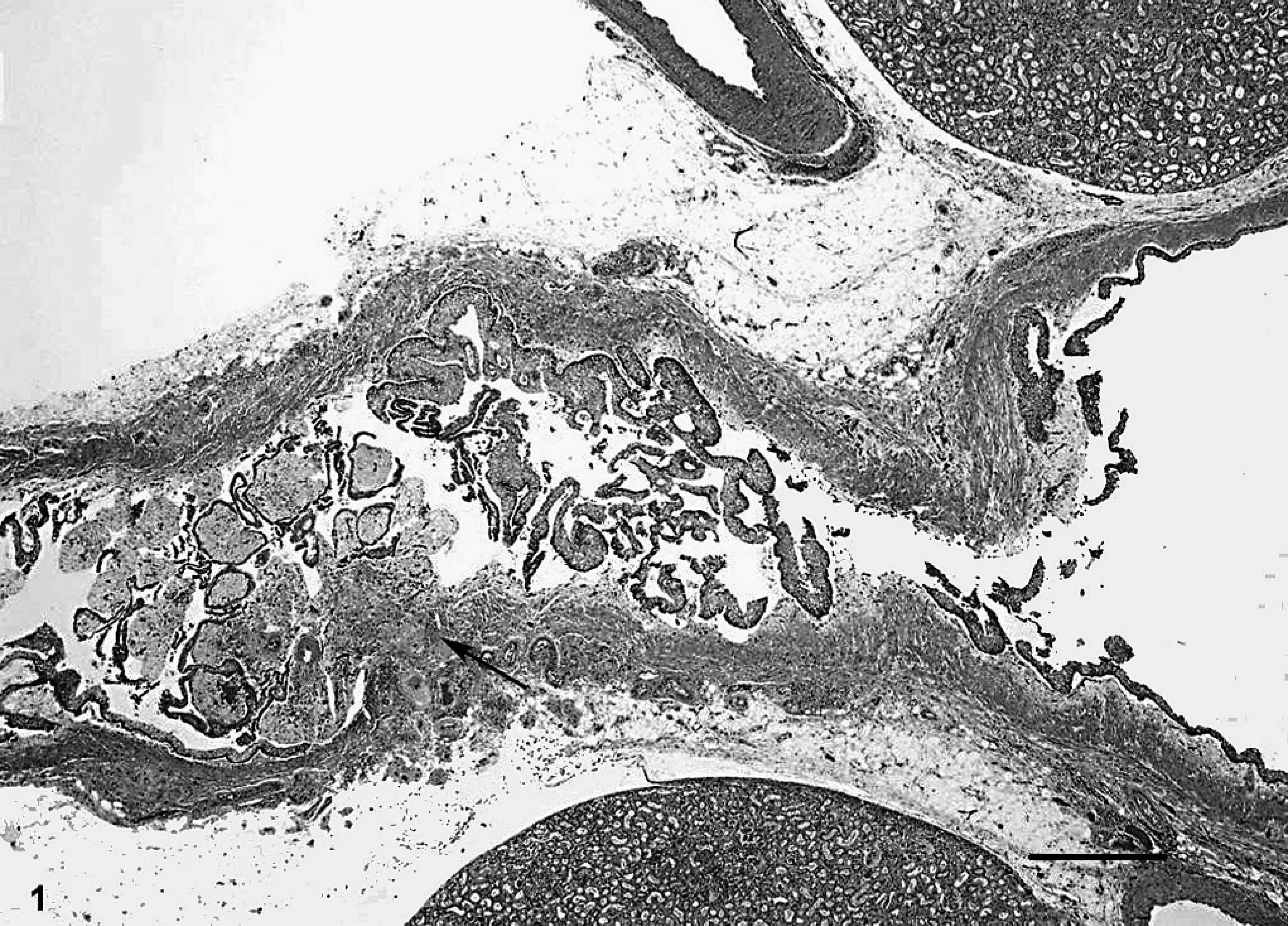
Ureter; Aotus nancymae. Left ureter containing a 2-mm diameter pedunculated mass with a narrow stalk at the base (arrow) projecting into the lumen just distal to the renal pelvis. HE. Bar = 500 µm.
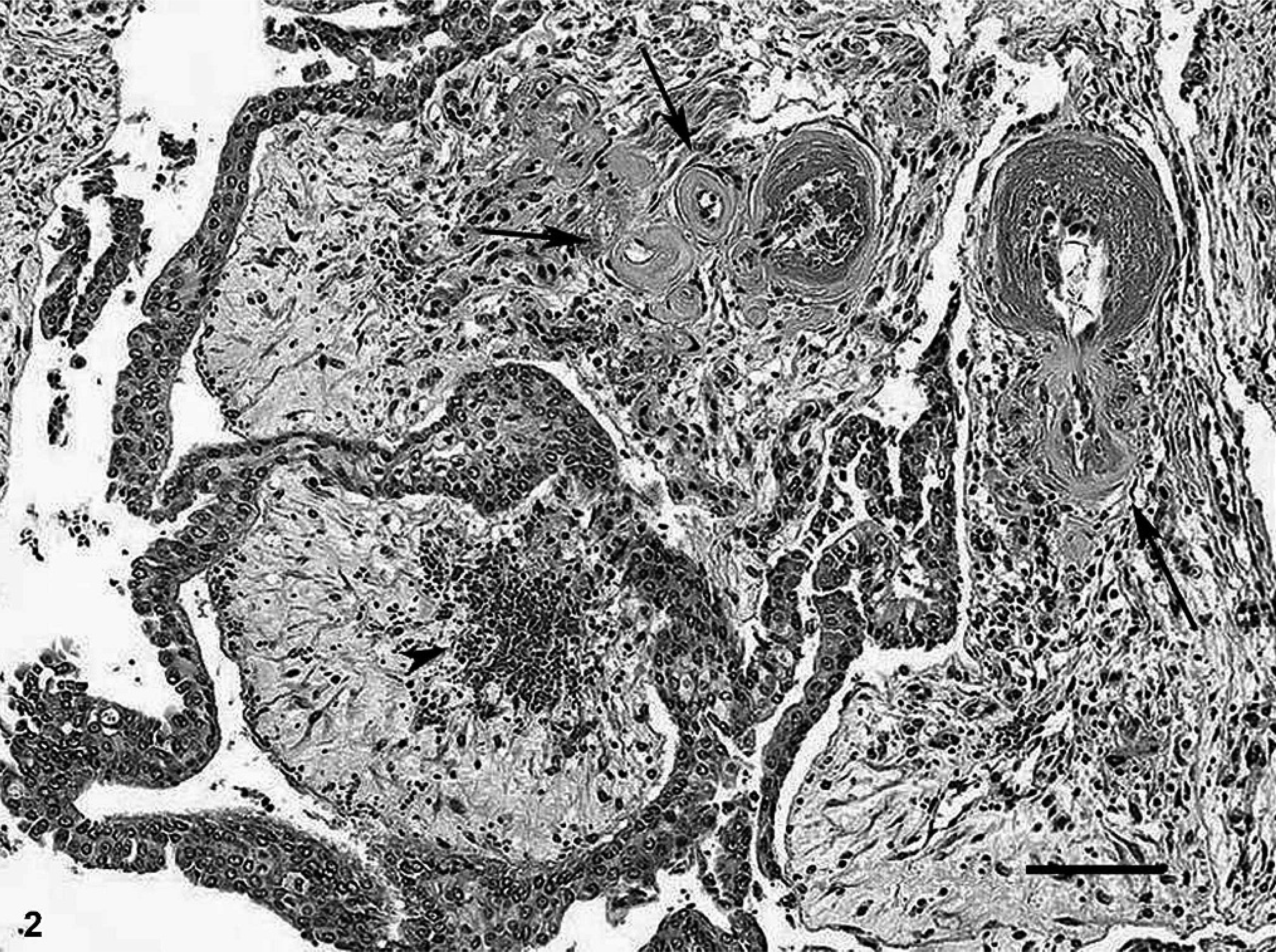
Ureter; Aotus nancymae. Ureteral mass with a layer of well-differentiated transitional epithelium, stroma consisting of loosely packed fibroblasts in a myxomatous matrix with small foci of hemorrhage (arrowhead), and blood vessels with hyaline change of the smooth muscle layer (arrows). HE. Bar = 100 µm.
The immediate cause of death in this owl monkey was the sudden blood loss resulting from the ruptured aorta. Aortic dissecting aneurysms, hypertrophic cardiomyopathy, arterial disease, and chronic renal disease are common findings in captive owl monkeys. 1, 5
Ureteral fibroepithelial polyps are unusual tumors of uncertain etiology. It is believed they are either of congenital origin owing to a developmental anomaly or acquired origin caused by infection, chronic inflammatory reaction, obstruction, or trauma. 3, 4, 7 The majority of fibroepithelial polyps occur in the region of the ureteropelvic junction or in the proximal ureter. The polyps appear as multiple finger-like projections arising from the submucosal connective tissue of the ureter. 2 In four dogs reported with ureteral fibroepithelial polyps, the animals were all adults and three were males. 8 The clinical signs were urinary incontinence, urinary tract infection, and/or polydypsia and pollakiuria. Findings included ureteral dilation proximal to the level of an intraluminal mass and ipsilateral hydronephrosis. The lesions in dogs appeared polypoid and were attached to the ureteral wall by a thin stalk. Histopathologically, they contained a superficial layer of well-differentiated transitional epithelial cells overlying a prominent fibrovascular stroma with varying degrees of lymphoplasmacytic inflammation. 8 The histopathologic characteristics, sex predisposition, and location, more commonly within the upper third of the ureter, were similar to the most common disease pattern in humans. 8
In this specific case, the fibroepithelial polyp location, histopathologic characteristics, sex, and age of the owl monkey were also consistent with the most common clinical and pathologic presentation observed in humans. The polypoid shape resembled that described for the dog. Some differences in our owl monkey are a double-layer of transitional epithelium and the lack of inflammatory cells. This is the first report describing a ureteral fibroepithelial polyp in a nonhuman primate.
Footnotes
Acknowledgements
This research was supported by the Intramural Research Program of the National Institutes of Health, National Institute of Allergy and Infectious Diseases/Comparative Medicine Branch, Malaria Vaccine Development Branch, the Office of Research Support; and SoBran Inc./Government Contractor. We thank Mr. Brad Fisher and Dr. Randy Elkins for constructive criticism and Dr. Jerrold Ward for microphotographic assistance.