
Abstract
A histopathologic, immunohistochemical, and ultrastructural study of the trachea and the bronchi of 6 rabbits experimentally intoxicated with the calcinogenic plant Solanum glaucophyllum was performed. Histologically, infiltration of the mucosa and the submucosa of the trachea and the bronchi by macrophages, multinucleated giant cells, a few lymphocytes and mast cells, and calcium deposits in the basal lamina of the epithelium and in elastic fibers were observed. Expression of osteocalcin, osteonectin, and osteopontin was detected in the mucosa, lamina propria, and epithelium. Electron microscopic study of the corresponding areas showed numerous macrophages in the process of fusion to form multinucleated giant cells, activated mesenchymal cells, and calcium precipitation in the basal lamina of epithelium and in elastic fibers. It is suggested that the high levels of 1,25-dihydroxyvitamin D3 (1,25(OH)2D3) in the plant induces macrophage proliferation, multinucleated giant-cell formation, mesenchymal cell activation, bone-protein synthesis, and calcification. In addition, the synthesis of 1,25(OH)2D3 by local macrophages may have contributed to the calcification.
During the last 2 decades, it was well demonstrated that the vitamin D endocrine system, in addition to its well-known role in calcium/phosphorus metabolism, modulates the differentiation, growth, and function of a broad range of cells, including cells of the mononuclear phagocytic system. 1, 6, 10, 17, 20, 25, 34 The active metabolite of vitamin D, 1,25(OH)2D3, mediates its effects via specific nuclear receptors that bind to DNA and alter the transcription of vitamin D response elements. 5, 6, 24, 34 Receptors for 1,25(OH)2D3 have been found in a variety of cells and tissues, including monocytes, macrophages, and smooth muscle cells (SMC). 33, 37
The participation of 1,25(OH)2D3 in the differentiation of monocytes to macrophages and fusion to form multinucleated giant cells has been extensively studied. 1, 11, 14, 15, 21 Infiltration of macrophages and multinucleated giant cells in lung, arteries, stomach, and heart were described in natural and experimental calcinosis induced by calcinogenic plants 2– 4, 18, 28, 37, 41 that contain high levels of 1,25(OH)2D3 as a glycoside derivative. 40
Until recently, ectopic calcification was believed to be a passive process that occurs as a nonspecific response to tissue injury and necrosis. Now, there is strong evidence to support the concept that arterial calcification is, at least in part, an active process associated with the expression of growth factors, matrix proteins, and other bone-related proteins. 23, 39 In fact, bone-matrix proteins influence not only mineralization of vasculature but also SMC differentiation. 12
The aim of the present study is to describe an unusual proliferation of macrophages and multinucleated giant cells in the trachea and the bronchi of rabbits experimentally poisoned by the calcinogenic plant Solanum glaucophyllum (synonym S. malacoxylon) to look for noncollagenous bone matrix proteins (BMP) and to discuss the possible role of these elements in the calcification process.
Materials and Methods
Seven young New Zealand male rabbits with a body weight near 1.5 kg were used. They were housed in individual cages and maintained on a standard commercial rabbit ration and tap water. Six rabbits were fed daily, by a stomach tube, with a water extract of S. glaucophyllum corresponding to 100 mg of dried leaves of the plant per body weight. One rabbit did not receive the extract and was kept as a control.
Seventy-two hours after the beginning of the experiment, all 7 animals were anesthetized by ether inhalation and killed by exsanguination. Samples of lung tissue, bronchi, trachea, aorta, stomach, intestines, kidney, and heart were fixed in 10% buffered formalin; routinely processed; and embedded in paraffin wax. The sections were stained by HE and the von Kossa method and examined by light microscopy.
Sections of bronchi for immunohistochemistry were mounted on slides coated with 3-aminopropyltriethoxy-silane (Sigma Diagnostics, St. Louis, MO), deparaffinized with xylene, passed through graded alcohol, and rinsed 3 times in deionized water and phosphate buffered saline solution (PBS).
The primary monoclonal antibodies used were as follow: osteocalcin (clone #OC1; Biodesign International, Kennebunk, ME) diluted 1 in 400, osteonectin (clone AON-1; Developmental Studies Hybridoma Bank, Iowa City, IA) diluted 1 in 800, and osteopontin (clone MPIIIB10; Developmental Studies Hybridoma Bank) diluted 1 in 500.
The immunohistochemical detection system was a dextran-polymer-based method (Universal EnVision System Peroxidase, DakoCytomation, Carpinteria, CA). Positively stained cells showed dark gold-brown in 3,3′-DAB tetrahydrochloride-H2O2 reaction product. After counterstaining with hematoxylin, the slides were dehydrated and mounted for examination.
Small samples of the bronchi were fixed in sodium cacodylate buffered 2% glutaraldehyde, 2% paraformaldehyde, postfixed in 1% osmium tetroxyde in cacodylate buffer, dehydrated in ethanol, and embedded in Epon. Thick sections were stained with methylene blue and selected thin sections were stained with lead citrate and uranyl acetate and examined with a transmission electron microscope.
Results
The animals who received the extract showed depression, reduced appetite, and diarrhea on the first day after treatment. The diarrhea persisted, with deterioration of body condition for the second and third days. The animals were euthanatized at 72 hours after the beginning of the experiment. At necropsy, the mucosa of the stomach was thick and red and, in 3 rabbits had areas of white finely granular deposits. White areas of calcification were observed mainly in the aorta, cortex of the kidney, and muscle layer of the stomach and intestines and, in 2 cases, in the myocardium. Small areas of emphysema were observed in the lungs. No significant alterations were observed in the trachea; however, there was mild thickness and white discoloration of the wall of some bronchi.
The mucosa and the submucosa of the trachea and some bronchi were infiltrated by macrophages, multinucleated giant cells, and a few mast cells and lymphocytes. Macrophages and giant cells were also infiltrating the epithelial layer (Fig. 1). Cellular infiltrates varied in severity: mild to marked in bronchi, mild in the distal trachea, and absent in the upper portion of trachea. Von Kossa stain demonstrated areas of calcification in the lamina propria, specially in bronchi, appearing as granular material near the epithelium. These areas of calcification could also be seen in thick sections stained by methylene blue (Fig. 1). In the lung parenchyma, small areas of emphysema were seen, but no calcification, macrophage proliferation, or giant cell formation was noted. The lamina elastica of the aorta lost the normal waviness and was calcified. Some SMC had an irregular branched aspect to the cytoplasm, enlarged and pale nuclei, and had lost the normal parallel orientation with the intima. Calcification of the tubules in the renal cortex and of the media of small renal arteries were observed. Interstitial mononuclear infiltration was mild in areas of calcification. The muscle layer of the stomach was heavily mineralized, and the SMC had alterations similar to those described in the aorta. The mucosa and the submucosa were infiltrated by macrophages, multinucleated giant cells, lymphocytes, and a few mast cells. In 3 rabbits, there was necrosis and calcification of the epithelial layer. The arteries of the stomach wall were strongly mineralized. Microscopic changes in the intestines were similar to those observed in the stomach, but no necrosis or calcification of the epithelial layer was seen. In the myocardium of 2 rabbits, the foci of mineralization of myocardial cells and interstitium were characterized by fine granular deposits. Calcification of the coronary arteries was also seen.
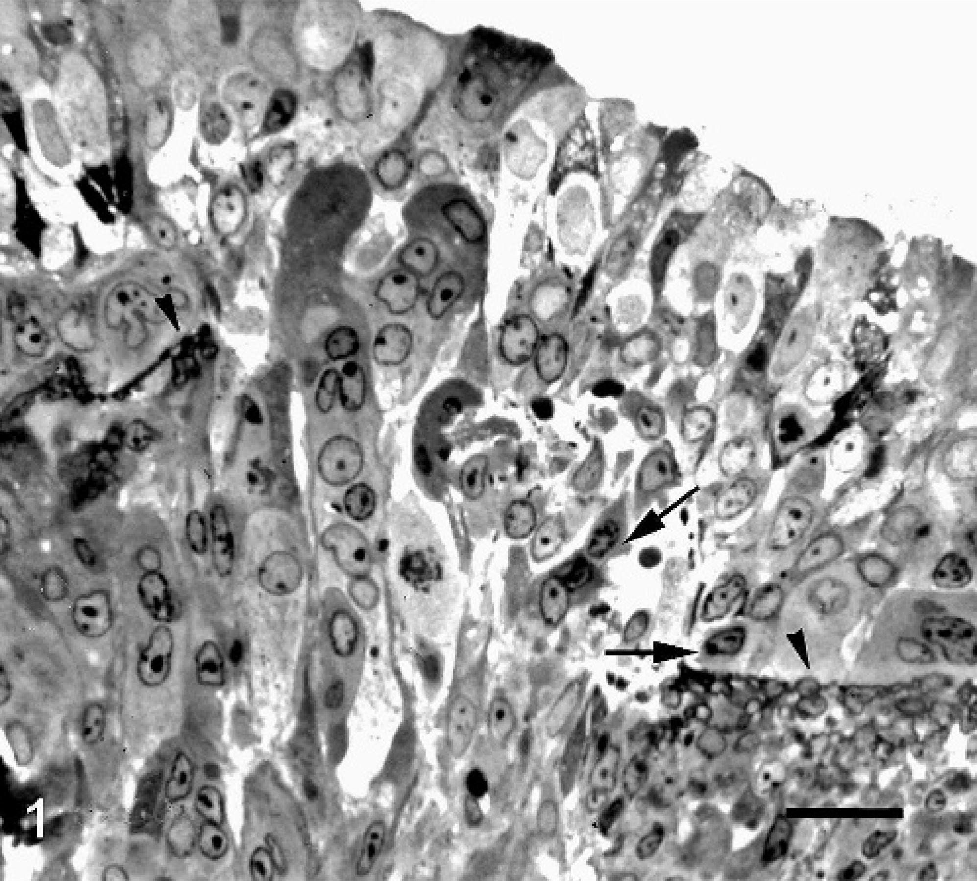
Bronchus; rabbit. Numerous macrophages and multinucleated giant cells infiltrate the lamina propria and the epithelial layer. Calcium deposits (arrow heads) are seen in the lamina propria near the basal lamina. Some activated mesenchymal cells are also seen (arrows). Epon, methylene blue. Bar = 35 µm.
In the bronchi, osteocalcin, osteonectin, and osteopontin were detected in the cytoplasm of macrophages, activated mesenchymal cells, and also in the extracellular matrix. The immunoreaction was observed in both the mucosa and the submucosa (Fig. 2), and on the SMCs of the arterial walls of the lung.
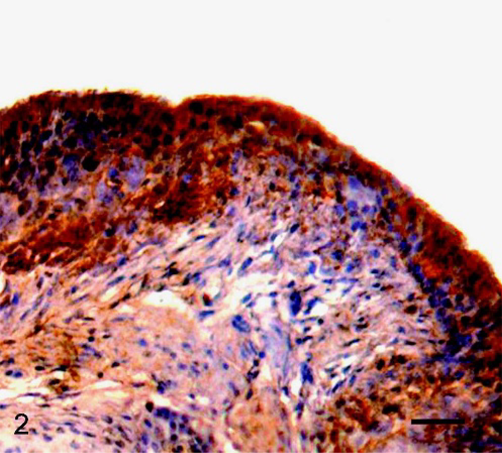
Bronchus; rabbit. Expression of osteocalcin in cells of submucosa, lamina propria, and epithelium. Bar = 70 µm.
Ultrastructurally, the mucosa and the submucosa of the bronchi were infiltrated by macrophages, multinucleated giant cells, a few lymphocytes, and mast cells. The fusion of macrophages to form giant cells was frequently observed (Fig. 3). Macrophages and giant cells were seen interspersed in the epithelial layer. The basal lamina of the epithelium exhibited lamellar crystalline bodies of electrodense and electrolucent bands of calcium precipitation (Fig. 4). Similar lamellar crystalline bodies were seen associated with elastic fibers. In the lamina propria, some activated mesenchymal cells (Fig. 4), characterized by hyperplasia of endoplasmic reticulum and an increase of free ribosomes, were seen. No alterations were observed in the control rabbit.
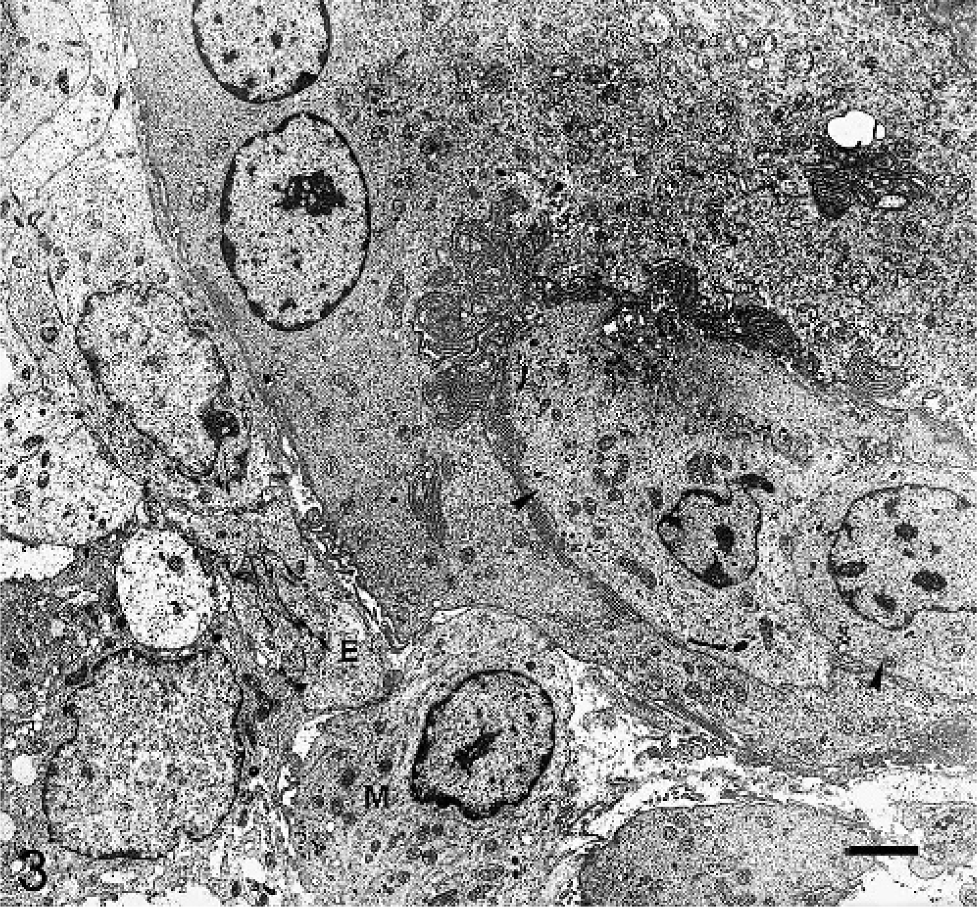
Bronchus; rabbit. Electron micrograph showing fusion of macrophages (arrow heads) to form a multinucleated giant cell. Giant cell and macrophages (M) infiltrate the epithelial layer (E). Bar = 1 µm.
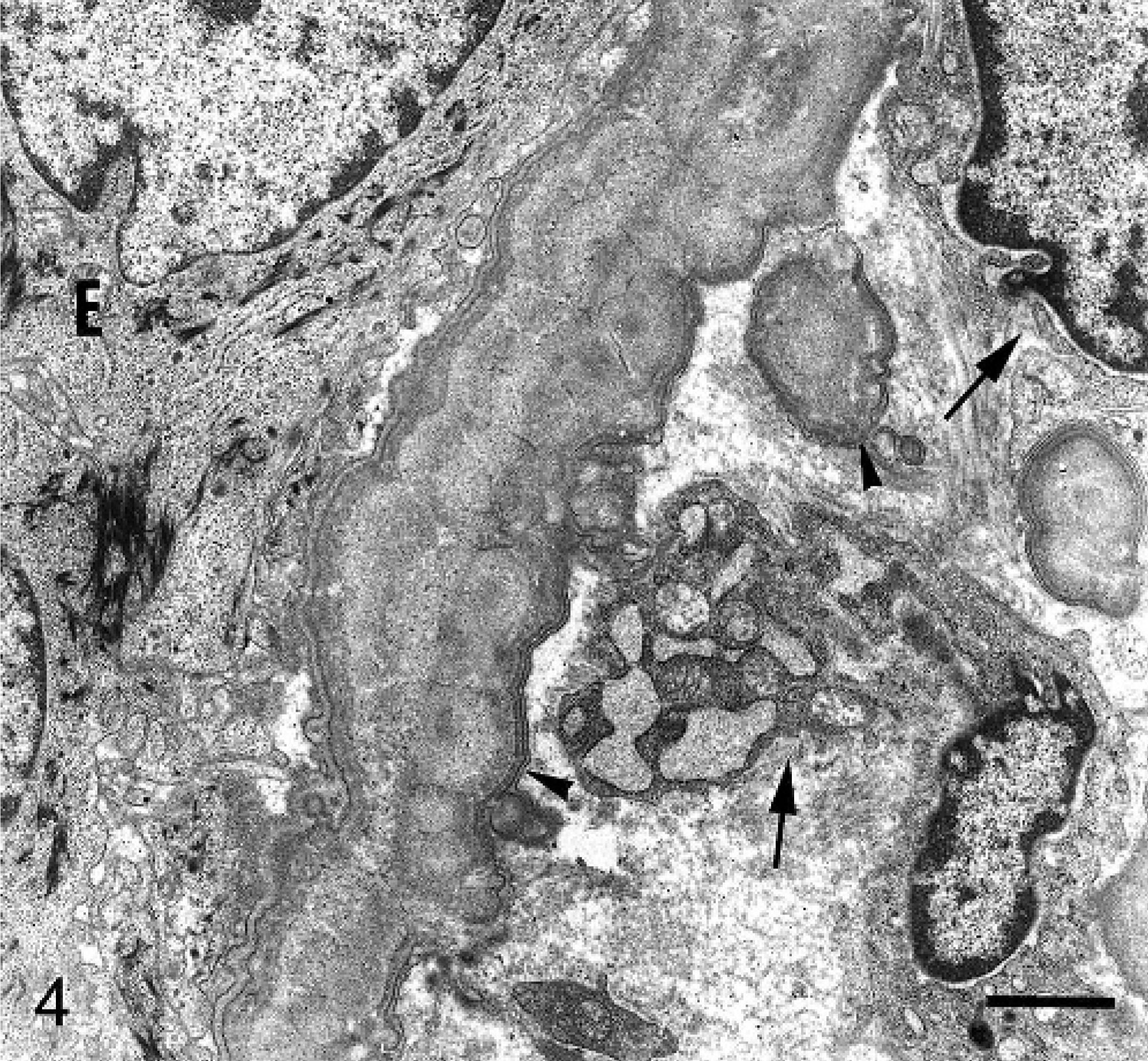
Bronchus; rabbit. Electron micrograph showing calcium deposits (arrow heads) with lamellar peripheral structure in the basal lamina and lamina propria. Epithelial cells (E). Activated mesenchymal cells (arrows). Bar = 3 µm.
Discussion
The microscopic changes found in aorta, heart, and stomach of rabbits in this experiment were similar to those described elsewhere. 4, 16, 18 The inflammatory changes associated with mineralization in bronchi and, to a lesser extent, in the trachea were more intense than those in other calcified tissues, although an explanation for this variation is not clear. A population of macrophages and giant cells comparable with that seen in some bronchi was found in the mucosa and the submucosa of the stomach of 3 rabbits.
Extracellular matrix mineralization is a normal physiologic process in skeleton, teeth, and hypertrophic cartilage, and is a pathologic process in other organs. 19 Pathologic calcification has been extensively investigated in vessels; nevertheless, the mechanisms underlying vascular calcification are still obscure. 35 Current data suggest that this process parallels many aspects of calcification in bone, as bone associated proteins have been detected in calcified vascular tissues. 7, 9, 19, 29, 32, 35, 38 The pathogenesis of calcification in other soft tissues is even more uncertain.
Vitamin D regulates the expression of several proteins that induces mesenchymal cell differentiation into osteogenic cells. 8, 22, 36 Activated mesenchymal cells, as observed in this study, were described in the lung of sheep in spontaneous enzootic calcinosis induced by the plant Nierembergia veitchii and it was shown that these cells synthesize noncollagenous BMP, such as osteopontin, osteonectin, and osteocalcin. 2 The activated mesenchymal cells might represent osteoblastic-like cells similar to those described in human atherosclerosis. 13, 35 The absence of calcification in the lung parenchyma, observed in chronic spontaneous cases of calcinosis of sheep, 2, 3, 28 probably was because of the short period of time of the experiment.
On the other hand, it is possible that an interaction among the macrophages and the activated mesenchymal cells was associated with local production of 1,25(OH)2D3 by macrophages. Such interaction among SMCs and macrophages has been proposed in the development of atherosclerotic calcification in humans. 31 The synthesis of 1,25(OH)2D3 by macrophages is related in vitro and in vivo. 26, 27, 30, 31
The differentiation of monocytes to macrophages and their fusion to form giant cells, induced by the 1,25(OH)2D3, has been well documented experimentally. 1, 14, 15, 20, 21 The proliferation of macrophages and multinucleated giant cells in the present study could be the response of these cells to the action of the 1,25(OH)2D3 present in S. glaucophyllum. Such action has been suggested in similar infiltrates in the muscle layer of the stomach of rabbits experimentally poisoned with the same plant 18 and in arteries and lungs of sheep with enzootic calcinosis induced by the calcinogenic plant Nierembergia veitchii. 2, 37
Footnotes
Acknowledgements
This work was supported by a grant from the Conselho Nacional para o Desenvolvimento Científico e Tecnológico (CNPq-PRONEX), the Fundação de Amparo a Pesquisa do Estado do Rio Grande do Sul (FAPERGS), Brazil, the Agencia Nacional de Promoción Científica y Tecnológica (ANPCYT), and the Consejo Nacional de Investigaciones Científicas (CONICET), Argentina. EJG is a Research Career Member of CONICET. The monoclonal antibodies to osteonectin and osteopontin, developed by J.D. Termine and M. Solursh/A. Franzen, respectively, were obtained from the Developmental Studies Hybridoma Bank developed under the auspices of the NICHD and maintained by The University of Iowa, Department of Biological Sciences, Iowa City, Iowa.
Part of this paper was presented as poster at the 9th National Meeting of Veterinary Pathologists, July 26–30, Belo-Horizonte, Brazil, p. 134, 1999. We thank Dr. Claudio S. L. Barros for help with the English manuscript.