
Abstract
An Appaloosa filly was born with a ventral midline, approximately 8 × 12 × 15 cm subcutaneous cervical mass. The nonencapsulated mass was composed of interlacing and haphazard bundles of spindle cells on moderate to abundant loose myxomatous stroma. A moderate number of cells showed cross striations with minor nuclear variation and a low mitotic rate. Immunohistochemical staining for myoglobin, desmin, actin, vimentin, and S-100 was positive and negative for glial fibrillar antigen and keratin. Rhabdomyomas are rare benign tumors of striated muscle. Rhabdomyomas described previously in the veterinary literature are analogous to the “adult form” of human rhabdomyoma. This is the first report of a veterinary case that 1) clinically and histologically parallels the “fetal form” in human rhabdomyoma and 2) describes a congenital extracardiac rhabdomyoma.
Benign tumors of striated muscle (rhabdomyomas) are rare in domestic animals and humans. 3 Rhabdomyomas have been reported in the dog, pig, cat, and horse at primary sites, including heart, larynx, diaphragm, and subcutis of the pinna, abdomen, and extremities. 2,6–8 Middle to old age animals are typically affected; however, reports of congential cardiac rhabdomyomas in pigs and a single case report of a diaphragmatic rhabdomyoma in a 2-year-old filly are notable exceptions. 3,7 Rhabdomyomas are usually well-delineated, pale, and fleshy masses that are 1–2 cm in size, but some as large as 30 cm in a 2-year-old filly have been described. Microscopically, rhabdomyomas have well-defined nonencapsulated margins, are arranged in sheets and rarely in lobules, and are composed of round to polygonal cells with a strongly eosinophilic, granular, and abundant cytoplasm. Cells may be vacuolated with small numbers of clear crisp-edged vacuoles. These vacuoles can be quite large with thin peripheral cytoplasmic processes that extend around the vacuole forming the so called “spider-web cells.” 2,7
In humans, rhabdomyomas are rare and case reports in the veterinary literature suggest a similar incidence in domestic animals. 8,9 Human rhabdomyomas are classified into three general forms, each with distinct microscopic and clinical features: adult, fetal, and genital. Rhabdomyomas described in the veterinary literature are clinically and histologically similar to the adult-type rhabdomyoma (AR) of human beings, but cases representative of the fetal rhabdomyoma (FR) are absent. 2 Herein, we describe a congenital rhabdomyoma in a filly that is clinically and histologically similar to human FR.
An 11-day-old Appaloosa filly that presented at the Iowa State University's Veterinary Teaching Hospital had a ventral midline cervical mass, approximately 4 cm caudal ventral to the ramus of the mandible and superficial to the trachea in the subcutis (Fig. 1). The mass had been present from birth, and no changes in size, shape, or consistency had been noticed. Fine needle aspirates were inconclusive, and surgery was elected. The 8 × 12 × 15 cm firm, pale mass was highly vascularized and appeared to arise from the sternothyroideus and sternohyoideus muscles overlying the trachea. The mass was placed in 10% buffered neutral formalin and processed routinely for microscopic examination.
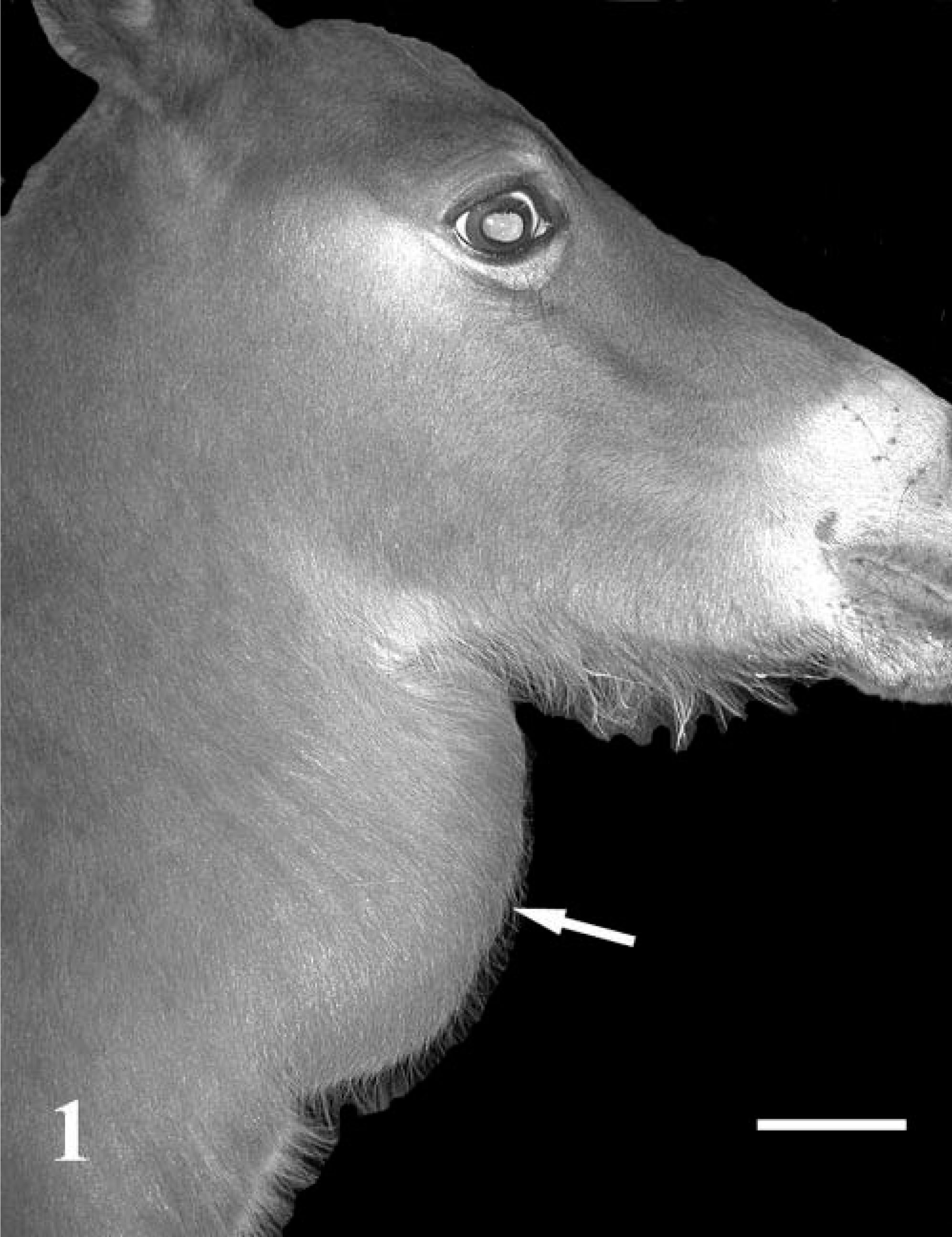
Filly. Lateral view. Note the subcutaneous ventral midline cervical mass (arrow). Bar = 5 cm.
Histologically, the mass was composed of intersecting and haphazardly arranged bundles of spindle to strap cells with a variably staining eosinophilic fibrillar cytoplasm and occasional cross striations in an abundant loose basophilic myxomatous stroma (Figs. 2, 3). The nuclei were small, hyperchromatic, oval to elongate, with clumped chromatin and low mitotic rate (< 1/10 of 40× fields). In cells having better striated muscle differentiation, the nuclei were slightly larger, had a fine chromatin pattern, 1–3 nucleoli, rarely binucleate to multinucleate cells, and the nuclei were peripherally oriented reminiscent of mature myofibers. The mass margins were well defined and bordered normal mature skeletal muscle and collagen. The tumor cells stained strongly positive immunohistochemically for actin, myoglobin, and desmin and had moderate staining for vimentin and S-100. Cells stained negative immunohistochemically for glial fibrillary acidic protein and keratin.
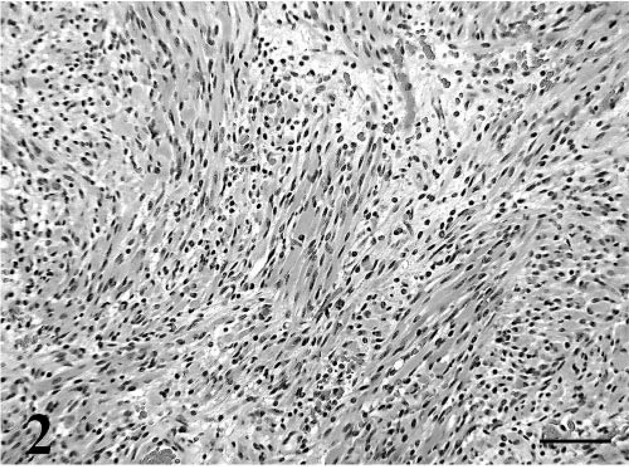
Cervical subcutaneous mass. Intersecting and haphazardly arranged bundles of spindle cells in a myxomatous stroma. HE. Bar = 90 µm.
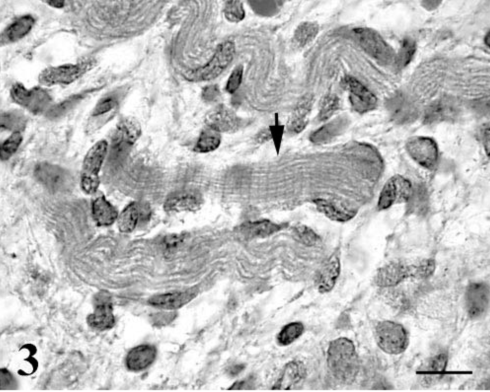
Cervical subcutaneous mass. Differentiated tumor cells have eccentric nuclei and cross striations (arrow). HE. Bar = 15 µm.
Human ARs are often present in the head or neck region, with an increased incidence in older men and are characterized by sheets of large ovoid to polygonal cells with abundant eosinophilic and granular cytoplasm in a fine fibrovascular stroma. 1 The fetal form is generally found in young children with a preference for occurrence in boys and up to 25% are congenital. The fetal form is generally characterized by primitive spindled cells with variable degrees of skeletal muscle differentiation in a myxoid to fibromyxoid stroma. The wide histologic variation in FR has prompted several attempts to subclassify FR, however, without significant success. Surgical excision is usually curative for FR, but rare recurrence has been reported. 5,9
In this case, the histologic appearance along with positive myoglobin, desmin, actin, vimentin and S-100 immunostains are consistent for a striated muscle tumor. 4,5 The lack of nuclear atypia, low mitotic rate, absence of necrosis or invasion, and advanced differentiation supported the diagnosis of rhabdomyoma. The clinical presentation of a congenital subcutaneous mass with the histologic pattern described for this case are similar to the features seen in human FR and are distinctly different from the previous AR-like rhabdomyomas described in animals. 5,6,8 This is to our knowledge the first veterinary description of a 1) fetal rhabdomyoma and 2) an extracardiac congenital rhabdomyoma.