
Abstract
Weissella confusa is a Gram-positive bacterium that has been identified in environmental and food samples from around the world. Rare cases of bacteremia in immunocompromised people have been reported. A 2-day-old foal was presented for weakness and suspected sepsis. Blood culture yielded pure growth of a Gram-positive coccobacillus, which was identified as W. confusa through sequencing of the 16S ribosomal DNA. Although the foal initially responded to antimicrobial therapy with ceftiofur and metronidazole, it later developed septic complications of the right tarsocrural joint and right digital flexor tendon sheath and was euthanized. Postmortem examination and histology revealed subcutaneous icterus, severe diffuse interstitial pneumonia, septic synovitis, necrotizing vasculitis with marked thrombosis and hemorrhage in the medial digital vessels of the right hind limb, and ischemic necrosis of the right hind hoof laminae. Gram-positive, coccobacilli were observed in the vascular lesion.
Weissella confusa, previously Lactobacillus confusus, is a Gram-positive coccobacillus found in fermented foods that is an occasional cause of septicemia and endocarditis in immunocompromised people.5,6,8,11,13,14 Weissella confusa has been identified in human, nonhuman primate, canine, porcine, and equine feces.1–4,7,17,18 In a case series from a human tertiary care facility, 5 of 10 patients with W. confusa bacteremia were infected solely with W. confusa. 8 The route of entry was speculated to be via a compromised gastrointestinal barrier either as a result of surgery or by chemotherapeutics such as antimicrobials. 8 Although found in equine feces, W. confusa has not previously been associated with disease in horses.3,4 To date, the only report of disease in a veterinary patient is a single case in a nonhuman primate. 18 In that case, a juvenile female mona monkey (Cercopithecus mona) was found dead with no prior evidence of disease, and W. confusa was isolated from the lung, liver, brain, and intestine. Congestion, edema, and petechiae were noted in the brain, liver, and spleen. 18
A 2-day-old full-term colt was presented to the equine emergency service at Texas A&M University Veterinary Medical Teaching Hospital (College Station, Texas) for weakness and suspected sepsis. The foal was born approximately 40 hr prior to presentation. Delivery of the foal was assisted due to dystocia. The foal did not nurse and was fed milk from its dam by bottle within 1.5 hr after birth. The dam’s milk was presumed to include colostrum but evaluation of serum immunoglobulin G by an enzyme-linked immunosorbent assay by the referring veterinarian indicated failure of passive transfer. The foal was treated with ceftiofur, metronidazole, and plasma infusion by the referring veterinarian. On presentation, the foal was weak, lethargic, and unable to stand or walk independently. The foal was severely dehydrated with icteric mucous membranes. A continuous heart murmur was detected, and petechiae were noted in both pinnae. On palpation, the umbilicus was mildly thickened. Laxity was noted in the flexor tendons of all 4 limbs, the coronary bands were hyperemic, and the right hind foot was cooler to the touch than the other 3 feet.
Diagnostic evaluation included complete blood cell count, serum chemistry panel, thoracic and abdominal ultrasound, urinalysis, and culture of the blood. The foal had neutrophilic leukocytosis with toxic changes in the neutrophils, elevated lactate, azotemia, elevated aspartate aminotransferase, elevated alkaline phosphatase, and elevated γ-glutamyl transferase. Abdominal ultrasound showed thickening of the walls of the right umbilical artery and umbilical vein, and thoracic ultrasound was suggestive of pneumonia. A clinical diagnosis of septicemia with right tarsocrural joint inflammation and pneumonia was made. Therapy included intravenous fluids, antioxidant administration, and antimicrobial therapy (metronidazole and ceftiofur). Orthopedic supports were placed on all limbs to stabilize tendon laxity. The foal initially responded favorably to therapy but 4 days after initial presentation, the foal was noted to be non–weight bearing on the right hind limb. The right tarsus was swollen and warm. Synovial fluid was collected from the right tarsocrural joint and the right digital flexor sheath. Cytology revealed a neutrophilic infiltrate, and synovial fluid culture was performed. No infectious agents were noted on cytologic examination, and no bacteria grew on the culture from the joint. No other synovial structures were cultured. Sepsis of the tarsocrural joint and right digital flexor tendon sheath was diagnosed. The umbilicus also became enlarged, a patent urachus was diagnosed, and an omphalectomy was performed. Six days after hospitalization, the right rear limb, from the mid-metatarsus distal was found to be cold and unresponsive to pain. Because of a poor prognosis, the foal was euthanized, and a necropsy was performed.
Blood was collected for culture at initial presentation and inoculated into a commercially available blood culture system that consists of a bottle with culture media. a The blood culture bottle was vented using a venting needle b and incubated at 37°C in an atmosphere supplemented with 5% carbon dioxide. A small amount of the culture media was subcultured onto trypticase soy agar supplemented with 5% sheep’s blood (BAP) c at 24 hr, 48 hr, and 168 hr of culture. Each subculture yielded pure growth of an α-hemolytic, catalase-negative, Gram-positive coccobacillus, which grew within 24 hr. No organisms grew from premortem and postmortem samples collected from the right tarsocrural joint nor did bacteria grow from cultures of the spleen and liver performed postmortem. Fecal samples collected premortem were tested by both direct and tetrathionate enrichment cultures and were negative for the presence of Salmonella spp. A commercially available kit identified the organism grown from the blood sample as Pediococcus pentosaceus (probability of 99.66%) or W. confusa (probability of 0.32%). d Sequencing was performed on an approximately 500–base pair segment of the 16S ribosomal DNA (rDNA) amplified from template DNA using polymerase chain reaction (PCR) assay. The DNA template was prepared using standard techniques. Briefly, after 24-hr culture on BAP, an isolated bacterial colony was transferred to 100 µl of a commercial reagent e in a 1.5-ml microcentrifuge tube. The lysate suspension was vortexed for 60 sec then heated for 15 min at 100°C. The suspension was centrifuged at 16,000 × g for 5 min, and 3 µl of supernatant was used for PCR assay. Part of the 16S rDNA was amplified using the primers fD1mod (5′-AGAGTTTGATCYTGGYTYAG-3′) and 16S1RR-B (5′-CTTTACGCCCARTRAWTCCG-3′) as previously described. 10 The PCR assays were performed in a 100-µl volume that included 3 µl of template DNA, 10 µl of 10× PCR buffer, 3 µl of a 10 µM stock solution of each primer, 10 µl of 4 mM deoxyribonucleotide triphosphate, f and 0.5 U of Taq DNA polymerase. g The PCR reactions were run in a thermocycler h with a temperature profile of 3 min at 94°C followed by 36 cycles of 30 sec at 94°C, 30 sec at 56°C, and 30 sec at 72°C. The 36 cycles were followed by 75 min at 72°C. An aliquot was checked on a 0.7% agarose gel and stained with ethidium bromide; the remaining reaction was cleaned with a commercial kit i then sequenced on both strands at the Texas A&M DNA Technologies Core Laboratory (College Station, TX). The sequence was used to search GenBank using the BLASTn algorithm (http://www.ncbi.nlm.nih.gov/), and results were sorted according to % identity, with identities ≥99% considered significant. The highest ranked hit corresponded to W. confusa (accession no. CAGH01000051.1 [100%]) followed by Weissella cibaria (AEKT01000037.1 [98%]).
A complete postmortem examination was performed. Marked jaundice was observed diffusely throughout the subcutaneous tissue. All fetlock and interphalangeal joints showed varying degrees of hyperflexion, hyperextension, and excessive lateral movement, indicative of marked laxity of the flexor tendons and collateral ligaments. The left front fetlock joint, the right hind fetlock joint, and the left front elbow joint were moderately swollen with markedly thickened synovial capsules and dark orange turbid fluid. These findings were consistent with septic synovitis. The left stifle joint was expanded by an increased amount of synovial fluid. The subcutaneous tissue in the right hind limb, distal to the fetlock, was dark red, edematous, and hemorrhagic (Fig. 1A). The sensitive lamina of the right rear hoof was diffusely necrotic and hemorrhagic with complete separation from the third phalanx (Fig. 1B). The lungs were diffusely expanded, dark pink to red, and slightly firm with a rubbery texture. The lungs also contained a few, multifocal, pale pink, soft, raised areas of emphysema and numerous multifocal to coalescing dark red to black foci (Fig. 1C).
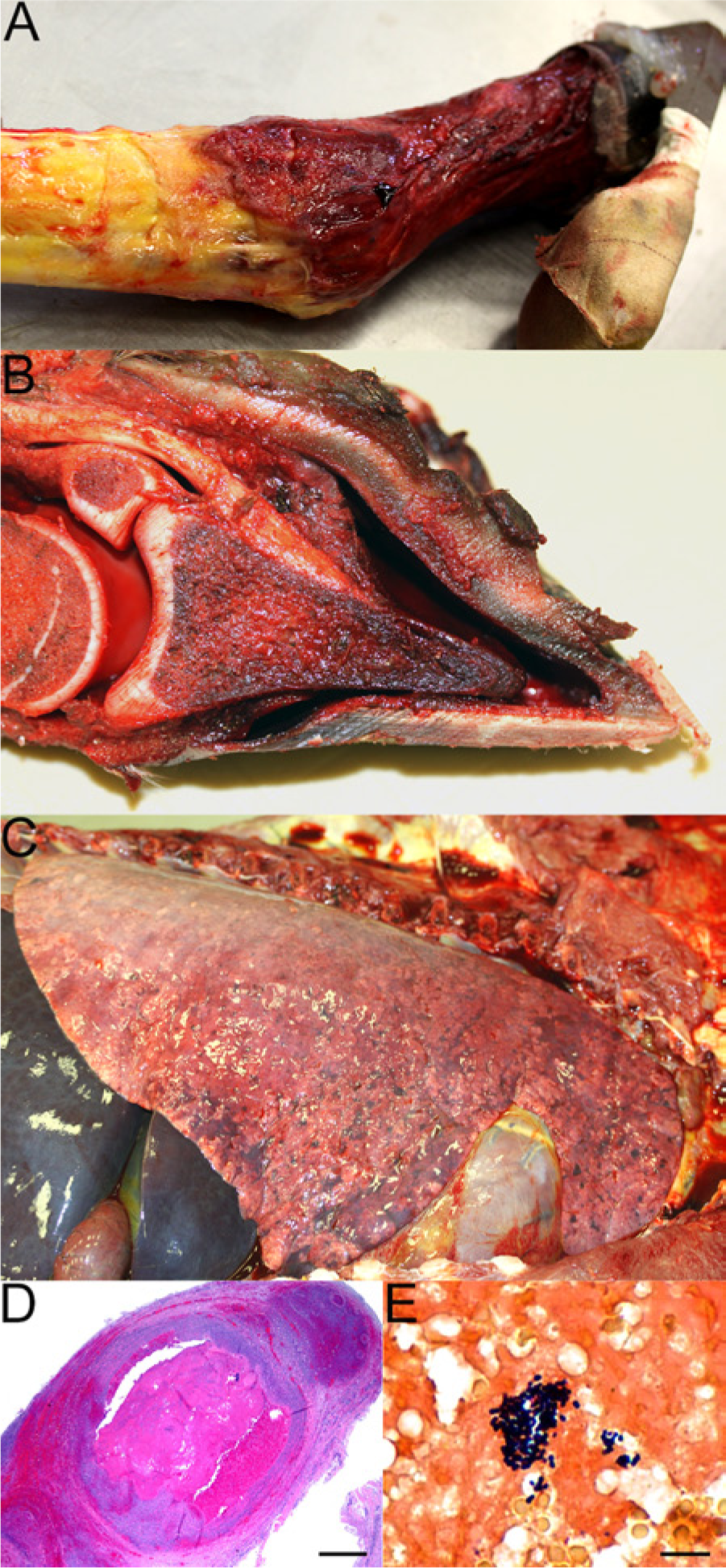
Foal.
Multiple tissue samples were collected and fixed in 10% buffered formalin, and sections of the medial digital vessels of the right hind limb, sensitive laminae of the hoof of the same limb, umbilical artery and vein, lung, spleen, liver, and urinary bladder were routinely processed, stained with hematoxylin and eosin, and subjected to histological evaluation. The medial digital vessels of the right hind limb had a diffuse necrotizing vasculitis with marked thrombosis and hemorrhage (Fig. 1D). A modified Brown and Brenn Gram stain was performed, and a few Gram-positive, coccobacilli were observed in the vascular lesion (Fig. 1E). The umbilical vein and arteries had a very mild neutrophilic perivasculitis. The hoof wall of the right hind limb had diffuse necrosis of the epidermal and dermal laminae. A marked, diffuse interstitial pneumonia was evident, characterized by expansion of the alveolar septa by macrophages and fewer neutrophils, moderate alveolar histiocytosis, few intra-alveolar neutrophils, abundant intra-alveolar fibrin, and scattered alveolar septal mineralization. Diffuse lymphoid follicular depletion was evident in the spleen. No significant histologic lesions were observed in other tissues.
The clinicopathological findings in this foal are consistent with septicemia. It is likely that endothelial damage led to necrotizing vasculitis in the right hind limb, resulting in hemorrhage throughout the limb and infarction of the sensitive laminae of the hoof. Septic synovitis in multiple joints and the diffuse interstitial pneumonia are consistent with hematogenous spread of a bacterial pathogen. The diffuse follicular lymphoid depletion of the spleen was probably secondary to the chronic debilitating disease process affecting the foal. Inflammation of the blood vessels and perivascular stroma of the umbilicus was very mild, therefore it is unclear if the systemic infection could have originated in the umbilicus or if there was a different, unidentified portal of entry. The possibility of an in utero infection could not be eliminated as no placental tissues were submitted.
Differential diagnoses for neonatal sepsis in foals include a variety of Gram-negative and Gram-positive bacteria. Prior studies have indicated that Enterococcus spp., Escherichia coli, and Actinobacillus spp., including A. equuli, and Salmonella spp. are commonly isolated from foals with sepsis.9,12,15,16 Blood culture is considered the gold standard for identification of sepsis in neonatal foals despite the potential for both false-positive and false-negative results.9,12,16 None of the above-mentioned microorganisms were cultured from any of the samples obtained pre- and postmortem, and the blood culture yielded a pure growth of W. confusa. In addition, histologic examination of tissues showed Gram-positive coccobacilli consistent with W. confusa associated with the vascular lesion in the right hind limb. No microorganisms grew on postmortem cultures, which is most likely the result of aggressive antemortem antimicrobial therapy.
The present case documents isolation of W. confusa from a neonatal foal with septicemia. Nucleic acid sequencing provided identification of this organism when phenotypic characterization was unsuccessful. Weissella confusa has previously been reported as an opportunistic pathogen, and the underlying failure of passive transfer may have contributed to the susceptibility of the foal to this organism. In addition to other bacterial pathogens, W. confusa should be considered as a possible cause of neonatal sepsis in foals.
Footnotes
Acknowledgements
The authors wish to acknowledge the staff of the Clinical Microbiology Laboratory of the Veterinary Medical Teaching Hospital for their efforts to identify this organism.
Declaration of conflicting interests
The author(s) declared no potential conflicts of interest with respect to the research, authorship, and/or publication of this article.
Funding
The author(s) declared that they received no financial support for their research and/or authorship of this article.
a.
BACTEC Plus Aerobic/F culture vials, BD, Franklin Lakes, NJ.
b.
Remel Inc., Lenexa, KS.
c.
BD, Franklin Lakes, NJ.
d.
RapID STR, Remel Inc., Lenexa, KS.
e.
PrepMan Ultra reagent, Applied Biosystems, Foster City, CA.
f.
Invitrogen Corp., Carlsbad, CA.
g.
AmpliTaq DNA polymerase, Applied Biosystems, Foster City, CA.
h.
DNA Engine, Bio-Rad Laboratories, Hercules, CA.
i.
QIAquick PCR Purification Kit, Qiagen Inc., Valencia, CA.