
Abstract
An 11-year-old male Collie was presented with a swelling of the face caused by tumor masses arising from the gingiva. Postmortem examination revealed metastases to the lymph nodes, lung, liver, and orbital cavity. Histologically, the tumor represented a combination of fibrosarcomatous proliferation, pulpal mesenchyme, and undifferentiated odontogenic epithelium, with a follicular or plexiform growth pattern. In addition, the follicular areas of the tumor showed a biphasic character, and there were numerous apoptotic cells in plexiform areas. Furthermore, acidophilic material resembling dysplastic dentine or enamel matrix was observed in the metastatic lesion in the lung. Based on the histological characters, the present case was diagnosed as malignant ameloblastic fibro-odontoma. This study is the first known description of a possible malignant ameloblastic fibro-odontoma in a dog with metastasis to distant organs.
Ameloblastic fibro-odontoma is a rare tumor in domestic animals, but it is the most common odontogenic neoplasm in cattle. 1 Histologically, it shows proliferation of both the odontogenic epithelium and pulpal mesenchyme. Gardner 2 reclassified odontogenic tumors in animals. However, there has been no report on malignant ameloblastic fibro-odontoma in dogs. In this study, we describe a malignant ameloblastic fibro-odontoma, which metastasized in a dog.
An 11-year-old male Collie was presented with swelling of the right side of the face and hypersalivation. A physical examination revealed swelling of the gingiva around the first premolar on the right upper jaw. Despite surgical excision of the mass, including the premolar, the mass recurred within a month, and after another month, the Collie died with multiple organopathy. Postmortem examination revealed that the oral cavity was occupied by multilocular masses (Fig. 1). Osteolysis was apparent. The tumor invaded the nasal cavity, the maxilla, and the base of the skull. Transverse sections of the masses were white, solid, or fibromatous and rich in serous effusion. Tumor masses of similar appearance were observed in the other organs including the mandibular lymph nodes, liver, lung and orbital cavity.
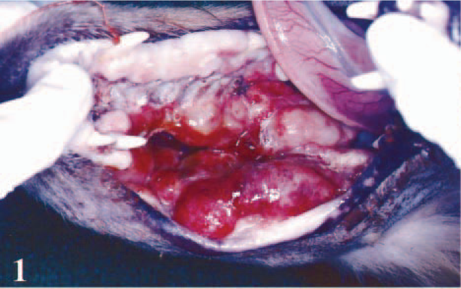
Gross appearance of the oral cavity; dog. Multilocular masses with apparent osteolysis.
Histologically, the tumor consisted of fibrosarcomatous proliferation, with mesenchymal tissue reminiscent of dental pulp, and undifferentiated odontogenic epithelium (Fig. 2). Microscopic evidence of metastases was found in the mandibular lymph nodes, liver, lung, and orbital cavity.
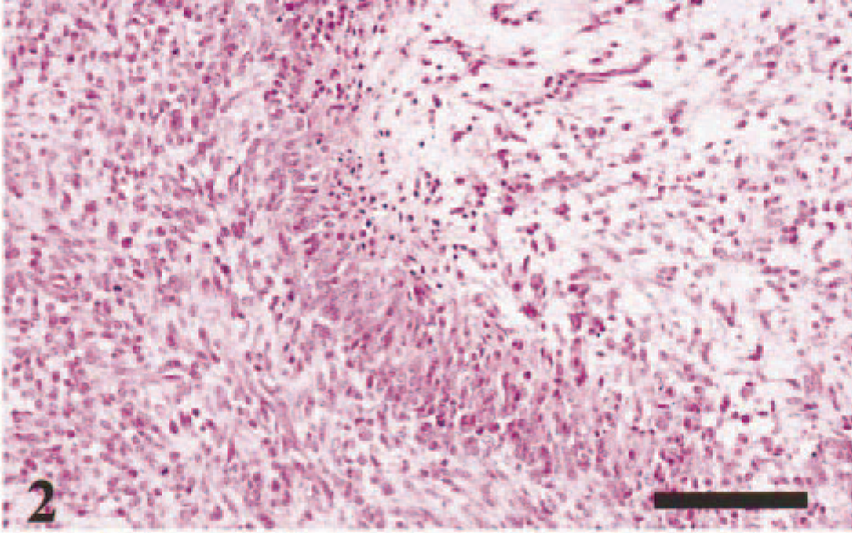
Maxillary mass; dog. Palisading-like appearance of undifferentiated odontogenic epithelium between fibrosarcomatous proliferation (left) and pulpal mesenchyme (right). HE. Bar = 100 µm.
In the area of fibrosarcomatous proliferation, fibroblastic spindle-shaped cells were highly atypical, with frequent mitoses. Small foci of bone formation were occasionally encountered in the lesion. The mesenchymal tissue was composed of loosely arranged angular cells resembling stellate reticulum. Between areas of fibrosarcomatous proliferation and mesenchymal tissue, and at the periphery of mesenchymal tissue, there were cords or islands of small, spindle to polygonal undifferentiated cells. Although these cells were not orderly, a palisading arrangement was evident. In some areas, tumor cells formed an epithelial layer, sometimes with follicular or plexiform growth patterns. We considered that these cells were derived from odontogenic epithelium.
Immunohistochemical analysis was performed using antibodies specific for actin (Biomeda Corporation, Foster City, CA), desmin, S-100 protein, and vimentin (all from Zymed Laboratories, San Francisco, CA). Most of the tumor cells were strongly positive for vimentin and negative for actin, desmin, and S-100 protein.
In areas with follicular growth pattern, some cavities contained small islands of tumor cells and were surrounded by a layer of cuboidal or columnar epithelial cells. Immunohistochemically, the epithelial cell layer showed specific immunoreactivity for cytokeratin AE1/AE2 (Nichirei Corporation, Tokyo, Japan), whereas the surrounding mesenchymal cells and the intracavity islands were positive for vimentin (Figs. 3, 4). The immunohistochemical contrast between vimentin and cytokeratin, i.e., biphasic, was characteristic of the follicular pattern. The cytokeratin-positive layer may reflect differentiation into internal dental epithelium or preameloblasts.
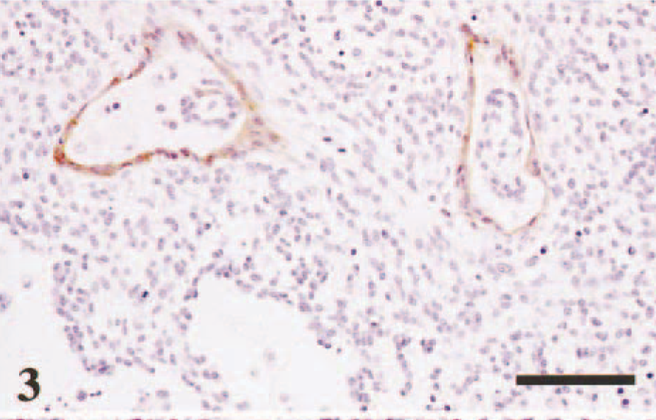
Maxillary mass; dog. Cytokeratin-positive layers in follicular growth pattern. Immunohistochemistry for cytokeratin AE1/AE3. DAB chromogen, haematoxylin counterstain. Bar = 80 µm.
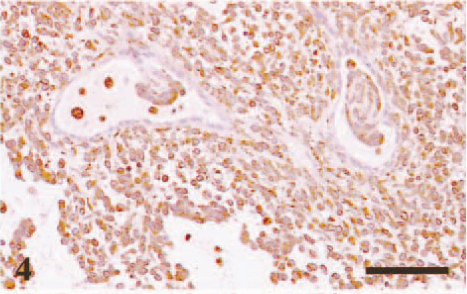
Maxillary mass; dog. Most of the tumor cells are vimentin positive. Immunohistochemistry for vimentin. DAB chromogen, haematoxylin counterstain. Bar = 80 µm.
In areas with a plexiform pattern, undifferentiated cells were arranged as a meshwork, partly palisading, and included cells resembling stellate reticulum of the dental lamina or collapsed cells (Fig. 5). In situ terminal deoxynucleotidyl transferase–mediated deoxyuridine triphosphate nick end labeling (TUNEL) method (ApopTaq™ Plus, Intergen) revealed numerous apoptotic cells in areas with this pattern (Fig. 6). During tooth formation, apoptosis is considered to play an important role in the atrophy or disappearance of the odontogenic epithelium, which becomes unnecessary after the completion of the tooth. 4 Moreover, apoptotic cell death is also considered to play an important role in oncogenesis or tissue differentiation (or both) in the odontogenic epithelium. 3 These reports would support our conclusion that the tumor originated from the odontogenic epithelium.
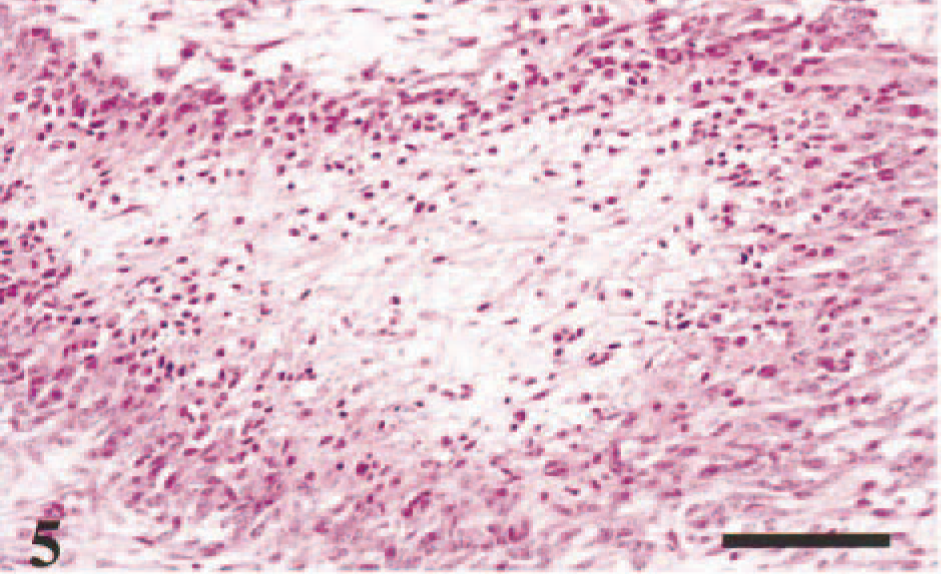
Maxillary mass; dog. Plexiform growth pattern involves cells resembling stellate reticulum. HE. Bar = 80 µm.
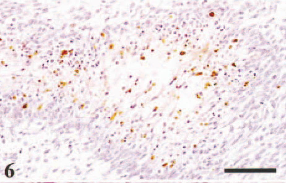
Maxillary mass; dog. Frequent apoptosis in the plexiform pattern. TUNEL method. Bar = 80 µm.
In the metastatic lesion in the lung, an acidophilic material was surrounded by loosely connected angular cells (Fig. 7). The material resembled dysplastic dentine or enamel matrix. The decisive features in naming odontogenic neoplasms are the presence or absence of a dental pulp–like mesenchyme, dentin, cementum, or enamel matrix. 1 Thus, in diagnosing the present case, the term odontoma should be included.
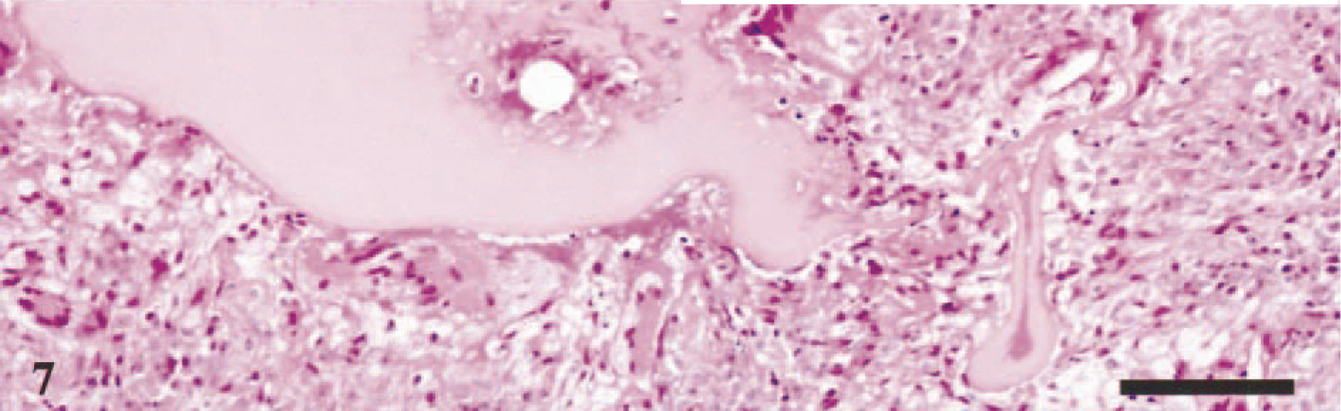
Lung metastasis; dog. Acidophilic material resembling dysplastic dentine or enamel matrix. HE. Bar = 100 µm.
In conclusion, taking together the findings of the fibrosarcomatous proliferation, mesenchymal tissue reminiscent of dental pulp, undifferentiated odontogenic epithelium, biphasic character, frequent apoptosis, and dysplastic dentine or enamel matrix formation, we diagnosed the case as malignant ameloblastic fibro-odontoma. This study is the first known report on a possible malignant ameloblastic fibro-odontoma in dogs with metastasis to distant organs.