
Abstract
A 10-year-old castrated male Standard Poodle presented with an acute onset of lethargy and abdominal pain. The animal had a history of traumatic splenic rupture requiring splenectomy 5 years previously. Surgical exploration revealed multiple cystic red nodules involving all liver lobes, several of which were submitted for histopathology. Microscopically, the cystic nodules were dilated bile ducts and lymphatics surrounded by ectopic splenic tissue. A diagnosis of intrahepatic splenosis was made.
Keywords
Intrahepatic splenosis has been reported rarely in humans and in one pig, 1 typically as an incidental finding at necropsy or during radiologic or nuclear imaging. 2,3 This case report details symptomatic intrahepatic splenosis in a dog several years after trauma to the spleen.
A 10-year-old castrated male Standard Poodle was presented to a veterinary hospital on an emergency basis with an acute onset of lethargy, weakness, and abdominal discomfort after jumping into an automobile and falling onto its abdomen. Five years prior to admission, a splenectomy had been performed after presumed trauma to the abdomen with rupture of the midbody of the spleen and continued abdominal hemorrhage during 5 days of medical management. At that time, histopathology of the spleen confirmed the presence of a hematoma with no evidence of neoplasia.
Upon examination for acute weakness, the animal's abdomen was painful. A large multinodular mass was palpable in the left cranial abdomen. The physical examination was otherwise normal. Radiographs were taken and confirmed the presence of an enlarged liver with an irregular silhouette. A decision was made to perform exploratory surgery the following morning. Preoperative hematologic and biochemical blood analyses were not performed.
Abdominal exploratory surgery was performed. The liver contained multifocal 1- to 8-cm cystic red nodules, which oozed serosanguineous fluid from cut surfaces (Fig. 1). Approximately 65% of all liver lobes were affected, although the left lobes were most severely involved. No other abdominal masses were observed, and splenosis within the mesentery and omentum was not present. A small amount of intraabdominal hemorrhage was noted. After an unsuccessful attempt was made to surgically excise the left medial and lateral liver lobes, euthanasia was elected on the basis of suspicion of widespread hepatic hemangiosarcoma. Several sections of liver were submitted for histopathology.
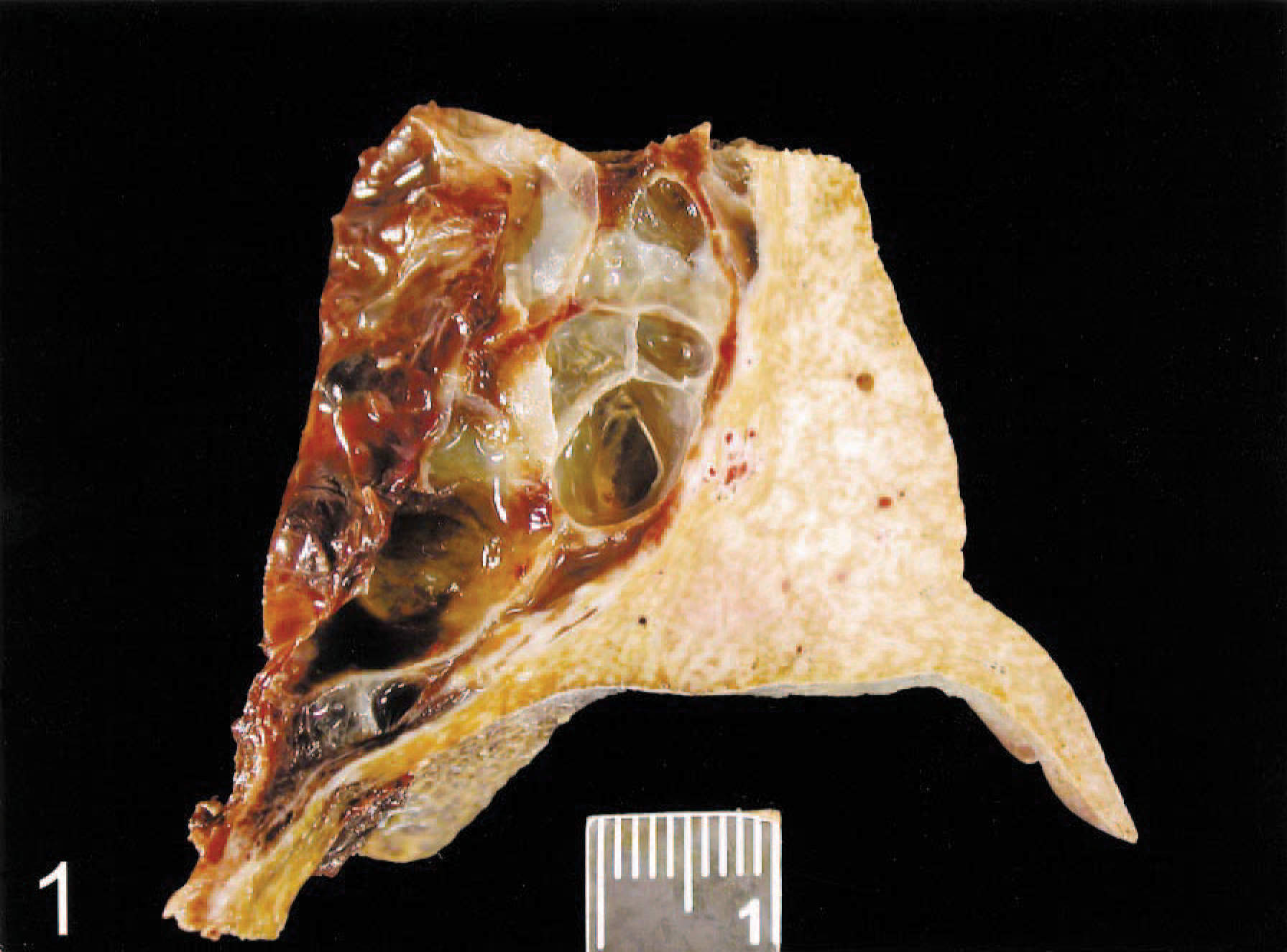
Liver; dog. Formalin-fixed specimen. Multiple cystic nodules within the liver with compression and fibrosis of adjacent hepatic parenchyma. Ruler = 1 cm.
Histologically, all sections of liver contained numerous cystic structures lined by flattened epithelium and containing clear lumens with small numbers of erythrocytes (Fig. 2). The adjacent parenchyma had multiple periportal foci of extramedullary hematopoiesis, stroma consistent with splenic red pulp, and lymphoid nodules. These foci were interpreted as ectopic splenic tissue (Fig. 3). Marked compression atrophy was present, and all portal triads had severe biliary hyperplasia and fibrosis (Fig. 4). Hepatocytes contained abundant yellow-brown cytoplasmic granules, interpreted as bile pigment. Multifocal congestion and acute hemorrhage were present.
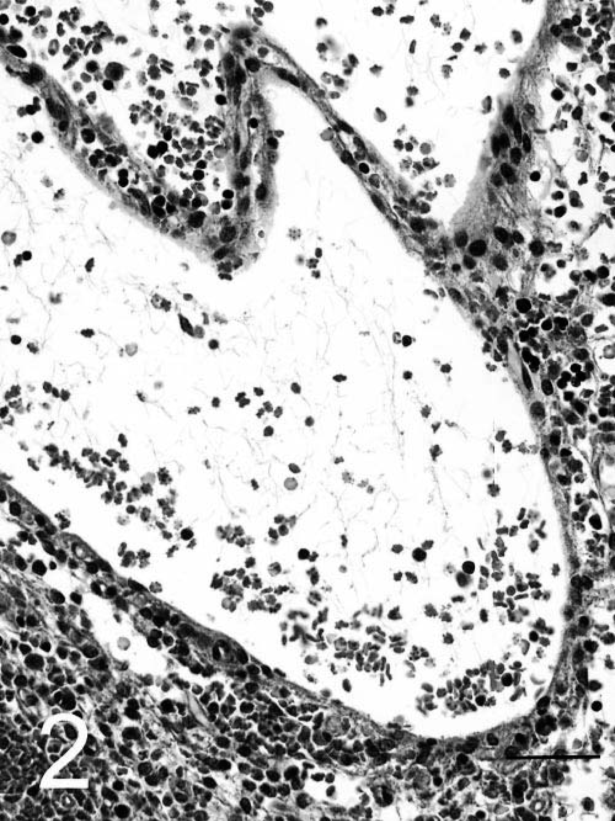
Liver; dog. Multiple cystic cavities filled with erythrocytes and low protein fluid. Hematoxylin and eosin. Bar = 100 µm.
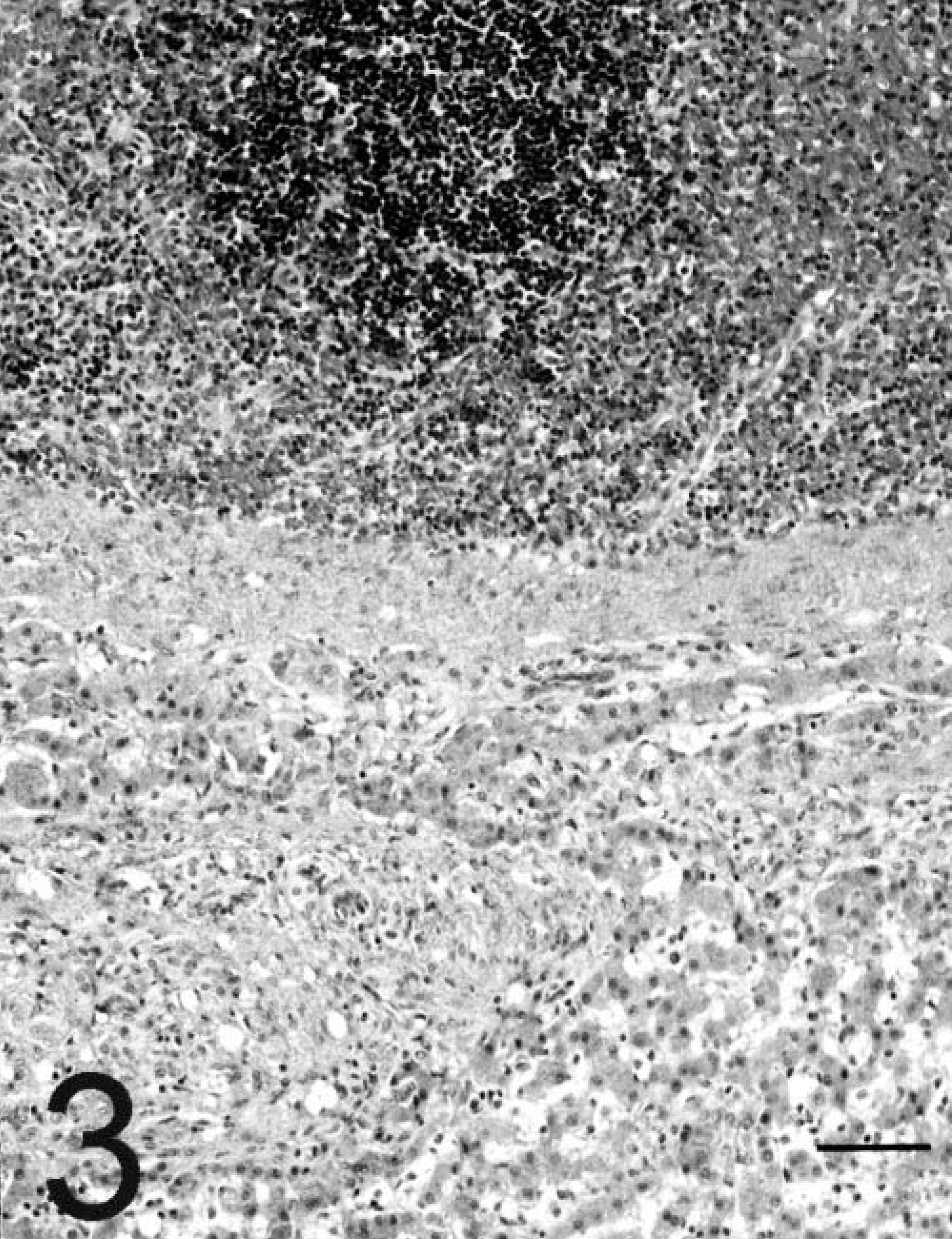
Liver; dog. Ectopic splenic tissue (top of photo) with compression and fibrosis of adjacent hepatic lobules (bottom of photo). Hematoxylin and eosin. Bar = 150 µm.
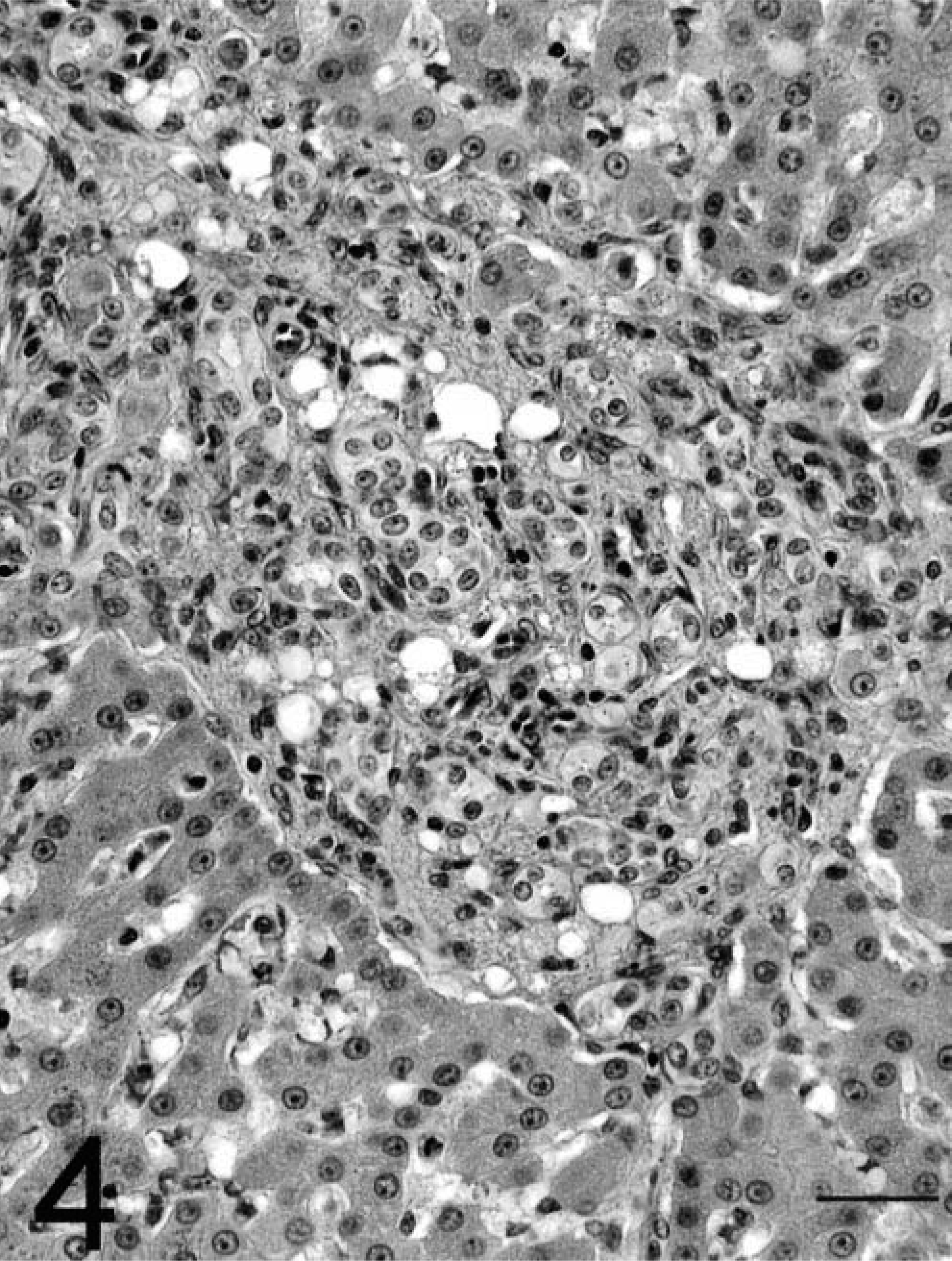
Liver; dog. Portal triad with severe fibrosis andbiliary hyperplasia. Hematoxylin and eosin. Bar = 30 µm.
It is presumed in this case that traumatic splenic rupture resulted in seeding of the liver with splenic tissue via embolization through the splenic vein to the gastrosplenic vein and into the portal vein. Streamlining of portal blood flow in the dog results in blood from the spleen perfusing the left lobes of the liver to a greater degree than the right lobes. 4 Concentration of the splenosis in the left liver lobes, near the location of the venous outflow of the spleen, is supportive of embolization of the liver by this route.
Ectopic splenic tissue has been reported to increase in size after splenectomy. 5 As a result, the autotransplanted splenic tissue in this dog compressed the adjacent hepatic parenchyma, resulting in cystic dilatation of lymphatics and bile ducts. Resultant cysts were susceptible to traumatic rupture and were painful to the animal. Biliary hyperplasia and portal fibrosis are not specific but might represent a response to obstruction of normal outflow by the expanding splenic tissue and cysts. Although liver enzymes were not evaluated preoperatively, the presence of marked accumulation of biliary pigment within hepatocytes suggests that increases in alkaline phosphatase and alanine aminotransferase might have been present in this case.