
Abstract
Proteases play important roles in modulating a wide range of cellular functions, in the regulation of biologic processes, and in the pathogenesis of various diseases. Several molecular techniques are available to identify and characterize proteases in cells and tissues. Most of these techniques do not provide information on the activity of proteases in tissues. In situ zymography (ISZ) is a relatively lowcost technique that uses specific protease substrates to detect and localize specific protease activities in tissue sections. Used in combination with other techniques, ISZ provides data that further our understanding of the role of specific proteases in various pathologic and physiologic conditions. This review describes the general principle of ISZ and highlights the past and future applications of this technique in molecular pathology.
Proteases are enzymes that cleave proteins by the catalysis of peptide bond hydrolysis. On the basis of their catalytic mechanism, proteases are further divided into five catalytic classes: aspartic, cysteine, metalloproteinase, serine, and unclassified. 1 There are more than 500 genes in the human genome that encode proteases or protease-like molecules, with the metalloproteinases and serine proteases comprising the largest classes. 33 Table 1 provides specific examples of proteases from each of the five catalytic classes. Initially only viewed as degradative enzymes that were associated with protein catabolism, it has become increasingly recognized that the highly specific hydrolysis of peptide bonds can regulate a wide range of biologic processes in all living organisms. This highly specific and limited substrate cleavage is referred to as proteolytic processing, a process that regulates the activity and localization of many proteins. 1 , 33 For instance, proteolytic processing controls the intracellular and extracellular localization of many proteins, activates or inactivates various enzymes, cytokines, growth factors or hormones, or generates cryptic neoproteins. 33 Through their catalytic activity, proteases regulate virtually all biologic processes, including, among others, cell proliferation and differentiation, cell migration, apoptosis, wound healing, and angiogenesis. 1 , 33 , 46 Thus, dysregulation of the temporal and spatial expression and activation pattern of these enzymes can result in various pathologic conditions, such as neurodegenerative and cardiovascular diseases, arthritis, and cancer. Consequently, there has been an increasing interest in the identification and functional characterization of proteases. In particular, several of these molecules and their substrates are considered attractive potential therapeutic targets by the pharmaceutical industry. For instance, the matrix metalloproteinases (MMPs) have been identified as important targets for the treatment of various pathologic conditions, such as cancer, arthritis, and cardiovascular diseases, and several MMP inhibitors have already been tested in clinical trials. 5
Catalytic classes of human proteases.
Several molecular techniques are available to identify and characterize proteases in cells and tissues. Northern blot analysis and reverse transcription–polymerase chain reaction (RT-PCR) can be used to quantitate protease messenger RNAs (mRNAs) in cell and tissue extracts, whereas in situ hybridization has been used to localize mRNA expression at the cellular level in tissue sections. A major limitation of evaluation of mRNA levels is that transcriptional activity does not necessarily reflect the amount and activity of the protein product of a particular gene. This is mostly due to variation in cellular location, regulation of translational activity, and complex and versatile protein regulation mechanisms, such as context-dependent posttranslational phosphorylation, sulphation, and glycosylation. 32 Conversely, immunohistochemistry and Western blot are used to evaluate expression at the protein level. However, these techniques do not provide any information on the activity of proteases in tissue because many enzymes are synthesized in an inactive or proenzyme form, called zymogen, which requires proteolytic processing for activation. In addition, ubiquitous protease inhibitors are present in tissues and can specifically or nonspecifically inhibit proteases in their active form. 13 MMPs are a perfect example to illustrate the limitations of immunohistochemistry to localize and quantify specific MMP activities in tissue sections. MMPs are produced and secreted as zymogens, which require proteolytic cleavage of a propeptide domain to be activated. Most antibodies available for immunohistochemistry cannot discriminate between the active and inactive forms of these enzymes. In addition, these antibodies are, in our experience, species-specific and do not cross-react with all species. Moreover, tissue inhibitors of metalloproteinases (TIMPs) can prevent matrix degradation by MMPs, even if the enzymes are in an active form. 13 Hence, other methods are necessary to assess the functional activity of MMPs and proteases in tissues. Biochemical techniques have been developed to detect protease activity in tissue extracts. In particular, gel enzymography, also referred to as substrate zymography, is an established technique for the routine detection and quantitation of various protease activities, such as gelatinase activity. 23 , 43 This technique provides reliable identification of proteases based on the molecular mass of their inactive and active forms after gel electrophoresis. For instance, gelatin zymography has been used to evaluate gelatinase activity in various neoplasms and normal tissues. 46 Other commercial or customized biochemical assays are also available to quantitate specific protease activity in tissue extracts. However, homogenization of tissues for these assays precludes the localization of enzyme activity. In addition, the extraction procedures can artifactually activate enzymes or result in the interaction of active enzymes with their respective inhibitors, which may have been localized in distinct compartments in the intact cells or tissues. 22 , 43 , 45 Likewise, if a specific protease activity is localized in a relatively small part of a diseased tissue, it may not be detected because of its dilution in the entire tissue extract. For these reasons, techniques to localize specific proteolytic activity in tissue sections may provide crucial additional information on the exact role played by certain proteases in various physiologic and pathologic conditions. One of these techniques is in situ zymography (ISZ), an adaptation of substrate zymography to frozen tissue sections. ISZ offers the ability to estimate various protease activities in combination with the localization of these activities in tissue sections. 17 The aim of this mini-review is to outline the general principle of ISZ, provide procedures to establish this technique in the laboratory, give an overview of its development and current research applications using specific examples from the literature, and discuss possible future uses for this technique.
ISZ Principle
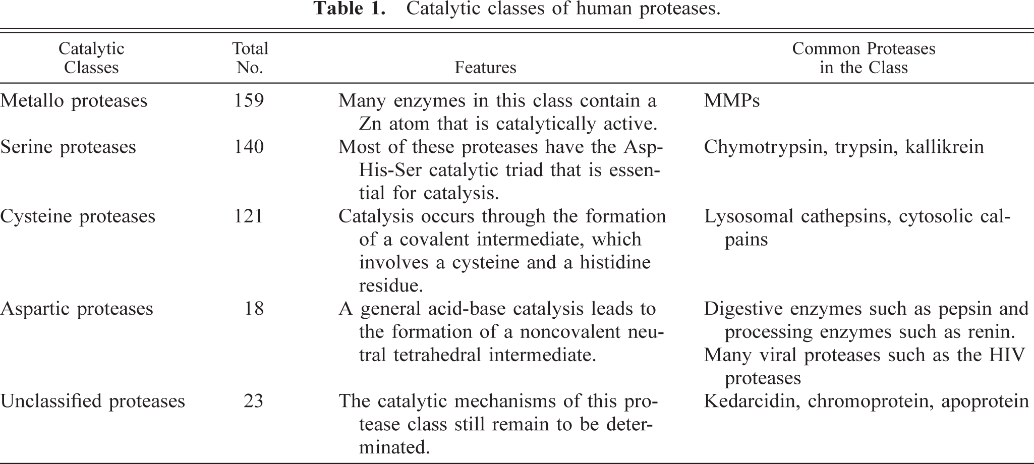
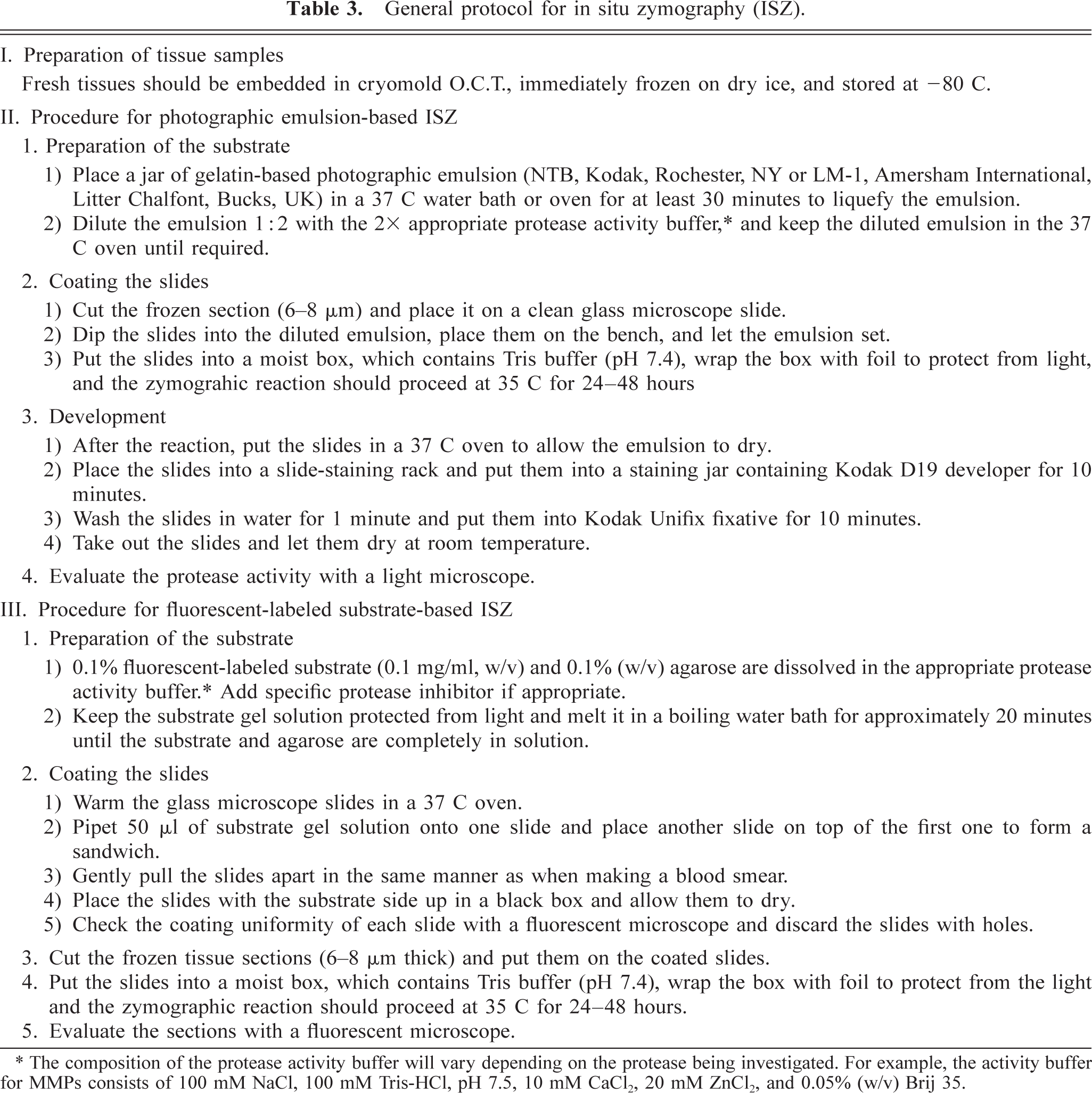
ISZ is a relatively low-cost technique that uses an enzymatic substrate-based support or overlay to detect and, more importantly, localize specific protease activities in tissue sections. ISZ was first used for detection of enzymatic activity released by explants of developing amphibian tissue. 20 Recently, its use has been expanded to localize specific protease activities, mostly MMP activity, in various other tissues. 3 , 6 , 10 , 13–16 , 18 , 19 , 26 , 27 , 30 , 37 , 49 , 52 The general principle underlying ISZ is illustrated in Fig. 1. In brief, an enzymatic substrate specific for certain protease(s) is deposited on or under a frozen section of an unfixed tissue. During the ensuing incubation period, the substrate will be digested in a time- and dose-dependent manner by the appropriate activated enzymes in their native location. After the incubation period, the lysis of the labeled substrate can then be detected by light microscopy or fluorescent microscopy, thereby permitting the precise localization of the specific protease activity in tissue sections. The nature of the substrate dictates which protease activity is to be detected. Table 2 provides a list of the substrates that have been commercialized and used for ISZ and the corresponding protease activity. Used as a complement with other molecular pathology techniques, such as immunohistochemistry, in situ hybridization and Western blotting, ISZ enhances the understanding of the role of proteases in physiology and pathology. It, however, is important to recognize that this technique has limitations regarding quantitation of protease activity; therefore, it does not replace, but rather complements, gel zymography.
Current commercially available protease substrates for in situ zymography.
Two broad approaches of ISZ can be followed on the basis of the nature of the substrates selected. General protocols for these two approaches are given in Table 3. The first approach consists in using a photographic emulsion overlay containing a substrate, such as gelatin. In the photographic emulsion-based ISZ, an unfixed frozen tissue section is mounted on an untreated histologic glass slide and then dipped in an autoradiography emulsion containing the substrate. The coated slides are then incubated at the optimal temperature in a humidity chamber to allow the protease reaction to occur. After incubation, protease activity can be detected using light microscopy as white holes on a black background of unlysed substrate caused by the enzymatic lysis of the substrate-containing photographic emulsion. 17 , 38 A second approach involves the use of a fluorescent-labeled substrate. In this approach, the frozen tissue section is mounted on a glass slide, which has previously been coated with a fluorescent-labeled substrate. After incubation in a dark humidity chamber, the enzymatic activity can be observed by fluorescent microscopy as black holes on a fluorescent background, the lysis of the fluorescent-labeled substrate resulting in the loss of fluorescence. 15
General protocol for in situ zymography (ISZ).
∗ The composition of the protease activity buffer will vary depending on the protease being investigated. For example, the activity buffer for MMPs consists of 100 mM NaCl, 100 mM Tris-HCl, pH 7.5, 10 mM CaCl2, 20 mM ZnCl2, and 0.05% (w/v) Brij 35.
Both types of ISZ have the same endpoint, i.e., the identification and microscopic localization of enzymatic activity within sections of unfixed frozen tissues. However, each approach has its pros and cons. In the photographic emulsion–based approach, the slides cannot be coversliped and are therefore more sensitive to variations in humidity during incubation. An important technical aspect of ISZ resides in the coating of the substrate because uneven thickness of the emulsion layer markedly influences the quality of the results. 15 Because the dipping into photographic emulsion takes places after the mounting of the tissue sections on the slide, it is very difficult to control the even distribution of the substrate overlay on the tissue. In addition, the dipping step requires an experienced person. In contrast, in the fluorescent-labeled substrate approach, microscopic inspection allows elimination of the poorly coated glass slides before placing the tissue sections on the slides. Examination of the slides under fluorescent light can be performed to ensure even coverage. If holes of any size are seen, these slides must be discarded. This limits the number of sections that needs to be incubated and evaluated. However, the sensitivity of the fluorescent-labeled substrate approach is lower because higher levels of enzymatic activity are necessary to produce lysis and holes large enough to be detected. Both approaches of ISZ are highly dependent on achieving an even layer of substrate; therefore, a great deal of user variation is typically observed. To overcome this problem, several groups have modified the initial protocols and have established an improved ISZ technique that has higher sensitivity and is easier to standardize. 19 , 31 , 42 This improved method uses a highly quenched fluorescent-labeled substrate, which is dissolved in protease activity buffer and added to an unfixed frozen tissue section. After incubation in a dark and humid chamber, the unbound substrate is removed by washing in water. Because the presence of protease activity results in the loss of quenching, an increase in enzymatic activity results in a corresponding increase in fluorescence. Under fluorescent microscopy, the enzymatic activity can then observed as a strongly fluorescent zone against a darker, low-fluorescent background (Figs. 2, 3).
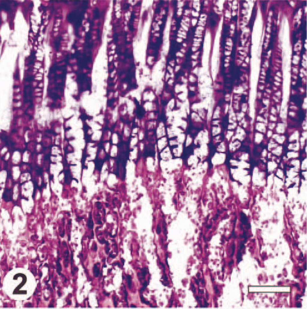
Tibia; rat, HE. Bar = 100 μm.
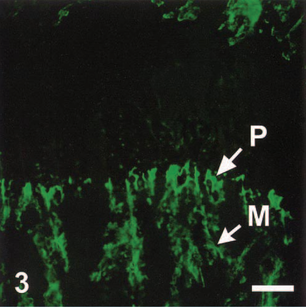
Tibia; rat, ISZ. The protease activity is identified as the strongly fluorescent areas (arrows). Using type IV collagen as a substrate, the protease activities are most likely from MMP-2 and MMP-9. Note that these protease activities are localized in the distal portion of the hypertrophic zone of the growth plate (P), as well as in the metaphysis (M). Bar = 100 μm.
Another limitation of ISZ is that it may be difficult to discriminate between the proteases responsible for the detected activity. This may be partly corrected by the use of complementary techniques, such as immunohistochemistry or in situ hybridization (Figs. 4, 5). However, it is also essential to use appropriate control slides to properly determine the nature of the protease activity present in tissue sections. This can be done by incubating serial sections with the appropriate inhibitors of each protease class. For instance, several MMP inhibitors have been used to confirm that the protease activity observed originated from specific MMPs. Such inhibitors include, for instance, ethylenediamine-tetraacetic acid (EDTA) (which by chelating bivalent ions inhibits the activity of the zinc-dependent MMPs) (Fig. 6), recombinant TIMPs, specific peptides, or various synthetic broad-spectrum or more specific MMP inhibitors, such as 1,10-phenanthroline or bamimastat. 15 , 36 , 37
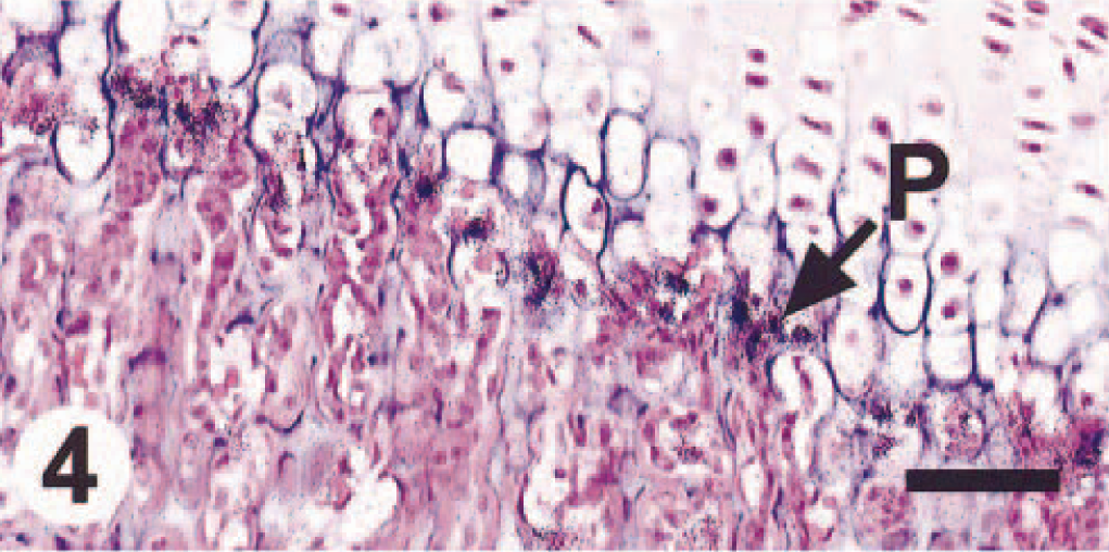
Tibia; rat, in situ hybridization (ISH). The protease activity is mostly due to MMP-9 because it correlates tightly with the signal pattern of MMP-9 mRNA expression as evaluated by ISH. Bar = 100 μm.
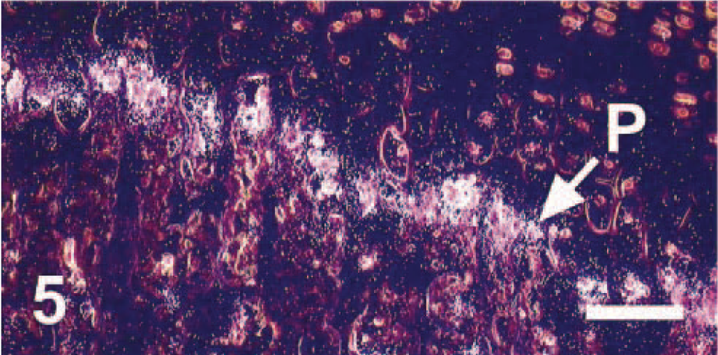
Tibia; rat, ISH. Dark-field microscopy of Fig. 4. Bar = 100 μm.

Tibia; rat, ISZ. When EDTA is incorporated during the incubation, the protease activities (in Fig. 3) can no longer be detected. Bar = 100 μm.
Specific Examples of ISZ Application
ISZ is applicable to virtually any protease, but to date ISZ has mostly been used for the localization of specific MMP activities, especially the gelatinases, MMP-2 and MMP-9. ISZ is becoming more widely used, with a steadily growing number of reports appearing in the literature. Most of the studies using ISZ focus on cardiovascular and neurologic diseases, physiologic studies, and cancer research. It would be beyond the scope of this mini-review to provide an extensive literature review of the use of ISZ. Therefore, we will limit our discussion to specific examples that illustrate the use of this technique. It is important to reiterate that most studies using ISZ also use complementary techniques, such as immunohistochemistry, in situ hybridization, Western blotting, Northern blotting, or gel zymography, to provide a more accurate evaluation of the role of specific proteases in various pathologic and physiologic states. Most of these studies confirmed the important role played by proteases, especially the MMPs, in various diseases and suggest that specific inhibitors of these proteases may be beneficial therapeutic alternatives.
The role of extracellular matrix–degrading proteases, such as the MMPs, in tumor invasion, progression, and metastasis is well established and further supported by the use of ISZ on tumor specimens. 21 , 47 For instance, ISZ has been used to demonstrate that, in most primary melanomas and virtually all melanoma metastases, gelatinase activity is located at the invading part of the neoplasms and especially at sites of tumor-stroma interactions, whereas only weak proteolytic activity is present in the centers of tumor nests. 29 In contrast, when the same neoplasms were evaluated by immunohistochemistry for MMP-2 and MT1-MMP, every tumor area was found to express these two proteases. 22 These results emphasize the importance of functional techniques, such as ISZ, to draw appropriate conclusions as to the role of proteases in cancer, where proteases and their respective endogenous activators are frequently found to be present in close proximity in vivo. In addition to melanomas, ISZ has been applied to various other types of tumors, including, among others, endometrial carcinomas, 4 , 53 dermatofibromas, malignant fibrous histiocytomas, 39 hepatocellular carcinomas, 28 ovarian and breast cancer, 12 , 13 , 25 , 30 and gliomas. 40 , 48 In general, these studies confirmed the role of various MMPs, especially the gelatinases, in cancer invasion, progression, and metastasis by demonstrating a correlation between MMP activity and tumor grade. This suggests that evaluation of the activity of specific MMPs may be a useful predictor of survival in certain cancer types.
ISZ has also been used to study vascular diseases, especially the development and progression of atherosclerotic plaques and aneurysm formation. In human atherosclerotic plaques, gelatinolytic and caseolytic activities were found to be localized and increased in the vulnerable regions of the plaques compared with normal arterial tissues and stable plaques, suggesting that enhanced MMP activity may contribute to plaque rupture. 9 , 14 Similarly, using a well-established mouse model of atherosclerosis, the ApoE knockout mouse, MMP activity was confirmed to be increased after TIMP-1 deficiency, and this increased MMP activity correlated with an increased number of aneurysms in the thoracic and abdominal aorta, suggesting that MMP activity may promote aneurysm formation. 11 The involvement of MMP activity in aneurysm formation is also supported by the demonstration of proteolytic activity in the medial smooth muscle cells of the inferior mesenteric artery or aorta collected from patients undergoing aneurysm repair. 9 , 19 In these studies, Goodall et al. were able to establish a highly sensitive and reproducible ISZ assay by using highly quenched fluorescent-labeled gelatin, which demonstrated increased MMP-2 activity in vascular smooth muscle cells of abdominal aortic aneurysms. 19 Other supporting evidence from ISZ studies for the role of activated proteases in vascular diseases include the increased activity of MMP-2 in rat aortas with aging, 51 the identification of caseinolytic and gelatinolytic activities in atherosclerotic plaques of hypercholesterolemic Syrian Golden hamsters, 8 an increased MMP-2 activity in pulmonary vessels during progression of pulmonary hypertension in rats, 11 and the increased caseinolytic and gelatinolytic activities in smooth muscle cells from surgically injured saphenous veins. 27
The activity of MMPs in various neurologic conditions has also been evaluated with ISZ, and these studies demonstrated the altered balance between multiple extracellular proteases and their inhibitors in diseased tissues. For instance, high caseinolytic and gelatinase activities were localized to areas of brain infarction in rat models of cerebral ischemia. 34 , 42 Increases in gelatinase activity accompanied the progression of neuronal cell death and glial reactivity, suggesting that MMPs are involved in cell viability and tissue remodeling in the ischemic brain. 34 Similarly, ISZ was used to demonstrate the concurrent induction of the plasminogen activator and MMP systems in a mouse model of experimental autoimmune encephalomyelitis 49 and to suggest a role for MMPs in spinal cord injury. 6 Although proteases in general, and MMPs in particular, are usually induced in pathologic conditions of the central nervous system, ISZ data also suggest that these proteolytic activities may, in certain circumstances, be beneficial, such as during the scarring of injured spinal cords. 7
In addition to their involvement in the pathogenesis of various diseases, proteases play critical roles in normal physiology. 41 , 50 In this respect, ISZ has been very useful to gain a better understanding of the role of various MMPs in various physiologic processes. In particular, ISZ has been used in several studies to establish the exact contribution of MMPs during the ovarian cycle. In conjunction with other techniques, such as immunohistochemistry and in situ hybridization, the MMPs and TIMPs have been shown to be major factors in the remodeling of the extracellular matrix as the ovarian follicle grows, ovulates, and forms the corpus luteum and during the structural luteolysis of corpora lutea. 3 , 35 , 44 Likewise, the critical involvement of MMPs in matrix degradation at menstruation was illustrated using ISZ to localize gelatinase and collagenase activities. 52
Potential Future Development and Applications
ISZ can already be used effectively for the localization of protease activity at the cellular and tissue level. However, like any other new technique, ISZ has some current limitations, including poor resolution and limited ability to accurately quantitate protease activity. Another main obstacle for the technique is the scarcity of specific highly quenched fluorescent-labeled protease substrates currently available. Therefore, there is room for improvement, and it is likely that, in the future, various modifications or new approaches will be introduced to increase the applications of ISZ. Among these improvements is the recent development of fluorescent labeling kits that permit the generation of a wide variety of highly quenched fluorescent-labeled substrates in the laboratory comparable with those that are now commercially available (Fig. 3). This should enable investigators to study an increased variety of proteases using more specific substrates and expand the use of ISZ in biological sciences.
Beyond the current applications discussed above, ISZ also has great potential in the validation of preclinical animal models used to test the efficacy of various experimental protease inhibitors. A critical step after the identification of a putative therapeutic target is to validate the target's relevance to the disease process. 2 An important part of this validation step can be achieved through the development of appropriate preclinical animal disease models. 2 It, however, is important to confirm that the target is expressed in these models in a pattern similar to the human disease being targeted. These preclinical models, if appropriate, can then be used to rapidly obtain proof-of-concept data using experimental compounds. In the case of proteases, where most molecular pathology techniques do not provide functional data, ISZ enables investigators to rapidly and inexpensively validate the preclinical models by demonstrating that the correct protease activity is present within the diseased tissues being targeted.
In addition, ISZ has potential use for the ex vivo screening and clinical evaluation of experimental protease inhibitors. When protease inhibitors are incorporated in the protease activity buffer during the incubation period, their inhibitory activity can be evaluated by the decrease in lysis of the appropriate substrate, provided that an appropriate test tissue is available. 15 Similarly, evaluation of protease activity in biopsy samples with ISZ after treatment with experimental compounds could provide important data to evaluate the clinical efficacy of compounds and determine the effective doses for these compounds. Proof of principle that this approach is indeed feasible has already emerged. In a study using the Ma44 human lung cancer xenografts model, oral administration of N-biphenyl sulfonyl-phenylalanine hydroxamic acid (BPHA), a synthetic MMP inhibitor, successfully inhibited gelatinolytic activity in tumor tissues in a dose- and time-dependent manner, whereas an inactive enantiomer of BPHA did not affect the net gelatinolytic activity in tissue sections. 24 Thus, ISZ has great potential to provide surrogate markers of efficacy in tissue specimens.
Conclusions
ISZ is a useful tool that can be used to localize protease activity in tissue sections in an inexpensive and rapid manner. When used in combination with other molecular pathology and molecular biology techniques, ISZ provides data that further our understanding of the role of certain proteases in various pathologic and physiologic processes. This technique also has potential applications in drug discovery and development to evaluate the efficacy and determine the proper dosing regimen of protease inhibitors. Future improvements and refinements of the technique are likely to emerge and should expand its use.
Footnotes
Acknowledgements
We thank Andrew R. Lisowski for providing the in situ hybridization data; Alan C. Opsahl and Steven D. Williams for help with the illustrations; and Dr. Michael J. Schlosser, Dr. Kyle L. Kolaja, and Mary C. Slater for their critical reading of the manuscript.