
Abstract
Three cases of bovine, ovarian epidermoid cysts were diagnosed as unilateral benign lesions in three out of 1,971 cows at slaughter. A stratified, cornifying, and squamous epithelium without associated skin adnexal structures or tissues from other embryonic tissue layers lined the cysts. The cysts were small, usually multiple, and contained keratin debris. Macroscopically, they resembled abscesses. These are benign, congenital lesions that are a separate entity from dermoids and teratomas, and they appear to have a higher incidence in cattle with Bos indicus breeding.
Epidermoid cysts are cysts lined by stratified, cornifying, and squamous epithelium without epidermal adnexal structures and filled with keratin debris or amorphous proteinaceous material.15,16 They usually have a fibrous wall but are not associated with teratomatous elements. In human beings they are described in the skin, brain, spleen, and gonads. Gonadal epidermoid cysts are intraparenchymal structures most commonly reported in prepubertal and postpubertal males.3,7,17 Because the cysts are benign, they are treated successfully with testis-sparing, enucleation surgery. A testicular epidermoid cyst has been described in a dog testicle, but it was classified as a dermoid.11 Ovarian epidermoid cysts are uncommon in women9,16,17 and unreported in the ovaries of domestic species.
In a study of granulosa cell neoplasia, both ovaries from 1,971 cows slaughtered over 3 days were examined at an abattoir. Approximately 98% of the cattle killed at this abattoir were beef or beef crossbreeds, and most included some Bos indicus lineage in their breeding. The ovaries from each animal were examined macroscopically both by palpation and by following one transverse section. Case No. 1 was from a heifer having a 5.5- × 4.0- × 3.0-cm left ovary with over a dozen 0.5- to 2.5-cm smooth-walled cysts containing liquid to pasty white material (Fig. 1a, b). The opposite ovary was normal but inactive. Case No. 2 was the right ovary from a mature cow. This ovary had three (2 × 1.5, 2.0 × 1.5, and 2.0 × 0.5 cm) smooth-walled cysts (Fig. 2) containing viscid, off-white fluid. The left ovary appeared normal and had developing follicles and a mature corpus luteum. Case No. 3 was the right ovary of a mature cow. This ovary contained a 4.2- × 2.5-cm thin-walled cyst filled with clear fluid and a 3.0- × 1.5-cm cyst containing caseous, gray material (Fig. 3). The left ovary contained developing follicles and a mature corpus luteum. The uterus contained a normal, 35-cm (crown to rump length) male fetus. Histology of the three abnormal ovaries was similar. Multiple sections of the ovaries demonstrated that the cysts, which macroscopically were seen to contain off-white, pasty or viscid material, were lined by stratified, keratinizing, squamous epithelium that surrounded uncompacted keratin (Fig. 4a, b). No difference in the thickness of the epithelium or the degree of keratinization was noted among the cysts, and most keratin debris was lost during tissue processing in all cases. Using Masson's Trichrome staining, no smooth muscle was demonstrable around the cysts, indicating that the cysts did not represent the remnants of mesonephric structures.11 The 5-cm cyst in case No. 3 was lined by a simple, cuboidal, occasionally ciliated epithelium typical of a cystic rete ovarii. In the affected ovaries, although subsurface tubular structures and granulosa cell islands were occasionally present, no follicles or oocytes were observed. The uterine tubes, uteruses, and contralateral ovaries were histologically normal.
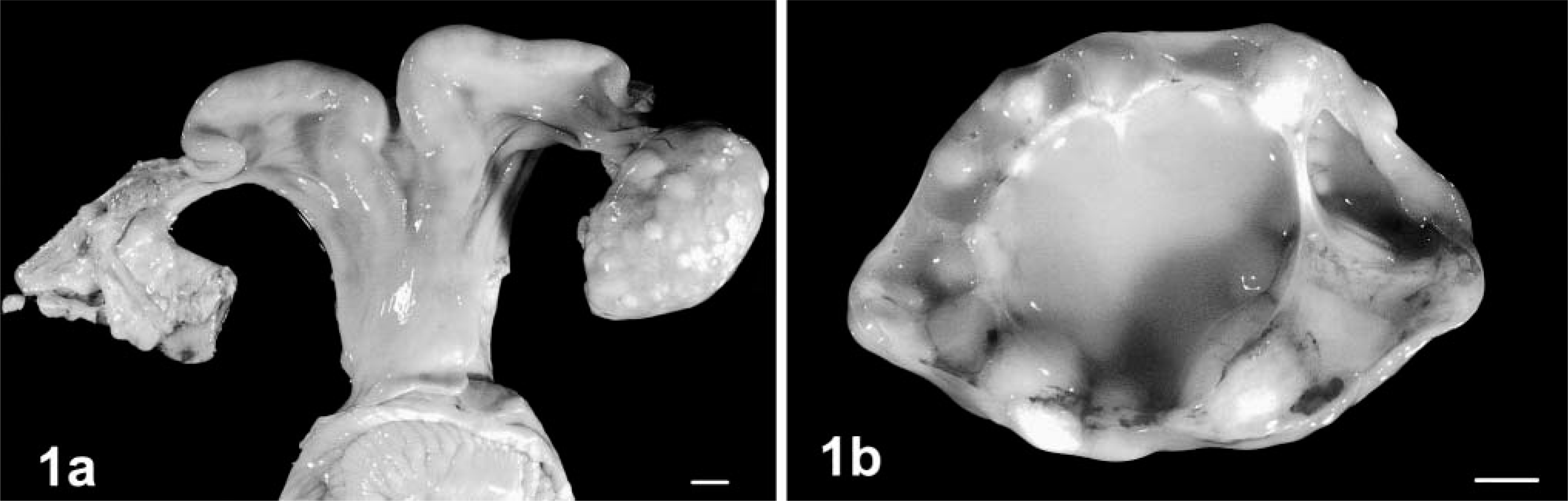
Reproductive tract of a heifer with epidermoid cysts; case No. 1.
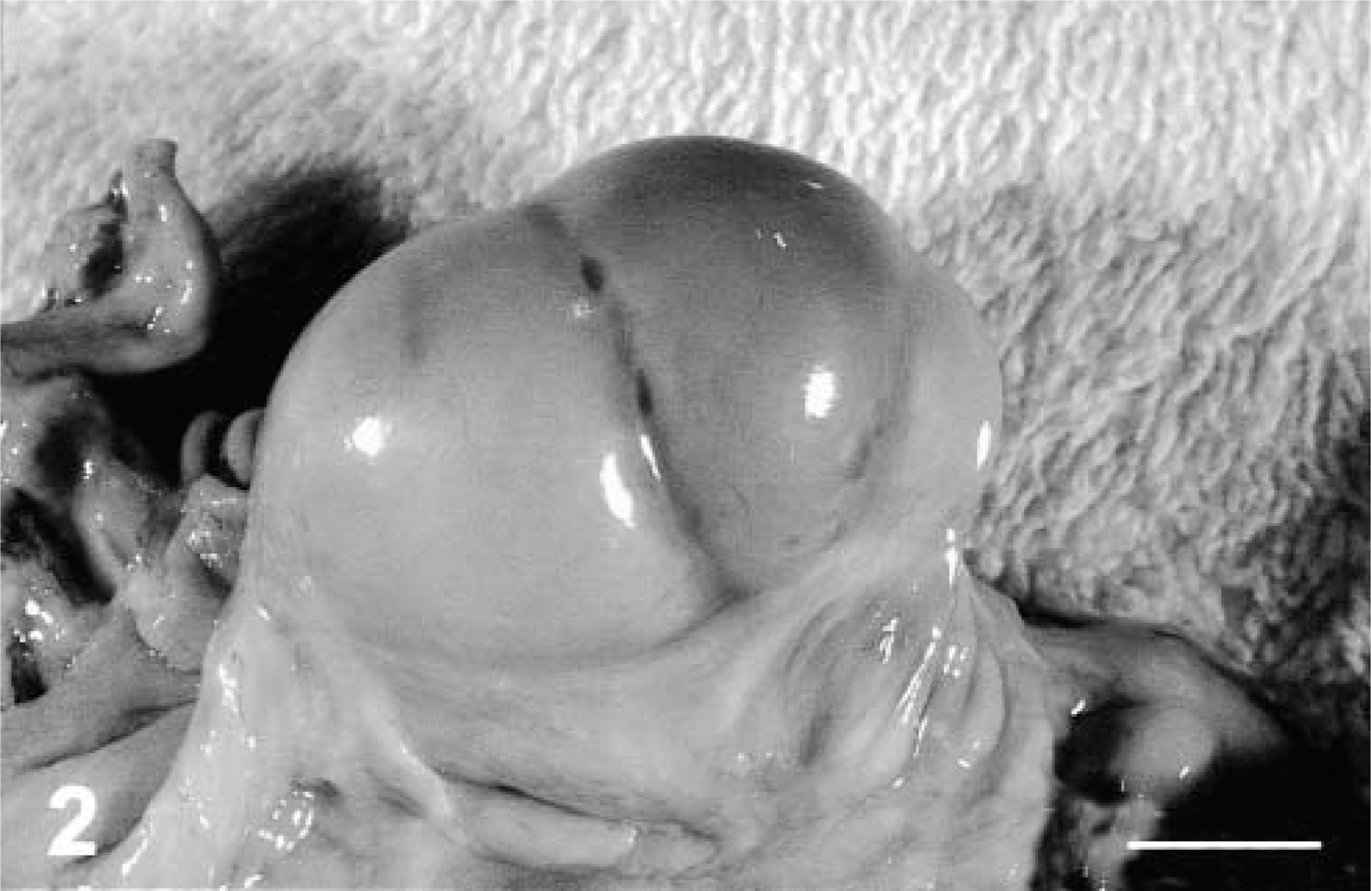
Bovine ovary with three, smooth-surfaced epidermoid cysts; case No. 2. Bar = 2 cm.
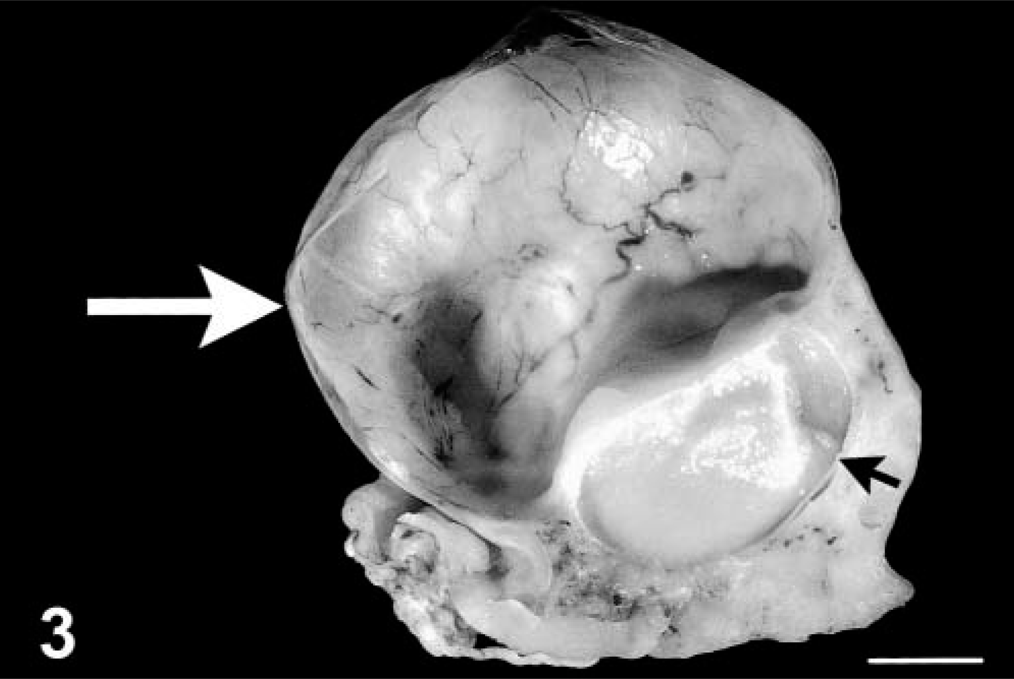
Formalin-fixed bovine ovary; case No. 3. On section, a large, cystic rete ovarii (large arrow) with its watery content removed and a small, epidermoid cyst (small arrow) containing caseous material are noted. Bar = 1 cm.
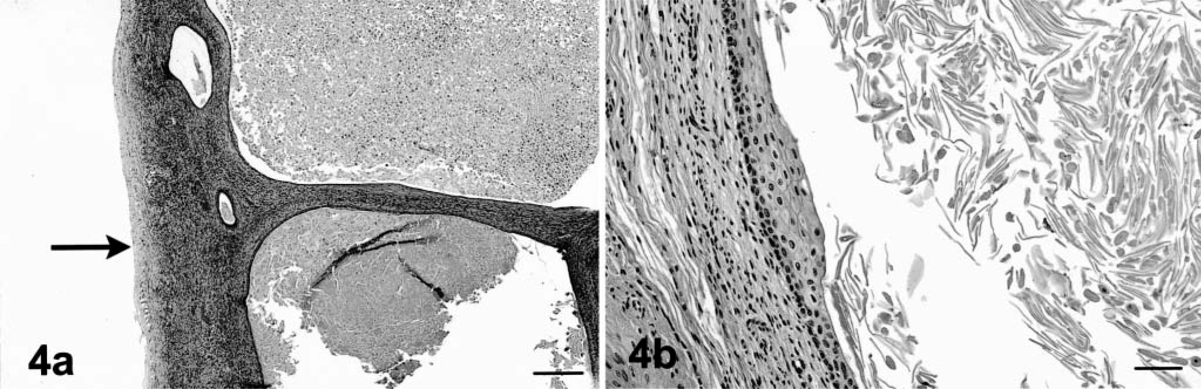
Histologic section of bovine epidermoid cysts; case No. 1.
Ovarian epidermoid cysts have not been reported in the veterinary literature. Human ovarian epidermoid cysts are rare, but one report suggested they may be more common in the ovaries of Chinese women.9 The authors of that report suggested that these cysts may be misdiagnosed as dermoids or teratomas. Dermoids and teratomas have different histologic features.14 In the human literature, dermoids are also called benign cystic teratomas, and besides stratified squamous epidermis with adnexa, they usually contain a variety of tissues, including neural tissue, thyroid gland, and mesenchymal proliferations. All three embryologic layers may be found in the dermoids of human beings. To find all the elements, examinations of multiple sections may be necessary because the extracutaneous elements may be limited to a small solitary nodule, the Rokitansky or embryonal nodule, in the wall of the cyst. In teratomas, tissues from three embryologic layers are observed as prominent features of the specimens, but they are often less organized. Dermoids and teratomas containing cysts lined by keratinizing, stratified, squamous epidermis are called complex or mixed epidermoid cysts.16 Simple epidermoid cysts do not contain epidermal adnexal structures such as hair and sebaceous glands, and they do not contain structures from the other embryonic germ layers.17 There is no report of the recurrence or metastasis of simple epidermoid cysts after removal. Metastasis is documented in some cases with complex, testicular, human epidermoid cysts.17 Human, testicular epidermoid cysts occurring as solitary nodules are regarded by some as a stage in the development of a teratoma.7,17 Although human teratomas are often malignant, teratomas rarely metastasize in domestic animals.7,14 In veterinary classifications, dermoids only contain cutaneous tissue derivatives, whereas a teratoma contains tissues of two or three embryologic layers.12,16 The term dermoid should not be used in reference to a neoplasm because the term is applied to nonneoplastic, congenital, and acquired lesions in the skin and other organs such as the eye and the brain. Epidermoid cysts are probably congenital malformations also and should not be considered as neoplasms.
In a study of ovarian cysts in Australian cattle,8 squamous metaplasia of the rete ovarii was mentioned in two cows with cystic Graffian follicles and nymphomaniacal behavior. No macroscopic images or histology were shown, and no endocrinologic data were provided. The metaplasia in those cases is presumed to be caused by hyperestrogenism. The normal rete ovarii is located near the ovarian hilus and is lined variably by simple cuboidal to columnar or ciliated columnar epithelium that may form papillary structures.11 Although data on hormone concentrations are not available, follicular or luteal cysts were not seen in the cases reported here, and rete ovarii with a normal lining were often noted in these cattle with epidermoid cysts. It is therefore felt that epidermoid cysts are formed by ectopic, ectodermal epithelium and not through abnormal differentiation of rete ovarian tubules. The frequent colocalization of epidermal cysts and rete ovarii in the medulla of the ovary is believed to reflect the common embryologic, hilar origin of both ovarian structures.
The relatively high incidence of ovarian epidermoid cysts in cows of this study is unusual given the absence of previous reports of this condition in the veterinary literature. It is probable that these lesions are misdiagnosed or are small and thus overlooked. Descriptions of some reported dermoids and teratomas are more compatible with those of a diagnosis of epidermoid cysts.2,12,13 In domestic animal species, teratomas are reported sporadically in male and female gonads, but they are reported most frequently in the equine testes.14 They occur in ovaries of all species. Water buffalo cows have a higher incidence of teratomas and dermoids than other domestic cattle.10 Comprehensive abattoir and archival surveys of reproductive tract lesions in B. taurus cattle infrequently document ovarian teratomas.1,6,12–14 Most of the cattle in this study were of B. indicus breeding, and it has been suggested that B. indicus cattle may have a higher incidence of dermoids and teratomas.4,5,12 Epidermoid cysts macroscopically resemble abscesses; thus, epidermoid cysts may be misdiagnosed as an ovarian abscess or a suppurative, tubo-ovarian cyst resulting from chronic salpingitis. Because the cysts can be small and located below the cortex near the hilus and because they do not necessarily result in infertility, most epidermoid cysts may not be submitted to the pathologist for identification.