
Abstract
A solitary mass approximately 1.5 X 2 cm located on the outer side of the third digit of the left forepaw of a 7-year-old male cross-breed cat was examined pathologically. The excised tumor mass was hard and white and located within the deep dermis and subcutis. Histopathologically, the mass consisted of a mixed population of small round epithelioid cells arranged in ribbon- or cordlike structures and spindle-shaped cells forming loose irregular bundles in a mucinous stroma. The epithelioid cells were often arranged around small blood vessels. Neoplastic cells were intensely positive for vimentin and alpha smooth muscle actin and negative for keratin, desmin, S-100 protein, and neuron-specific enolase. Based on these pathologic features, the tumor was diagnosed as a glomus tumor, a neoplasm not previously reported in cats and extremely rare in animals.
Glomus tumors are neoplasms arising from the glomus body, a specialized form of arteriovenous anastomosis.7 In humans, the pathologic features of this unique neoplasm are well documented by many case reports and review articles.7 This tumor commonly develops in the deep dermis and subcutis in the subungual region of the finger, although it sometimes arises in other regions, including the bone, stomach, and nose.7 Histologically, this tumor is characterized by perivascular proliferation of plump, round epithelioid glomus cells, which are closely associated to vascular smooth muscle cells. Human glomus tumors have several morphologic variants based on the relative proportion of glomus cells, vessels, and smooth muscle cells and are typically classified into three major subtypes: glomus tumor proper, glomangioma, and glomangiomyoma.
Some information on glomus tumors in animals is available, and previous reports have documented a glomus tumor in a dog5 and glomangiomas in nonhuman primates, including four irradiated macaques.3,4 Shinya et al.5 reported a canine case of glomus tumor that developed in the medial aspect of the right arm and emphasized its morphologic similarity to human glomus tumors. Here, we describe the pathologic features of a feline digital tumor having morphologic characteristics similar to those of human and canine glomus tumors.
A solitary mass on the outer side of the third digit of the left foreleg was found in a 7-year-old male cross-breed cat. When the owner first recognized the mass, it was approximately 2 cm in diameter. At the owner's request, the mass was surgically excised with an insufficient surgical margin so that the third digit could be preserved. The excised mass, located within the deep dermis and subcutis, was hard, white, and 1.5 × 2 cm. The whole mass was fixed in 10% formalin for routine histologic examination. Paraffin sections, 2–4-μm thick, were prepared and stained with hematoxylin and eosin (HE), periodic acid–Schiff (PAS), and Gomori's methenamine silver (GMS). For a control tissue containing normal glomus bodies, cutaneous tissues from the forelimb and hind limb digits of a 5-year-old cross-breed cat were taken at necropsy. Immunohistochemical stains were performed using the Envision polymer reagent (Dako-Japan, Kyoto, Japan). Primary monoclonal antibodies were against vimentin (1:100 clone V9, Dako-Japan), alpha smooth muscle actin (SMA, prediluted; Dako-Japan), neuron-specific enolase (NSE, prediluted; Dako-Japan), desmin (prediluted D33; Dako-Japan), and factor VIII–related antigen (prediluted; Dako-Japan). Primary polyclonal antibodies were against keratin (prediluted, wide range; Dako-Japan), and cow S-100 protein (prediluted; Dako-Japan).
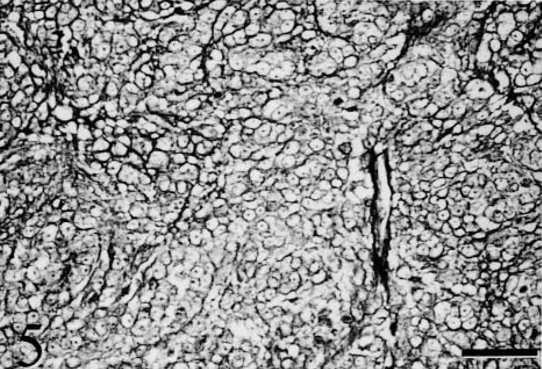
Normal feline glomus bodies were occasionally found in the deep dermis of the digital cutaneous tissues examined and glomus cells appeared as plump, round epithelioid cells around blood vessels (Fig. 1). Immunohistochemically, the normal glomus cells were positive for SMA and vimentin and were negative for keratin, desmin, S-100, NSE, and factor VIII–related antigen. Histopathologically, the digital mass was located within the subcutis and loosely demarcated from the dermis by collagenous tissues. The neoplastic foci in the subcutis consisted of small, round epithelioid cells with hypochromatic round nuclei and scant amphophilic indistinct cytoplasm. These small, round epithelioid cells were frequently arranged in ribbon- or cordlike structures (Fig. 2). In some areas, spindle-shaped neoplastic cells were present, forming loose irregular bundles within a mucinous stroma (Fig. 3). Neoplastic cells were frequently centered around small and middle-sized blood vessels (Fig. 4). Individual neoplastic cells were encircled by a well-developed network of thin PAS- and GMS-positive fibrous material (Fig. 5). Immunohistochemically, almost all neoplastic cells were intensely positive for vimentin and SMA (Fig. 6). The spindle-shaped neoplastic cells usually showed more intense immunoreactivity for SMA than did the epithelioid cells. All neoplastic cells were negative for keratin, desmin, factor VIII–related antigen, S-100, and NSE.
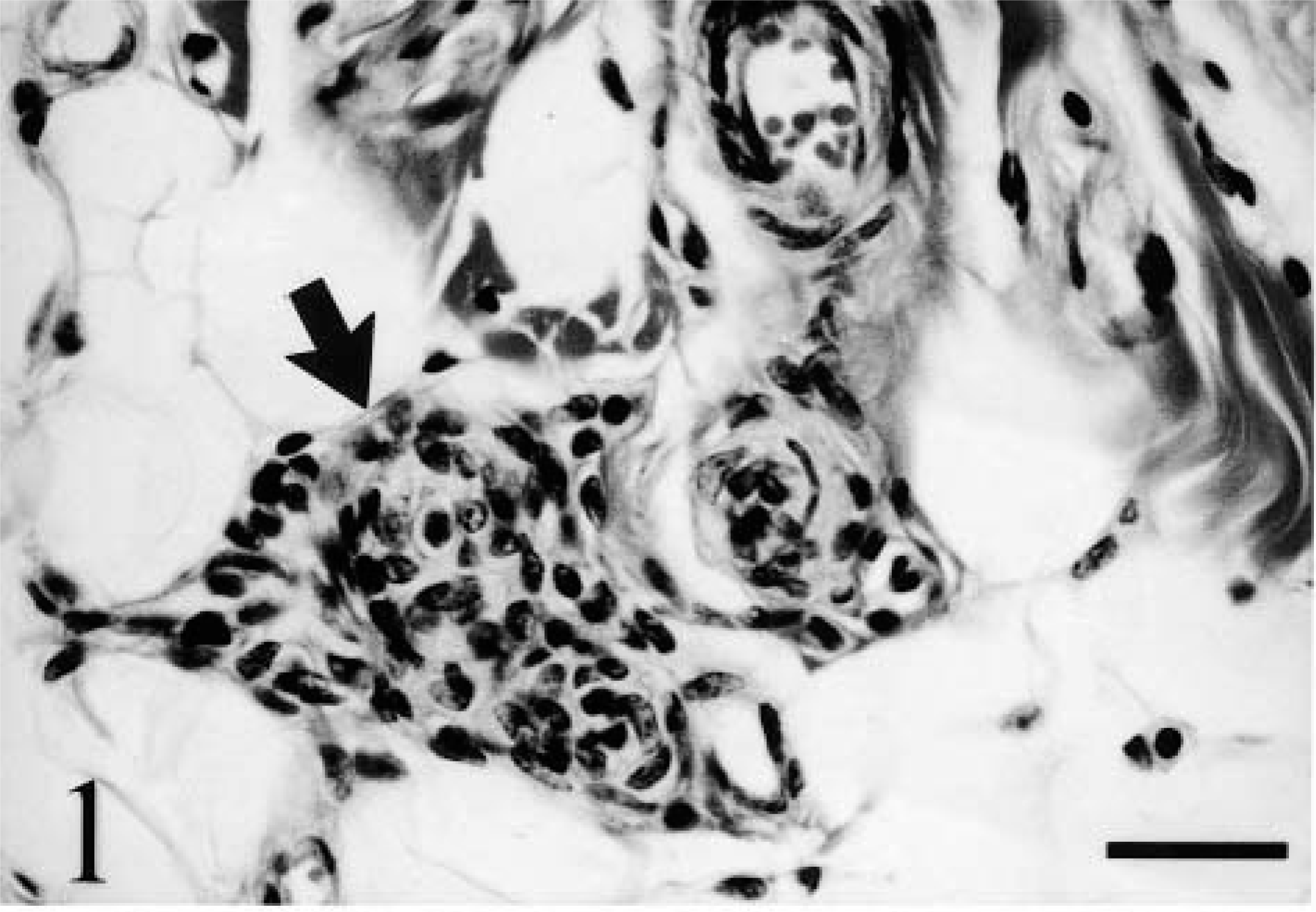
Normal digit; cat. Subcutaneous tissue has a glomus body. Note the plump round epithelioid cells (arrow) around a blood vessel. HE. Bar = 20 μm.
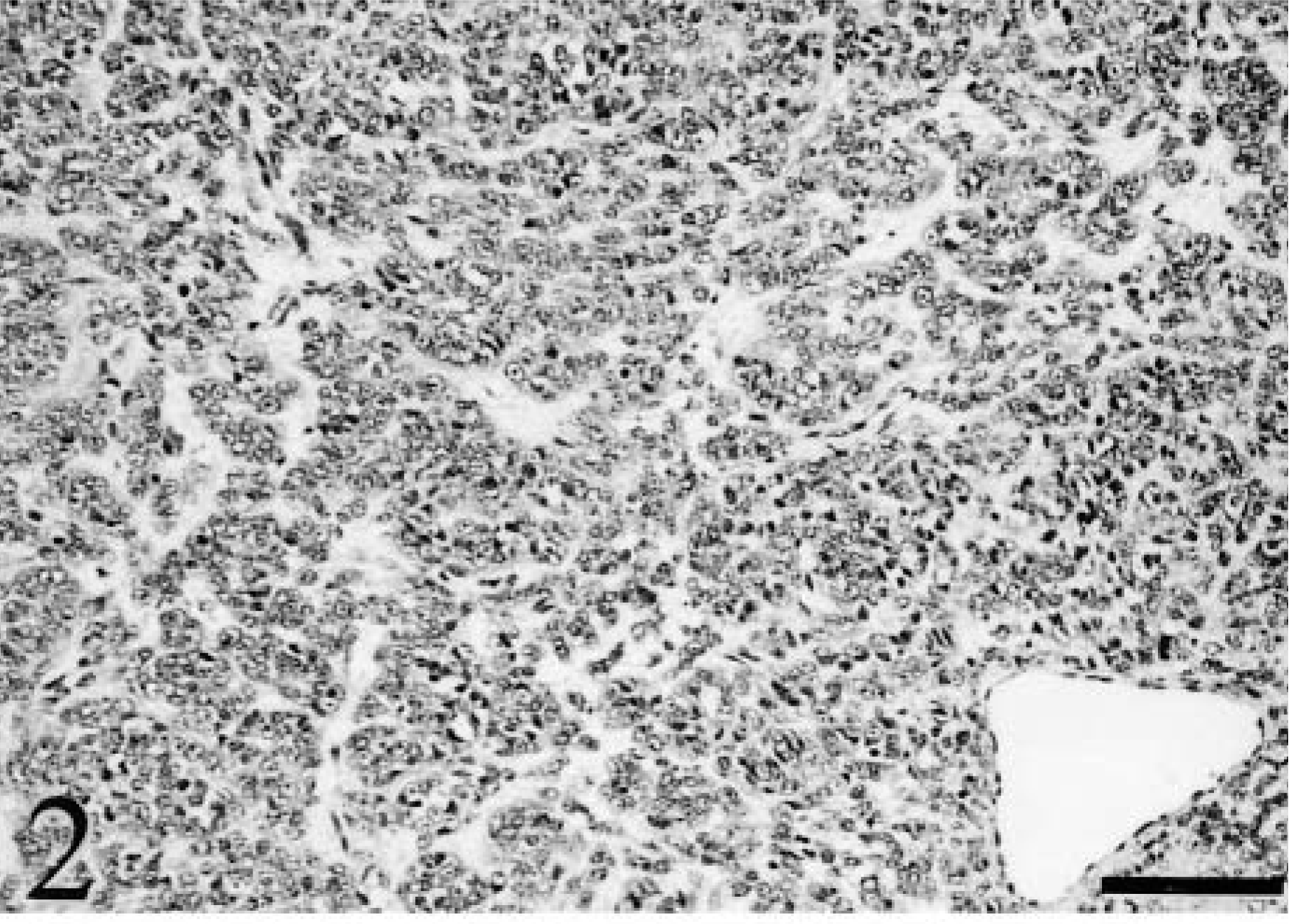
Digital mass; cat. Most of the neoplastic mass consisted of small, round epithelioid cells arranged in ribbon- or cordlike structures similar to a basal cell tumor pattern. HE. Bar = 80 μm.
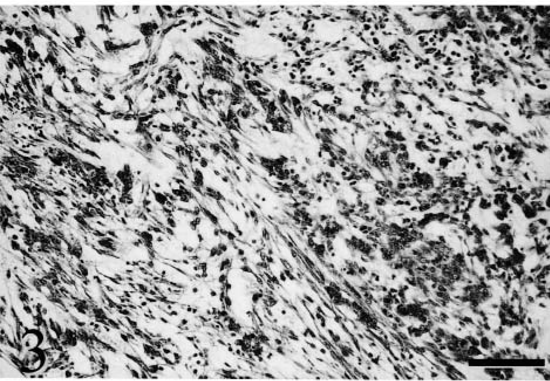
Digital mass; cat. Scattered areas consist of spindle-shaped cells forming loose irregular bundles within a mucinous stroma. HE. Bar = 80 μm.
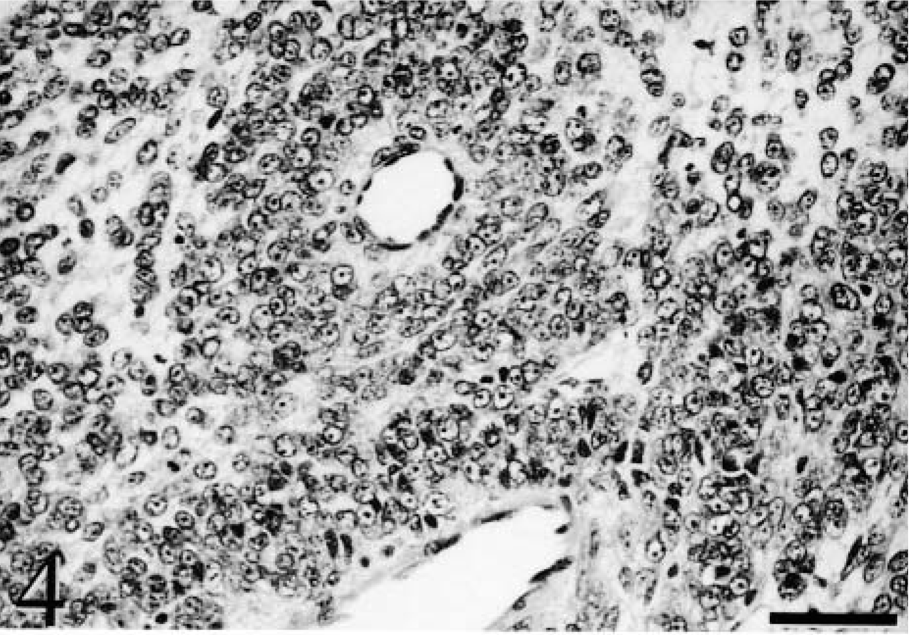
Digital mass; cat. Note the small round epithelioid cells around large and small blood vessels. HE. Bar = 40 μm.
Digital mass; cat. Note the well-developed network of silver-positive fibers surrounding individual tumor cells. GMS. Bar = 40 μm.
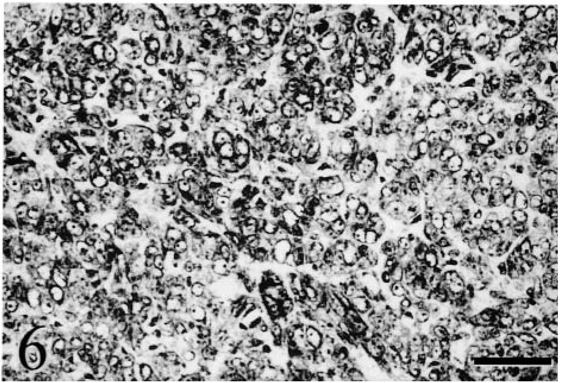
Digital mass; cat. Note the intense cytoplasmic immunoreactivity of neoplastic cells against SMA. SMA immunohistochemistry. Bar = 40 μm.
These histopathologic and immunohistochemical findings indicated that this neoplasm had originated from smooth muscle cells or their derivatives. The anatomic location and morphologic features of this neoplasm were consistent with those of human glomus tumors, especially the glomus cell proper type, although some areas had glomangiomyoma-like foci characterized by spindle-shaped cells forming loose irregular bundles within a mucinous stroma.7 The foci consisting of epithelioid cells arranged in cord- and ribbonlike structures were also similar to some epithelial tumors, such as basal cell tumors; however the immunohistochemical features of the neoplastic cells were inconsistent with an epithelial origin.
Shinya et al.5 determined that the closest differential diagnosis in their case of canine glomus tumor was epithelioid leiomyoma, although this tumor is not a well-identified neoplasm in animals.2 The cellular morphology, some aspects of the growth pattern, and the immunohistochemical characteristics of human epithelioid leiomyomas are similar to those of glomus tumors,1 and these tumors occur frequently as large masses within the human gastrointestinal tract, abdominal cavity, and uterus.1,6 In contrast, the present tumor developed in the digit, which is the most common anatomic location of glomus tumors in humans. In addition, the unique growth pattern around small blood vessels suggested that this neoplasm arose in a glomus body.
In animals, hemangiopericytoma, rather than epithelioid leiomyoma, might be the closest entity to consider in the differential diagnosis. Recently, Weiss and Goldblum7 classified glomus tumors and hemangiopericytomas in the same group because of their anatomic similarity and suggested that glomus cells were more closely related to smooth muscle cells than to pericytes. In animals, hemangiopericytomas are the most common soft tissue tumors in dogs but are unusual in cats.2 The morphologic features of the present tumor did not resemble those of “canine” hemangiopericytomas but were similar to those of “human” hemangiopericytomas.7 It is uncertain whether canine and human hemangiopericytomas are completely equivalent, although both tumors show unstable immunoreactivity for SMA, S-100, and NSE.2,7 Recently, CD34 expression has been reported to be the most specific immunohistochemical feature of human hemangiopericytomas.7 In contrast, vimentin and muscle actin isoforms can be identified in almost all human glomus tumors, whereas the immunoreactivity for desmin is variable.7 The distinct immunoreactivity for SMA and vimentin and the lack of S-100, NSE, and desmin expression did not support a diagnosis of hemangiopericytoma but were more consistent with a glomus tumor. Because the affected cat did not show recurrence or metastasis 8 months after surgical excision in spite of the insufficient surgical margin, the tumor appeared to have a rather benign behavior, as has been described for human glomus tumors.