
Abstract

Keywords
Ceroid-lipofuscinoses are a complex group of inherited neurodegenerative storage diseases that occur in humans and animals and are characterized by the progressive accumulation of a pigment similar to lipofuscin and ceroid within neurons and other cells of the body. In humans, there are four forms of the disease: infantile, late infantile, juvenile (Batten's disease), and adult (Kuff's disease). 8 A comparable spread was found in animals, especially in the dog. 8
Ceroid-lipofuscinosis has been reported in several breeds of dogs, including English Setters, 4 Border Collies, 9 Cocker Spaniels, 2 5 Dachshunds, 10 Salukis, 1 and Chihuahuas. 7 Similar disease has been studied in Siamese cats, South Hampshire and Rambouillet sheep, Devon cattle, Nubian goats, and primates. 8
Although the stored pigment has similar staining and fluorescent properties similar to those of ceroid and lipofuscin, the major constituent of this pigment is a lipid-binding protein that is a component (subunit c) of mitochondrial ATP synthase. 3 Studies on the ovine, bovine, canine, and human (late infantile and juvenile diseases) pigments have shown storage of subunit c. Patients with the infantile form of ceroid-lipofuscinosis and Miniature Schnauzer dogs store a different protein, identified as sphingolipid activator protein (or saposins). 6 There are two families of ceroid-lipofuscinoses: Those characterized by storage of subunit c and those in which saposins accumulate. 6 Here, we describe ceroid-lipofuscinosis in an adult Cocker Spaniel dog. This is the first reported case of ceroid-lipofuscinosis in Argentina.
A 4-year-old female Cocker Spaniel dog was presented to the referring veterinarian with progressive difficulty in walking. The clinical examinations showed no abnormalities except the gait. Neurologic examinations revealed hypermetric ataxia and proprioceptive deficits in all four limbs. The dog was euthanatized because of the progressive nature of the disease and the lack of response to treatments, and the body was submitted to the Department of Pathology at the Buenos Aires Faculty of Veterinary Science for postmortem examination.
At necropsy, no significant gross abnormalities were found, except a mild cystitis. The brain and spinal cord and samples of urinary bladder were fixed in 10% formaldehyde neutral solution. Representative blocks were processed for paraffin embedment, sectioned at 5 μm, and stained with hematoxylin and eosin (HE). The following special stains were applied to paraffin-embedded sections: periodic acid–Schiff (PAS), Sudan black B, luxol fast blue, and thionine. Unstained frozen sections of the spinal cord were examined with fluorescence microscopy. Small formalin-fixed blocks from the spinal cord were postfixed in 2% osmium tetroxide in phosphate buffer and processed for electron microscopic examination.
Microscopic examination of slides stained with HE revealed storage of yellow-brown granules in the cytoplasm of some neurons of the brain and spinal cord. The amount of stored pigment varied in different anatomic areas. Deposits were most pronounced in neurons of the spinal cord (both dorsal and ventral horn) and basal bodies of the cerebellum (Fig. 1) but were less evident in neurons of the cerebral cortex and brain stem and in Purkinje cells. The most affected neurons were swollen, and their nuclei and Nissl bodies were displaced to the periphery; others looked normal except for small amounts of stored material. The aggregations of granules were commonly at one pole of the neuronal body, often near the axon hillock (Fig. 2). Neuronal necrosis with astrocytosis and aggregation of gitter cells were found in basal bodies of the cerebellum and spinal cord, but loss was not a common feature. The pigment was also evident in the cytoplasm of glial cells. Similar but fewer yellow-brown granules were found in the cytoplasm of the smooth muscle cells of the urinary bladder (Fig. 3) and in the wall of small muscular arteries.
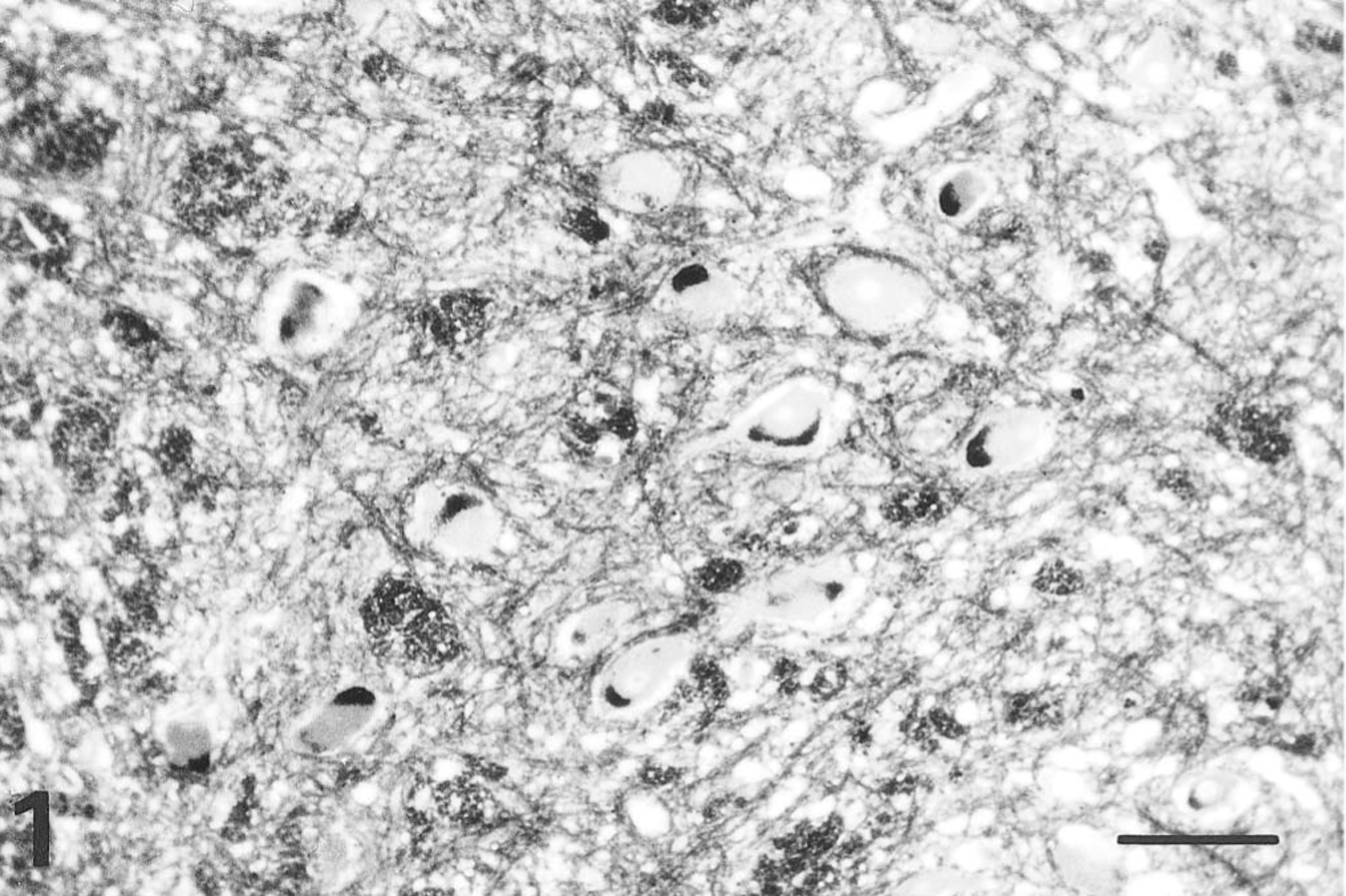
Cerebellum; dog. Neurons of the basal bodies contain stored pigment in the cytoplasm. Sudan black. Bar = 100 μm.
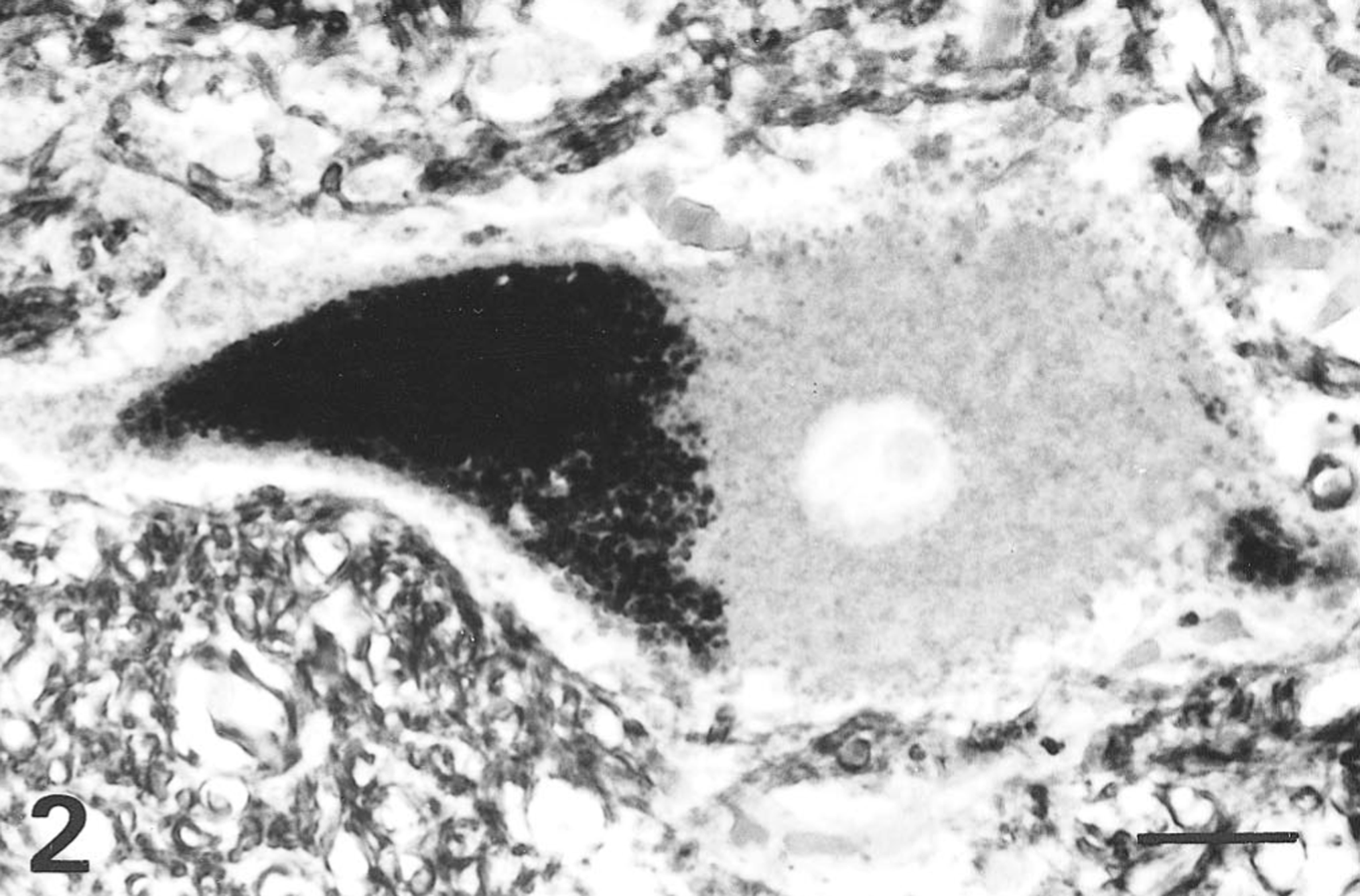
Cerebellum; dog. Heavy aggregation of granular material at one pole of a neuron. Sudan black. Bar = 16.5 μm.
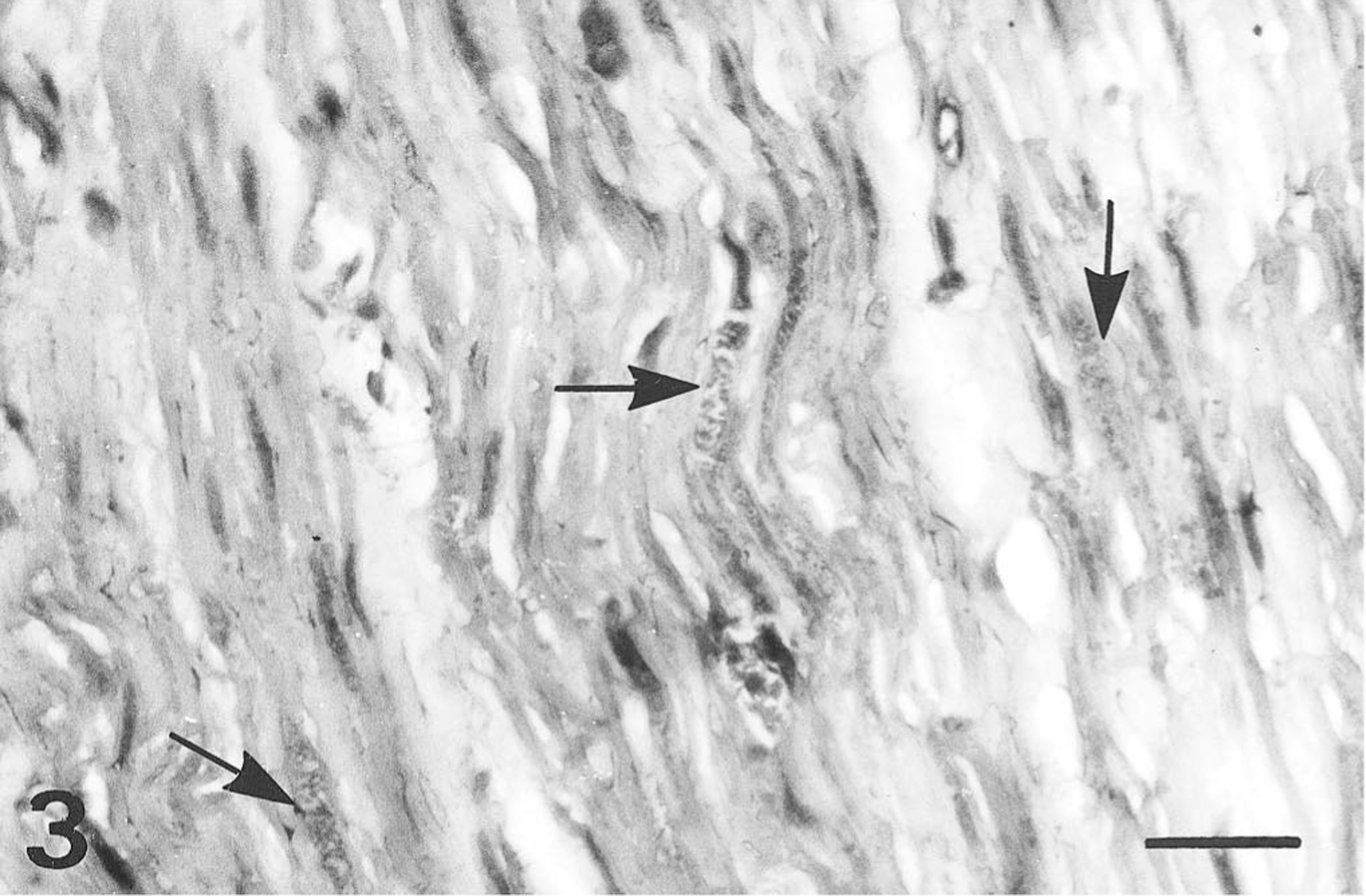
Urinary bladder; dog. Mild accumulation of granular material in the cytoplasm of smooth muscle cells (arrows). PAS, Mayer's hematoxylin counterstain. Bar = 25 μm.
The stored granules stained magenta with PAS, black with Sudan black B, blue with luxol fast blue, and green with thionine. Fluorescence microscopic examination showed masses of yellow-green autofluorescent cytoplasmic granules.
Electron microscopic examination of affected neurons revealed a variable number of cytosomes containing some granular and amorphous but mostly membranous material (Fig. 4).
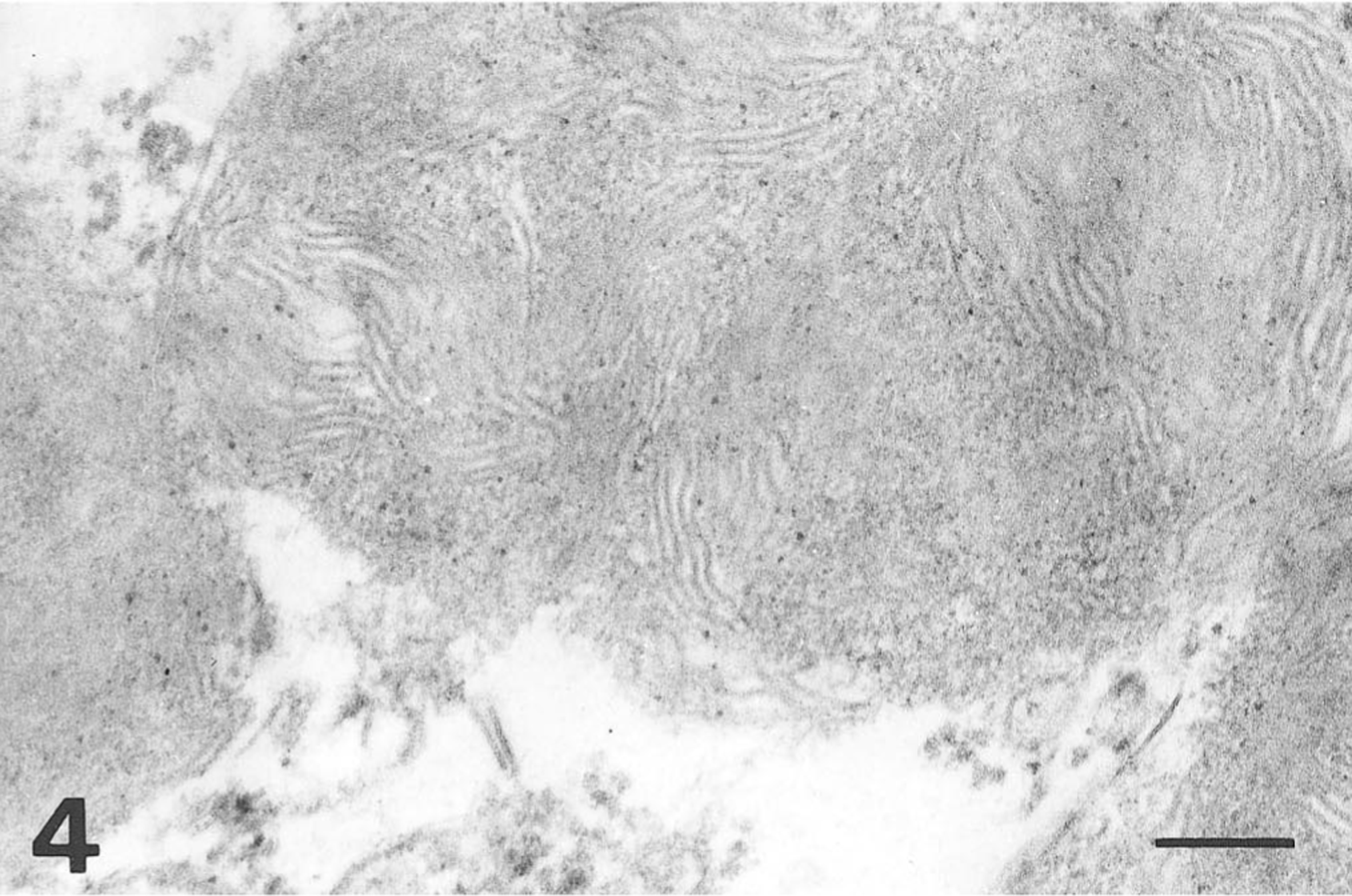
Electron micrograph. Spinal cord; dog. Laminated membranous material of neuronal cytosomes. Bar = 0.2 μm.
The light microscopic findings, autofluorescence, and appearance of the inclusions on electron microscopy correspond closely with previous descriptions of ceroid-lipofuscinosis. Six cases of this disease have been reported in Cocker Spaniel dogs. 2 5 All of them had the same feature: a generalized storage of a lipofuscinlike pigment, with a heavy accumulation in smooth muscle. Moreover, all cases had the same gross lesion, yellow-brown discoloration of the intestines. These characteristics defined a clinical entity in Cocker Spaniel dogs named brown bowel syndrome. 2 In the present however, stored material was found within smooth muscle cells of the urinary bladder and small muscular arteries, but the major gross lesion of this syndrome was not found. Intestines of this dog were not available for pathologic study.
There are three possible reasons for the lack of this characteristic gross lesion: 1) the amount of pigment stored in smooth muscle cells of the intestine was not enough to give the viscera a yellow-brown discoloration, 2) the severity of neurologic signs precipitated euthanasia before appreciable amounts of pigment could accumulate in smooth muscle cells, and 3) discoloration of the intestines was so mild that the lesion was missed. The small amount of pigment found in smooth muscle cells of the urinary bladder support the first explanation.
Blindness and behavioral changes were reported in Cocker Spaniel dogs with ceroid-lipofuscinosis. 2 5 None of these signs were noticed in the present case.
The microscopic findings in this dog are in accord with those found in the other six Cocker Spaniel dogs with generalized ceroid-lipofuscinosis. However, because brown bowel was not found, gross discoloration of the intestines should not be expected in all cases.
Footnotes
Acknowledgements
We thank Dr. Daniel H. Farfallini, Buenos Aires, Argentina, for submitting the case.