
Abstract

DECLARATIONS
Written informed consent to publication was obtained from the patient or next of kin
RS wrote the report, researched for the discussion, and assimilated the figures; SM-W helped with the writing of the discussion; JG and PR reviewed the article
John Meyrick-Thomas
It is necessary to think outside of the box when dealing with patients presenting with initially straightforward surgical problems.
Background
Common variable immunodeficiency (CVID) is the commonest primary immunodeficiency disease, which is characterized by frequent bacterial infections and hypogammaglobulinaemia. The prevalence of CVID is estimated at 1:25,000 in the Western world. 1 Men and women are affected equally with a peak onset between 20 and 40 years. There is an average of 4–6 years between onset of symptoms and diagnosis. The commonest manifestation of CVID is recurrent bronchitis. 1 We report a case of a patient whose first presentation of CVID was an inguinal swelling, which was secondary to reactive lymphadenopathy.
Case report
A 61-year-old Greek man presented to the Surgical Department with a firm, tender and erythematous right inguinal swelling. He was not taking any regular medications and had a history of chronic bronchitis for the past 20 years. He was an ex-smoker with a history of 20-pack years. The patient underwent a groin exploration under general anaesthetic, which revealed an enlarged infected lymph node that was excised and sent for histology. The histology report indicated reactive follicular hyperplasia. Ten weeks later, he presented again with a right inguinal abscess (Figure 1) which was drained and treated with antibiotics. He then represented two weeks after this with a left inguinal swelling. An ultrasound scan of the groin identified several reactive nodes. The recurrent lymphadenopathy prompted referral to the haematology department. Tuberculosis and sarcoidosis were excluded following a chest X-ray, mantoux test and serum angiotensin converting enzyme (ACE) measurements (51 iU/L). HIV, toxo-plasmosis, cytomegalovirus, hepatitis B and syphilis were all considered and excluded following negative serology results. Serum immunoglobulin levels were found to be low; IgG <2.7 g/L (6–16 g/L) and IgA <0.40 g/L (0.8–4 g/L) but with normal IgM 1.57 g/L (0.5–2 g/L). Serum protein electrophoresis found <1 g/L of paraprotein in the gamma region (Figure 2). The patient was diagnosed with common variable immunodeficiency.
Right inguinal abscess of patient in question Serum protein electrophoresis of patient in question (lane 2) showing less than 1 g/L of paraprotein in the gamma region. The arrows correspond to the gamma region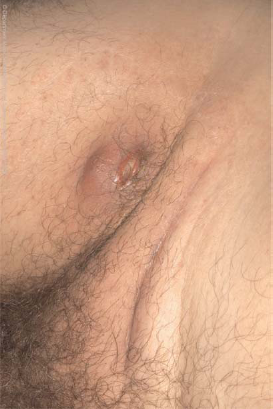

Discussion
A ‘lump in the groin' is a very common surgical presentation, with many possible causes; where the swelling is found to be an inguinal lymph node, there is a myriad of diseases that may be responsible. CVID is a rare cause – infections and malignancies are perhaps more important differentials to consider, and the history and examination should be tailored as such. Painful nodes are usually secondary to inflammation from infection, and can be secondary to a number of microbes including Mycobacterium tuberculosis. Painless, firm nodes should raise the clinician's suspicion of malignancy. 2 Testicular, penile, vulvar, urethral carcinomas, melanoma and lymphoma can all present with inguinal lymphadenopathy.3–6 CVID should be considered as a diagnosis of exclusion, 7 and investigations for it should not be initiated until infection and malignancy have been ruled out. Other important causes of hypo-immunoglobulinaemia include nephrotic syndrome, chronic lymphocytic leukaemia and myeloma. These need to be excluded before pursuing the diagnosis of CVID, 1 as do other secondary causes of antibody deficiency, 8 e.g. drug-induced.
The underlying pathophysiology of CVID is poorly understood, but is believed to be due to disruption of the B cell differentiation process, leading to decreased immunoglobulin secretion. 9 It is a group of conditions characterized by this feature, rather than an individual disease. 10
Patients with CVID typically present with respiratory tract infections usually caused by Streptococcus pneumoniae and Haemophilus influenza. 1 Chronic respiratory infections can cause bronch-iectasis, emphysema and fibrosis. It is likely the patient's history of chronic bronchitis in this report is related to his previously undiagnosed immunodeficiency. Clinicians should include CVID among their differentials where recurrent respiratory tract infections with lymphadenopathy is present (bearing in mind that haematological malignancies can present like this, and thus need to be investigated).
Gastrointestinal symptoms are the second commonest manifestation, namely malabsorption, and chronic diarrhoea as a result of secondary infection. 11 Typical causative organisms are Giardia lamblia and Campylobacter jejuni. Bone and joint inflammation may be caused by infection with mycoplasmas or chronic rheumatoid inflammation.
Approximately 20% of individuals with CVID have autoimmune manifestations: in particular autoimmune haemolytic anaemia and autoimmune thrombocytopenia. There is also an increased incidence of cancer, especially lympho-mas and gastric cancer. 11 Non-malignant lympho-proliferation also occurs in these patients causing splenomegaly, lymphadenopathy and follicular nodular lymphatic hyperplasia of the gastrointestinal tract, but it is unusual for patients to present in this way. 12
Diagnosis of CVID is achieved through testing serum immunoglobulin levels with reduced IgG, IgA and/or IgM in the absence of any other clear cause for immunodeficiency. 13 Full blood count and film and protein electrophoresis can exclude haematological malignancies; biopsy of lymph nodes can rule out solid organ cancer.
Management of CVID currently consists of immunoglobulin replacement,14–16 and active treatment of the associated manifestations described above. Prophylactic antibiotics covering both Streptococcus pneumoniae and Haemophilus influenzae may help prevent associated recurrent sinopulmonary infection and control small bowel bacterial overgrowth. Infections need to be treated early with higher doses and longer courses of antibiotics.
The prognosis is affected by the presence of structural lung damage, malignancy and autoimmune disease. The 20-year survival rate is approximately 65%. 17
Conclusions
Inguinal swellings are common presentations to surgical departments. This case demonstrates a possible presentation of CVID to surgical departments and should be considered in patients with persistent or infected lymphadenopathy.