
Abstract
Due to non-productive infections, mice are not a good model to study some human adenoviruses. However, mice provide an excellent model to study the metabolic effects of human adenovirus, Ad36. Research interest in Ad36 is increasing rapidly, and consequently an increase in the use of mice as a model is anticipated. However, little is known about the transmission potential of Ad36 from infected mice to other laboratory animals or personnel. While underestimating the infectivity could promote inadvertent spread of Ad36, overstating it could drain valuable laboratory resources and animals. Therefore, we determined the duration of infectivity in female C57BL/6J mice that were experimentally infected with human adenoviruses Ad36 or Ad2. Other uninfected mice were co-housed for one week with the experimentally-infected animals, four or eight weeks postinfection. Additionally, uninfected mice were housed in the cages of mice that were infected with Ad36, 12 weeks earlier. The presence of viral DNA in tissues was used to indicate infection of mice. Although experimentally-infected mice harboured viral DNA at least up to 12 weeks, the horizontal transmission of infection was observed in co-housed mice only up to four weeks postinfection. Thus, Ad36-infected mice should be considered potentially infective for eight weeks and appropriate handling and barrier containment should be used. After eight week postinfection, horizontal transmission appears unlikely. This information may provide guidelines for animal handling, and experimental design using Ad36, which may increase safety for laboratory personnel and reduce the number of mice required for experiments.
Mice are an excellent animal model to investigate metabolic efffects of various viruses. While the infectivity of a virus and its spread to various organs within an animal would be important to an investigator, its inadvertent spread to other laboratory animals or personnel would be a concern for a laboratory animal facility. Various levels of biological safety barriers are used to prevent such inadvertent horizontal transmissions from experimentally-infected animals to other animals or humans. However, beyond the infectivity period, barrier housing and handling of animals may be unnecessary or too restrictive in some cases, and a burden on valuable laboratory resources. Most importantly, because certain commonly shared equipment should not be used for infected animals, biosafety barrier housing markedly increases the number of animals required in a study. For instance, due to the concerns of horizontal transmission to uninfected animals, nuclear magnetic resonance (NMR) spectroscopy – a commonly shared equipment for determining body composition – may not be available for infected animals. This necessitates determining body composition by physicochemical methods upon necropsy and increases the number of animals to be used at each time point, due to the terminal nature of the assay. Whereas, if the duration of infectivity is known, NMR studies could be used for monitoring body composition changes periodically in the same animals once they cease to be infective. Thus, determining transmissibility of an infectious agent under investigation is an important welfare measure for laboratory animals and personnel.
We use animal models to study the effect of human adenoviruses, which are very commonly used in research as viral vectors or wild-type viruses. Particularly, we study the effect of human adenoviruses Ad36 and Ad2. Unlike many other human adenoviruses, Ad36 infects many species, including chickens, mice, rats, hamsters and marmosets. 1–6 Importantly, unlike many human adenoviruses, which have abortive infection of murine cells, Ad36 can efficiently infect murine cells. 7–9 While Ad36 has been a major research focus of our group, 1–16 interest of other research groups in Ad36-related research is rapidly increasing globally, as indicated by several recent publications about Ad36 from other groups. 17–27 Therefore, in anticipation of increased use of animal models to investigate the effects of Ad36, we conducted the following experiments to determine the duration of infectivity of mice infected with Ad36 or Ad2. We determined infectivity of experimentally-infected mice, by tracking the duration of horizontal transmission to cagemates. Our data indicate that mice experimentally infected with Ad36 or Ad2 are infective earlier, but do not infect cagemates at and after eight weeks postinoculation. Thus, the risk of accidental Ad36 infection of other animals due to sharing common equipment should be negligible eight weeks post-experimental infection of mice. The details of the experiment are described below.
Materials and methods
Overview
Mice were experimentally infected with Ad36 or Ad2. Other mice were co-housed with the experimentally-infected animals at the durations outlined below. Conventional polymerase chain reaction (PCR) as well as realtime PCR were used to determine viral DNA in various tissues of mice. We expected experimentally-infected animals to harbour viral DNA in tissues. The presence of viral DNA in naïve co-housed animals would indicate horizontal transmission from experimentally-infected animals.
Animals
The protocol for animal experiments was approved by the Pennington Biomedical Research Center's (PBRC) Animal Investigations Committee, which has a training programme for certification in animal handling for all personnel who work with animals. All experiments in this study were performed according to the provisions of the Animal Welfare Act of 1966 (Public Law 89–544), plus its subsequent amendments, as well as the standards set forth in the document entitled ‘The Guide for the Care and Use of Laboratory Animals’ (NIH Publication No. 85–23). The comparative biology department of PBRC has highly experienced, full-time veterinary care and management of its research including veterinary direction for the appropriate use of anaesthetic and analgesic drugs, supervision of preparation of sterile surgical packs and the performance of aseptic surgery, and veterinary care of any animal which may be ill or injured.
Female C57BL/6J mice were purchased (Stock number 000664; The Jackson Laboratory, Bar Harbor, ME, USA). All animals were housed in microisolator cages under biosafety level two containment, with a separate air supply to each room. Ad libitum access to water and chow (Opensource rodent diets # D12450B, protein 20% of total energy, CHO 70% of total energy) was offered.
At appropriate times, animals were euthanized by CO2 inhalation followed by cervical dislocation.
Experiment 1
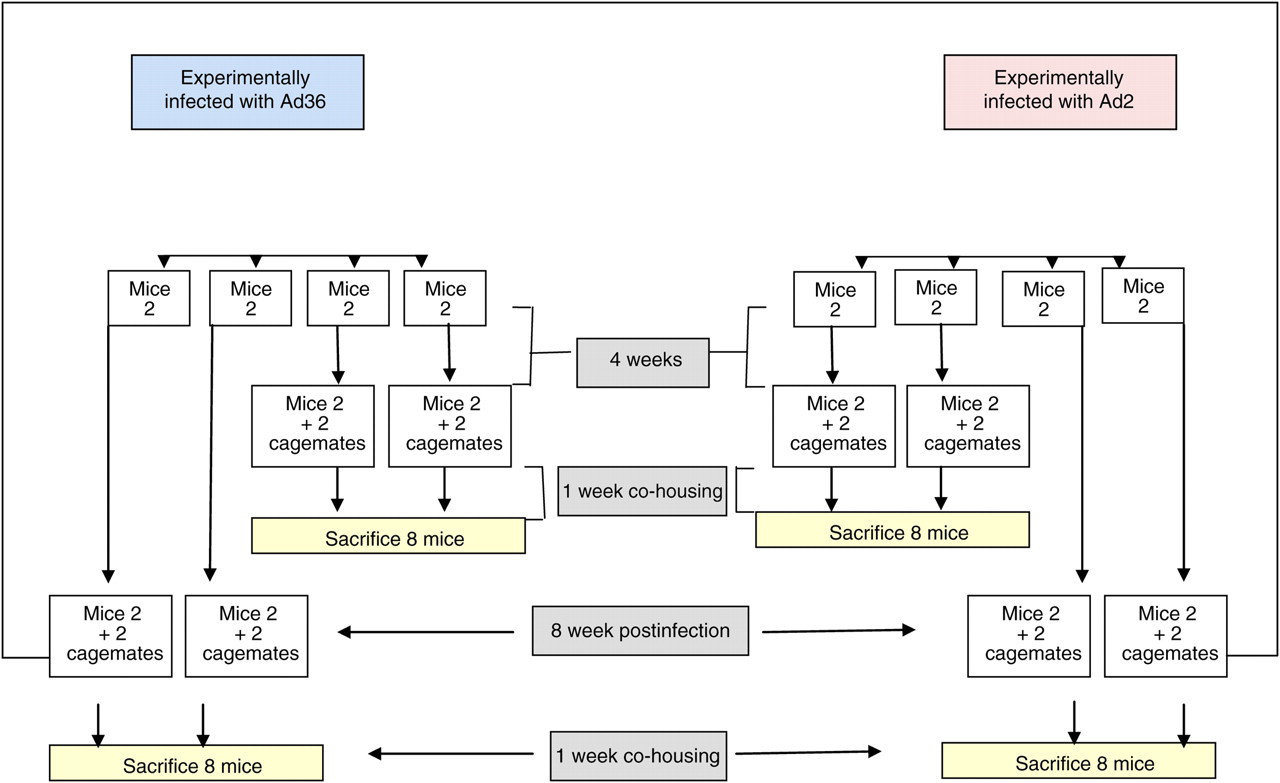
Figure 1 shows an outline of the experimental design. Sixteen female C57BL/6J mice (4 weeks old) were intranasally, orally and intraperitoneally inoculated with 107 plaque forming units (PFU) of Ad36 or Ad2 (n = 8 mice per virus). Multiple routes of inoculation were used to assure infection. Two animals were housed per microisolator cage, with individual and ad libitum food and water supply. Four weeks postinoculation, two littermate females (cagemates) were added per cage to two cages of Ad36-infected mice and two cages of Ad2-infected mice. After one week of group housing, experimentally-infected animals and their cagemates were sacrificed. Livers and lungs – main target tissues of adenovirus infection 6 – were screened for the presence of Ad36 or Ad2 DNA.
Experiment 1 design
Eight weeks postinoculation, two littermate females (cagemates) were added per cage to the remaining two cages of Ad36-infected mice and two cages of Ad2-infected mice. After one week of group housing, experimentally-infected animals and their cagemates were sacrificed and their livers and lungs were utilized in screening for the presence of Ad36 or Ad2 DNA.
Experiment 2
Experiment 2 was conducted as an additionally supporting study for Experiment 1. The aim was to test whether the soiled bedding of mice 12 weeks postinfection was capable of transmitting infection. Two uninfected littermate C57BL/6J female mice were housed individually for two weeks in two cages previously resided in individually by two mice infected with Ad36 12 weeks ago. Thus, the uninfected mice were exposed for two weeks to dirty bedding of the two mice that were infected with Ad36 12 weeks ago. Two weeks after housing, all four mice were killed and their livers and kidneys were screened for the presence of Ad36 DNA.
Techniques and assays
Virus preparation
Ad36 was obtained from American Type Culture collection (ATCC cat # VR913) plaque purified and propagated in A549 cells (human lung cancer cell line) as described and used previously. 1,2 Ad2 was also obtained from ATCC (cat # VR846) and propagated in A549 cells. Viral titres were determined by plaque assay 1 and inoculations expressed as PFU.
Screening of tissue for viral DNA
Conventional PCR
DNA isolation: Genomic DNA (gDNA) was isolated using a QIAMP DNA mini kit (cat # 51306). Primers were designed to E4 genes of Ad36 or Ad2 and also for mouse β-actin. DNA was amplified by PCR.
Primer sequence
Ad36 E4 forward primer: 5′-GGCATACTAACCCAG TCCGATG-3′,
reverse primer: 5′-AATCACTCTCAGCAGCAGCAGG-3′;
Ad2 E4 forward primer: 5′-CCTAGGCAGGAGGGTTTTTC-3′,
reverse primer: 5′-ATAGCCCGGGGGAATACATA-3′;
mouse β-actin forward primer: 5′-GAT CTTCATGGTGCTAGGAG-3′,
reverse primer: 5′- ACGTTGACATCCGTAAAGAC-3′.
Negative PCR control: water
Positive PCR control: DNA from Ad36 or Ad2-infected A549 cells.
DNA was denatured for 2 min at 95°C and subjected to 35 cycles of PCR (94°C for 1 min, 55°C for 1 min, 72°C for 2 min followed by incubation at 72°C for 5 min). PCR products were visualized on a 2% agarose gel with a 100 bp DNA ladder.
Realtime PCR (qPCR)
DNA isolation: DNA was isolated as described above for conventional PCR.
Primers and probes
Primers and probes were designed for Ad36e4orf1 using IDT PrimeTime qPCR Assay tool.
Ad36 E4 probe:
5′-/56-FAM/AAC CCT GCT /ZEN/GCT GGA GAG AGT GAT TT/3IABkFQ/-3′
Ad36 E4 forward primer: 5′-ACC AGG GTG GCT ATT CTC ACT GAA-3′
Ad36 E4 reverse primer: 5′-TGC TCT TTA ACC ACA CGG ACC GAT-3′
Final primer concentration per reaction was 250 nm primer and 125 nm probe. Taqman 2X master mix from Applied Biosystems (Carlsbad, CA, USA) was used (cat # 4304437). DNA was amplified on the ABI prism 7900HT by qPCR using the standard curve method using pooled samples of both gDNA from liver tissue from Ad36-infected mice, uninfected mice and gDNA isolated from infected A549 cells. Standard curve used 100, 20, 4, 0.8, 0.16, 0.032, 0.0064, 0.00013 and 0.00026 ng of DNA. gDNA from liver tissue was loaded at concentrations of 30, 50 and 100 ng, which is the upper limit of template load for detection. A549 cells infected with Ad36 served as a positive PCR control. To determine a range of concentrations of gDNA that Ad36 is detectable in infected A549 cells, a serial dilution of 1:5 of gDNA was used. Loading DNA concentrations of infected A549 cells were: 3, 0.6, 0.12, 0.24, 0.0048 and 0.00096 ng. Negative PCR control was water. DNA was denatured for 2 min at 95°C and subjected to 35 cycles of PCR (94°C for 1 min, 55°C for 1 min, 72°C for 2 min followed by incubation at 72°C for 5 min).
Results
Experiment 1
Amplification of β-actin demonstrated the presence of amplifyable DNA. Positive and negative control demonstrated satisfactory PCR assay. Mice inoculated with Ad36 or Ad2 showed the presence of respective viral DNA in either lungs or liver tissues five weeks postinoculation (Table 1 and Figure 2). These mice horizontally transmitted the infection to their cagemates, as indicated by the presence of DNA of respective adenoviruses in lungs and liver tissues of the cagemates. The viral DNA continued to be present in inoculated mice at nine weeks postinoculation (Table 1 and Figure 3). Yet, these mice did not infect their cagemates, as indicated by the lack of viral DNA in lungs or livers of their cagemates.
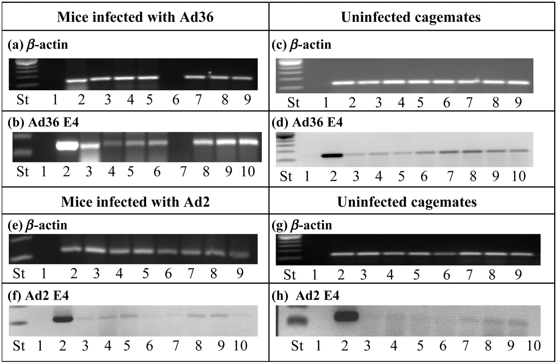
Presence of viral DNA in mice infected four weeks earlier, and in their co-housed cagemates. PCR assay for viral DNA detection in mouse liver and lung tissues. (a) β-actin from mice infected with Ad36. Key: St: DNA ladder, 1 – negative control, 2 to 5 – liver and 6 to 9 – lung tissue DNA from the Ad36-infected mice. (b) Ad36 E4 DNA in Ad36-infected mice. Key: St: DNA ladder, 1 – negative control, 2 – positive control for Ad36-infected A549 cells DNA, 3 to 6 – liver and 7 to 10 – lung DNA tissue from Ad36-infected mice. (c) β-actin from UICM of Ad36 group. Key: St: DNA ladder, 1 – negative control, 2 to 5 – liver and 6 to 9 – lung tissue DNA from UICM. (d) Ad36 E4 DNA in UICM of Ad36 group. Key: St: DNA ladder, 1 – negative control, 2 – positive control for Ad36-infected A549 cells DNA, 3 to 6 – liver and 7 to 10 – lung DNA tissue from UICM of Ad36. (e) β-actin from mice infected with Ad2. Key: St: DNA ladder, 1 – negative control, 2 to 5 – liver and 6 to 9 – lung tissue DNA from the Ad2-infected mice. (f) Ad2 E4. Key: St: DNA ladder, 1 – negative control, 2 – positive control of Ad2-infected A549 cells DNA, 3 to 6 – liver and 7 to 10 – lung DNA tissue from Ad2-infected mice. (g) β-actin from UICM of Ad2. Key: St: DNA ladder, 1 – negative control, 2 to 5 – liver and 6 to 9 – lung tissue DNA from UICM of Ad2. (h) Ad2 E4. Key: St: DNA ladder, 1 – negative control, 2 – positive control of Ad2-infected A549 cells DNA, 3 to 6 – liver and 7 to 10 – lung DNA tissue from UICM of Ad2. PCR: polymerase chain reaction; UCIM: uninfected cagemates
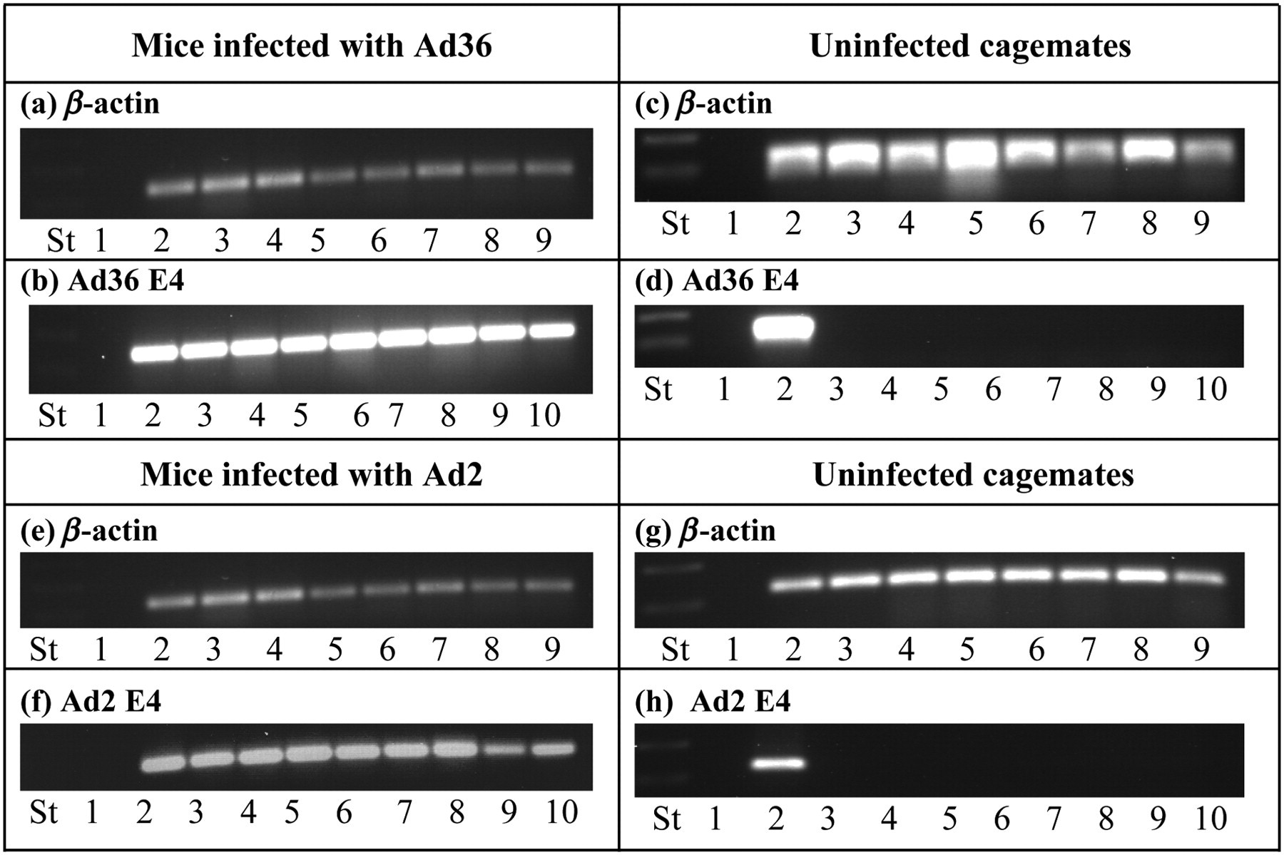
Presence of viral DNA in mice infected eight weeks earlier, and in their co-housed cagemates. PCR assay for viral DNA detection in mouse liver and lung tissues. (a) β-actin from mice infected with Ad36. Key: St: DNA ladder, 1 – negative control, 2 to 5 – liver and 6 to 9 – lung tissue DNA from the Ad36-infected mice. (b) Ad36 E4 DNA in Ad36-infected mice. Key: St: DNA ladder, 1 – negative control, 2 – positive control for Ad36-infected A549 cells DNA, 3 to 6 – liver and 7 to 10 – lung DNA tissue from Ad36-infected mice. (c) β-actin from UICM of Ad36 group. Key: St: DNA ladder, 1 – negative control, 2 to 5 – liver and 6 to 9 – lung tissue DNA from UICM. (d) Ad36 E4 DNA in UICM of Ad36 group. Key: St: DNA ladder, 1 – negative control, 2 – positive control for Ad36-infected A549 cells DNA, 3 to 6 – liver and 7 to 10 – lung DNA tissue from UICM of Ad36. (e) β-actin from mice infected with Ad2. Key: St: DNA ladder, 1 – negative control, 2 to 5 – liver and 6 to 9 – lung tissue DNA from the Ad2-infected mice. (f) Ad2 E4. Key: St: DNA ladder, 1 – negative control, 2 – positive control of Ad2-infected A549 cells DNA, 3 to 6 – liver and 7 to 10 – lung DNA tissue from Ad2-infected mice. (g) β-actin from UICM of Ad2. Key: St: DNA ladder, 1 – negative control, 2 to 5 – liver and 6 to 9 – lung tissue DNA from UICM of Ad2. (h) Ad2 E4. Key: St: DNA ladder, 1 – negative control, 2 – positive control of Ad2-infected A549 cells DNA, 3 to 6 – liver and 7 to 10 – lung DNA tissue from UICM of Ad2. PCR: polymerase chain reaction; UCIM: uninfected cagemates
Results of screening for DNA by PCR
+ and − indicate presence or absence of DNA screened. Water was used as a negative control, and DNA from Ad36-infected A549 cells was used as a template for positive control. (a) Liver and lung samples from experimentally-infected and the cagemates show viral DNA, which indicates infection of the experimentally-infected group, and its horizontal transmission to the cagemates. (b) Experimentally-infected animals continued to show the presence of viral DNA in livers and lungs. Absence of viral DNA in cagemates at this point indicates the inability of experimentally-infected animals to infect the cagemates. (c) Experimentally-infected animals continued to show the presence of viral DNA in livers and lungs. However, the absence of viral DNA in cagemates indicates that the soiled bedding of the experimentally-infected aimals is not likely to infect the cagemates at this time point
Livers of mice infected with Ad36, and those of the four- and eight-week cagemates were also screened for Ad36 DNA by qPCR assay. Ad36 DNA in A549 cells was detectable in as little as 0.0048 ng of DNA, indicating the appropriateness of the primer probes and a functioning assay. However, qPCR assay could not detect Ad36 DNA in liver tissues, probably due to template loading limits for this system.
Experiment 2
Although no horizontal transmission was observed at eight weeks postinfection, Experiment 2 was conducted to provide additional support. Ad36-inoculated mice continued to show the presence of Ad36 DNA 12 weeks later. Despite this, the cages used by these mice for the last two weeks during 12 weeks postinoculation were not able to transmit Ad36 to naïve mice housed in the dirty cages for two weeks, as indicated by a lack of Ad36 DNA in their tissue samples (Table 1 and Figure 4).
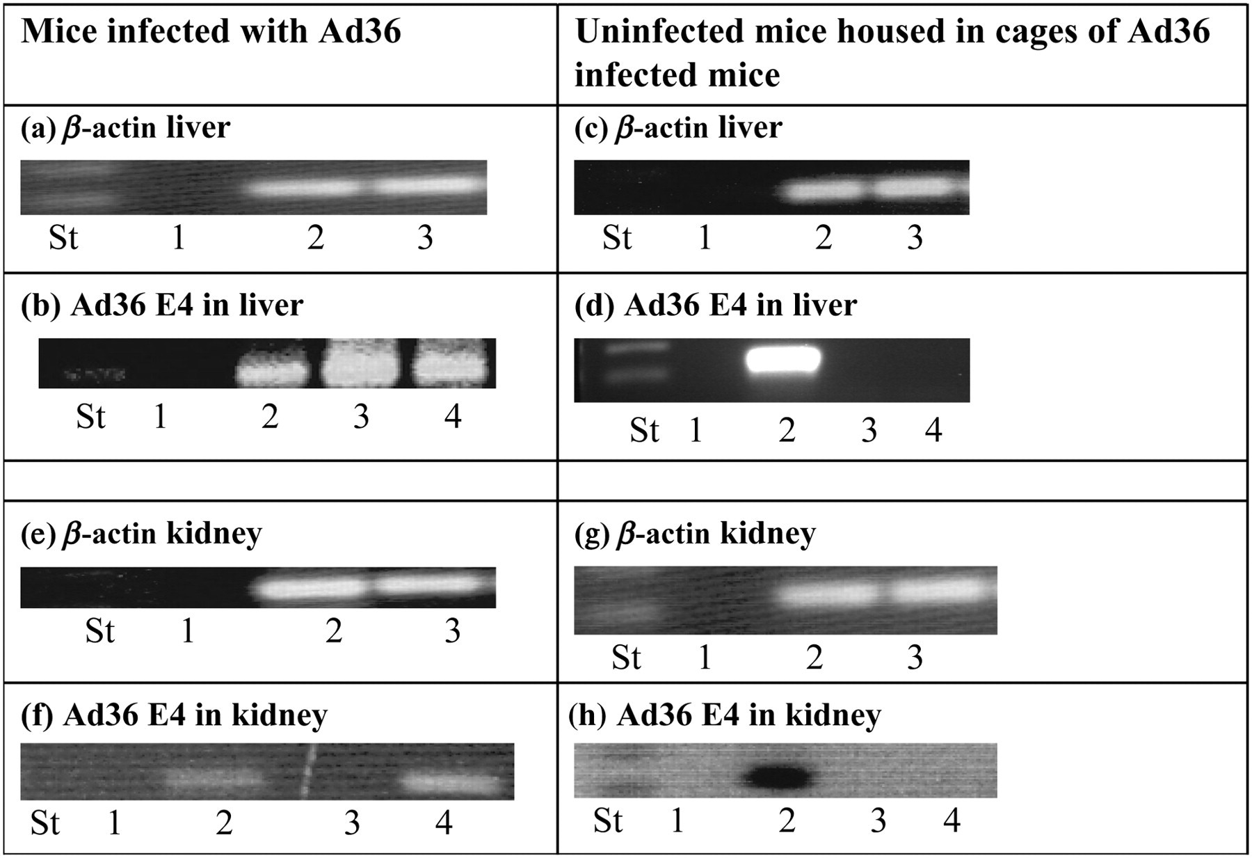
Presence of viral DNA in mice infected 12 weeks earlier, and in mice housed in their cages. PCR assay for viral DNA detection in mouse liver and kidneys. (a) β-actin from mice infected with Ad36. Key: St: DNA ladder, 1 – negative control, 2 to 3 – liver DNA from Ad36-infected mice. (b) Ad36 E4 DNA. Key: St: DNA ladder, 1 – negative control, 2 – positive control – Ad36-infected A549 cells DNA, 3 to 4 – liver DNA from Ad36-infected mice. (c) β-actin from uninfected housed mice. Key: St: DNA ladder, 1 – negative control, 2 to 3 – liver from uninfected mice. (d) Ad36 E4 DNA in uninfected housed mice. Key: St: DNA ladder, 1 – negative control, 2 – positive control – Ad36-infected A549 cells DNA and 3 to 4 liver of uninfected housed mice. (e) β-actin from mice infected with Ad36. Key: St: DNA ladder, 1 – negative control, 2 to 3 kidney DNA from Ad36-infected mice. (f) Ad36 E4 DNA. Key: St: DNA ladder, 1 – negative control, 2 – positive control – Ad36-infected A549 cells DNA, 3 to 4 kidney from Ad36-infected mice. (g) β-actin from uninfected housed mice: Key: St: DNA ladder, 1 – negative control, 2 to 3 kidney DNA from uninfected mice. (h) Ad36 E4Orf1 from uninfected housed mice key: St: DNA ladder, 1 – negative control, 2 – positive control of Ad36-infected A549 cells DNA, 3 to 4 – kidney of uninfected housed mice. PCR: polymerase chain reaction; UCIM: uninfected cagemates
Discussion
Human adenoviruses are generally considered to induce semi-permissive or abortive infections in animal models. 28,29 This view is challenged by recent reports that some human adenoviruses can modulate animal metabolism and may also result in productive infection in animal models. 25,30–34 Ad36, in particular, expresses mRNA in an animal host model and is also shed by various species of experimentally-infected laboratory animals. 1–3 This suggests a productive infection and active excretion of Ad36 from mice. If Ad36 can develop a productive infection in animals, it is a potential safety concern to other laboratory animals and human workers. This necessitates the determination of infectivity duration.
The results of this study indicate that mice experimentally infected with adenoviruses continue to harbour viral DNA in their livers, kidneys or lungs at least up to 12 weeks postinoculation. Also, these experimentally-infected mice can transmit Ad36 or Ad2 infection to co-housed cagemates at four weeks postinoculation. However, there is no horizontal transmission to cagemates at eight weeks postinfection. This observation was further strengthened by Experiment 2. The used dirty bedding of mice experimentally inoculated 12 weeks earlier did not transmit Ad36. This suggests that the virus is probably not shed by mice at this time. This finding conforms with an earlier study that showed continued presence of viral DNA in Ad36-infected marmosets for six months postinoculation, but the virus was shed in faeces only for two months postinoculation. 3
A comparison of conventional versus qPCR provided useful practical information. It appears that to detect Ad36 DNA in tissues of mice, conventional PCR method is more suitable and sensitive. This may be because it can use up to 1 µg DNA template for the assay, whereas the upper limit of DNA template of qPCR is 100 ng. This difference in DNA template amounts probably makes it harder to detect small quantities of viral DNA that may be present in the tissue. Therefore, qPCR successfully detected Ad36 DNA in A549 cells infected with Ad36, but would have yielded false-negative results in mouse tissue samples. We recommend the use of conventional PCR for screening mouse tissues for Ad36 DNA.
It should be noted that our conclusions are applicable to our specific experimental conditions, including the species and strain of animals used. Overall, these data indicate that mice experimentally infected with Ad36 or Ad2 by intranasal, oral or intraperitoneal route are not likely to be infectious to other mice eight weeks postinoculation. This also suggests that Ad36-infected mice should be considered potentially capable of infecting other animals or laboratory personnel, till eight weeks postinfection and appropriate handling and barrier containment should be used during this period. On the other hand, considering that even one week of co-housing could not infect the cagemates, use of commonly shared equipment such as the NMR machine for body composition determination or the metabolic chambers for determination of oxygen consumption, which are thoroughly cleaned after use, should not pose a threat to other users. We believe this information will help in planning guidelines for animal handling, and in designing experiments using Ad36, which may increase safety for laboratory personnel and reduce the number of mice required for experiments.
Footnotes
ACKNOWLEDGEMENTS
Research funded in part by the American Diabetes Association grant # 1–09-IN-13. This work used Genomics core facilities that are supported in part by COBRE (NIH P20-RR021945) and NORC (NIH 1P30-DK072476) centre grants from the National Institutes of Health.