
Abstract
Precocity and efficiency of ultrasonography for pregnancy diagnosis and determination of litter size in mice were assessed on a total of 46 adult mice of different lines (19 BALB/c, 15 C57BL/6 and 12 CD1) from Day 4.5 after vaginal plug. Different commercial ultrasound machines and probes (linear versus sectorial; 7.5 MHz versus 10 MHz) were compared. The best images were obtained by the use of 10 MHz linear transducers. The first visualization compatible with pregnancy, specifically with the implantation sites, was observed at Day 4.5 in three animals. Presence of embryonic vesicles was differentiated at Day 5.5 in all the females. The embryos and remaining gestational structures inside the vesicles were clearly distinguished at Day 8.5. Data were validated not only after delivery but also by comparison with postmortem findings on crucial days (Days 4.5 and 8.5). The efficiency for counting the exact number of embryos was very low, due mostly to underestimation in highly prolific females. Conversely, the estimation of the range of the number of conceptuses, instead of the total number, was more accurate. Sensitivity, specificity and total efficiency reached 100% at Day 8.5. Ultrasonography can be accurately used as an alternative non-invasive technique for pregnancy diagnosis and determination of litter size in the mouse from very early stages of gestation, replacing other procedures currently used and increasing efficiency in animal management and research.
The mouse is widely used as a model for research purposes worldwide. 1,2 Control and planning of reproduction, either for management in the animal units or for experimental purposes in the research units, is routinely required. Assessment of pregnancy in mice is usually made by direct observation after laparotomy or through abdominal palpation. 1 While the first method impedes the progression of the pregnancy, the reliability of the second method depends on the ability of the operator, but its precocity is never earlier than 12–14 days of gestation, depending on litter size. 3 Thus, the need for an early, non-invasive and reliable method for pregnancy diagnosis, capable of determining the number of conceptuses, is important.
In large animals and humans, the diagnosis of pregnancy and the determination of litter size are routinely performed by realtime ultrasound imaging. Ultrasonography is widely recognized as a reliable non-invasive method for the diagnosis of pregnancy and embryo development. Its application to mice would be beneficial for the management of both the general population and the experimental groups in research centres. Moreover, it would contribute to the improvement of animal conditions in research, in agreement with the philosophy of the 3Rs model of Russell and Burch (refinement, reduction and replacement). 4 The possibility of non-invasive pregnancy diagnosis would reduce the number of experimental animals and refine the studies, using only pregnant animals with a known litter size.
However, ultrasonography in the mouse has been limited to ultrasound microscopy with probes between 40 MHz and 70 MHz, 5–7 which are technically complex, very expensive and not commonly available. The use of lower frequencies (15 MHz or lower) for pregnancy assessment in mice has been described only very recently, 8,9 but commercial probes usually have a maximum of 10–11 MHz, frequency only used in a study published during the development of our work. 9 However, because both studies started after the first week of pregnancy, a serial study of the precocity of ultrasound diagnosis and its reliability were not analysed.
The objective of the present study was to determine the precocity of ultrasonography for pregnancy diagnosis and to assess its accuracy for the estimation of the number of conceptuses in pregnant mice. For this purpose, we used different commercial ultrasound machines and probes (linear and sectorial varying from 7.5 MHz to 10 MHz) on different mouse lines used in research (BALB/c, C57BL/6 and CD1). The quality of the ultrasound imaging depends on the transducer used (type and frequency). Linear probes offer a better image resolution but need a larger area for exploration, whereas sectorial probes are very adequate for small animals. On the other hand, resolution is higher when using higher frequencies, but the price of the equipment is also increased. Thus, we have compared different linear and sectorial probes varying from 7.5 MHz to 10 MHz. Results were validated both by assessment of the deliveries and by comparison with macroscopic findings.
Material and methods
Animals
The study was performed using 46 adult mice of different lines (19 BALB/cAnNCrl, 15 C57BL/6J and 12 Crl:CD1[ICR]) in breeding age. BALB/c and CD1 were obtained from Charles River Laboratories Inc (Wilmington, MA, USA), and C57BL/6 were obtained from The Jackson Laboratory (Bar Harbor, ME, USA). All the animals were maintained at the facilities of the CNIC Animal Laboratory Unit in Madrid, Spain, which meets the requirements of the European Union for Scientific Procedure Establishments. The experiment was carried out under Project Licence 156/07 from the CNIC Scientific Ethic Committee, and animal manipulations were performed accordingly with the Spanish Policy for Animal Protection RD1201/05, which meets the European Union Directive 86/609 about the protection of animals used in experimentation.
Ultrasonographic study
The females used in the trial were individually placed with males until the morning of the day when a vaginal plug was found as a result of overnight mating. This day was considered Day 0.5 for estimation of gestational age. Ultrasound observations started at Day 3.5 (reported to be the day of embryo implantation). 3 For examination, the mice were manually restrained in dorsal recumbence. Anaesthesia was avoided to prevent any effect on pregnancy. 10 To reduce animal distress, abdominal hair was not clipped, and the presence of air between the skin and the transducer, which makes the quality of the images worse, was avoided by wetting the abdomen profusely with gel.
Observations were performed using three different ultrasound machines: an Aloka 500 SSD fitted to a 7.5 MHz linear array transducer (Aloka Co, Tokyo, Japan), an Aloka 2500 equipped with a multifrequency (7.5–10 MHz) sectorial array probe (Aloka Co) and a Siemens Acuson Antares connected to a multifrequency (7.5–10 MHz) linear array probe (Siemens Medical Solutions, Erlangen, Germany). Ultrasound scanning was done by placing the transducer on the abdominal wall and moving it in different directions for viewing the uterine horns and their content; when the presence of gestational structures was observed, the number of embryos/fetuses was determined by placing the transducer on one side of the abdomen and moving it to the opposite.
Validation of the technique
The accuracy for very early pregnancy diagnosis was validated in two animals that, after ultrasound scanning, were anaesthetized with isoflurane vapours and killed by cervical dislocation at Days 4.5 and 8.5. The genital tracts were immediately removed and the characteristics of gestational structures and the number of embryos were checked.
Data analysis
Data on pregnancy diagnosis and number of conceptuses were grouped according to the day of gestation and compared with those obtained at delivery for determining the accuracy or predictive value of ultrasonography. Accuracy was estimated both for the total number of embryos and for different ranges of number of embryos (less than 5 embryos, between 5 and 10 and more than 10). Accuracy for both pregnancy diagnosis and litter size was estimated in terms of sensitivity (percentage of mice correctly diagnosed as pregnant and percentage of females correctly diagnosed as having a certain number/range of conceptuses, respectively), specificity (percentage of mice correctly diagnosed as non-pregnant or not having a certain number/range of embryos) and total efficiency (total percentage of individuals correctly classified as pregnant or not and total percentage of mice correctly classified as having a certain number/range of conceptuses or not). Relationship between predictive values and gestational age was assessed by Pearson correlation procedures, considered to be statistically significant at P < 0.05.
Results
The overview of the main pregnancy features observed by ultrasonography and times of observation is highlighted in Figure 1. The first visualization compatible with pregnancy, specifically with the implantation sites, was observed at Day 4.5 in three different mice, one of every line, by using both the 10 MHz linear and sectorial transducers (Figure 2). The scanning with 7.5 MHz was hindered by the scarce amount of amniotic fluid in the embryonic vesicle, which made it difficult to distinguish the uterine horns within the abdominal content. Thereafter, using 10 MHz, the presence of embryonic vesicles and embryos was differentiated at Day 5.5 in all the animals (Figure 3). The embryo and remaining gestational structures were clearly distinguished at Day 8.5 (Figure 4), whereas their viability could be determined by assessing heart beats at Day 9.5. In later stages of pregnancy, it is possible to differentiate phenotypical characteristics and to evaluate normality or abnormality in fetal development (Figure 5).
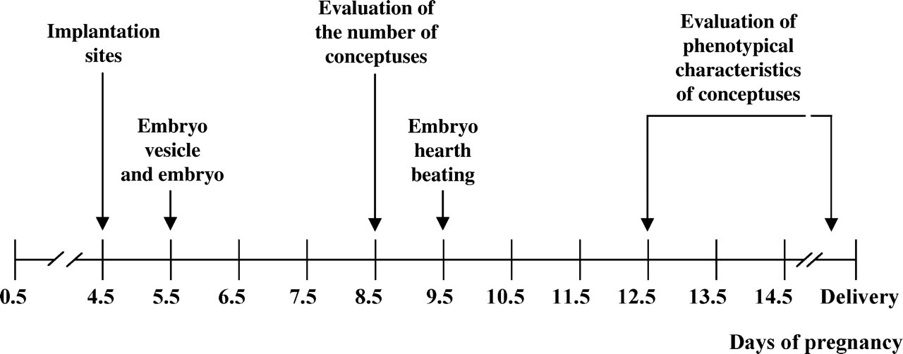
Overview of the main pregnancy features observed by ultrasonography and times of observation in the mouse
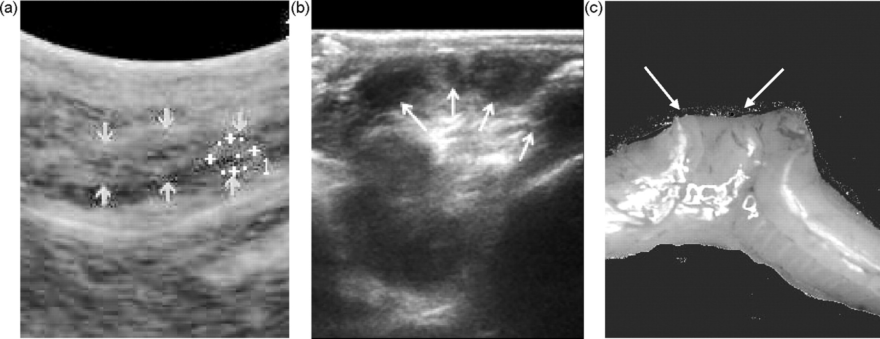
Visualization of implantation sites in mice at Day 4.5 post-coitum (marked by arrows). Ultrasonographic images obtained with (a) 10 MHz sectorial and (b) linear probes, and (c) macroscopic finding
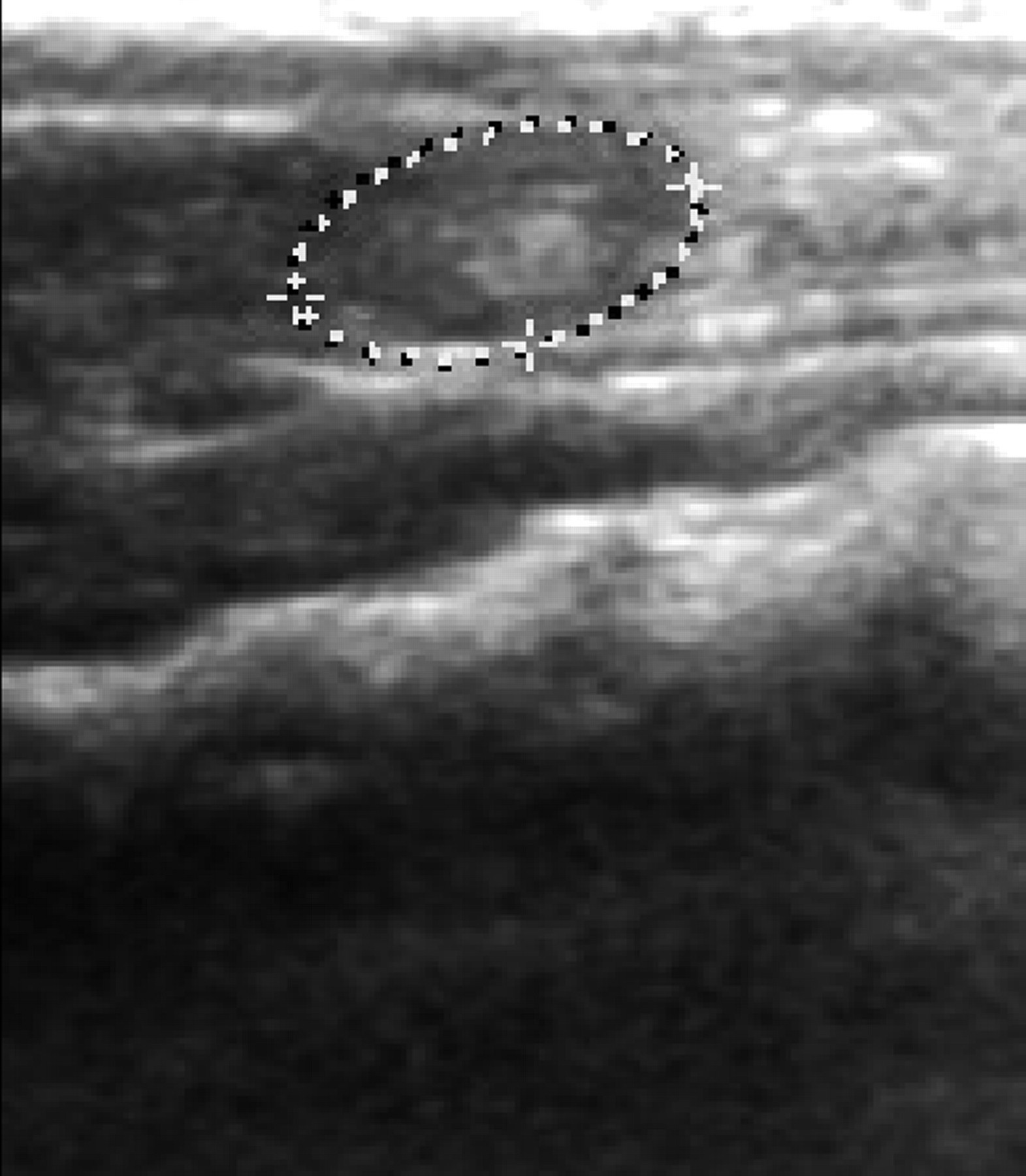
Determination of gestation by ultrasound imaging of embryonic vesicles in mice at Day 5.5 post-coitum
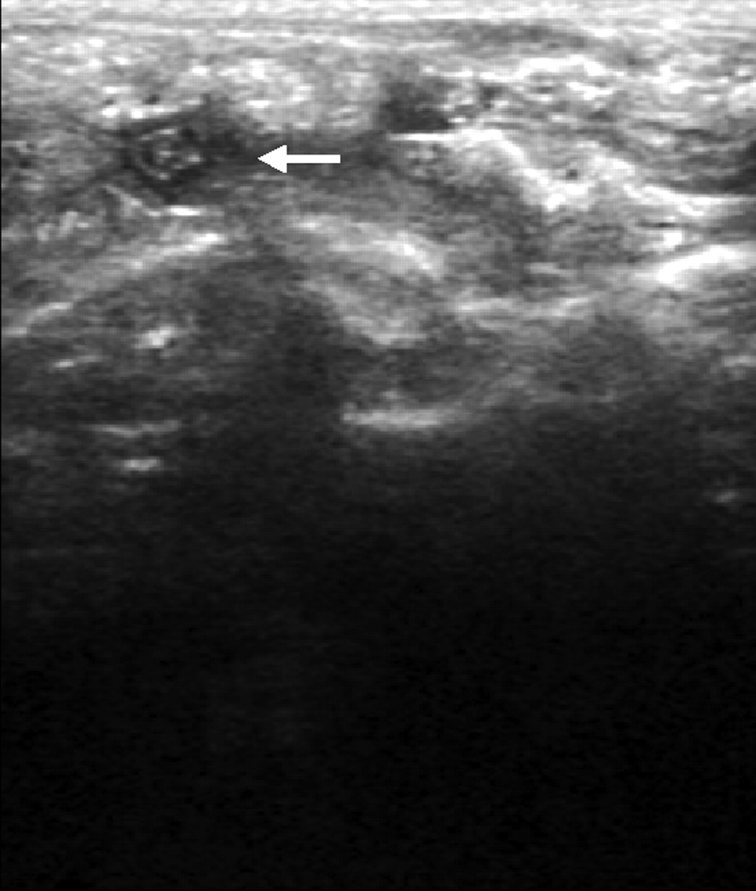
Ultrasonography of the mouse gestational sac and embryo inside (white arrow) at Day 8.5 post-coitum
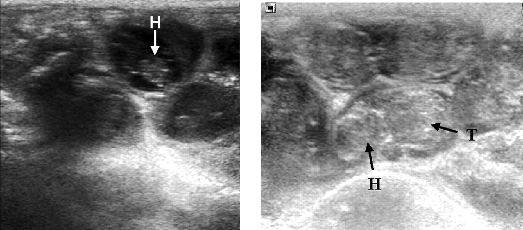
Ultrasonographic images of mouse embryos at Days 12.5 (left hand) and 14.5 (right hand) of pregnancy (H: head; T: trunk)
The number of embryos could be determined from Day 6.5, but the efficiency for counting the exact number was very low due mostly to underestimation in highly prolific females. The evaluation of the range of conceptuses, instead of the total number, was however more accurate, with sensitivity, specificity and total efficiency reaching 100% at Day 8.5 (Figure 6).
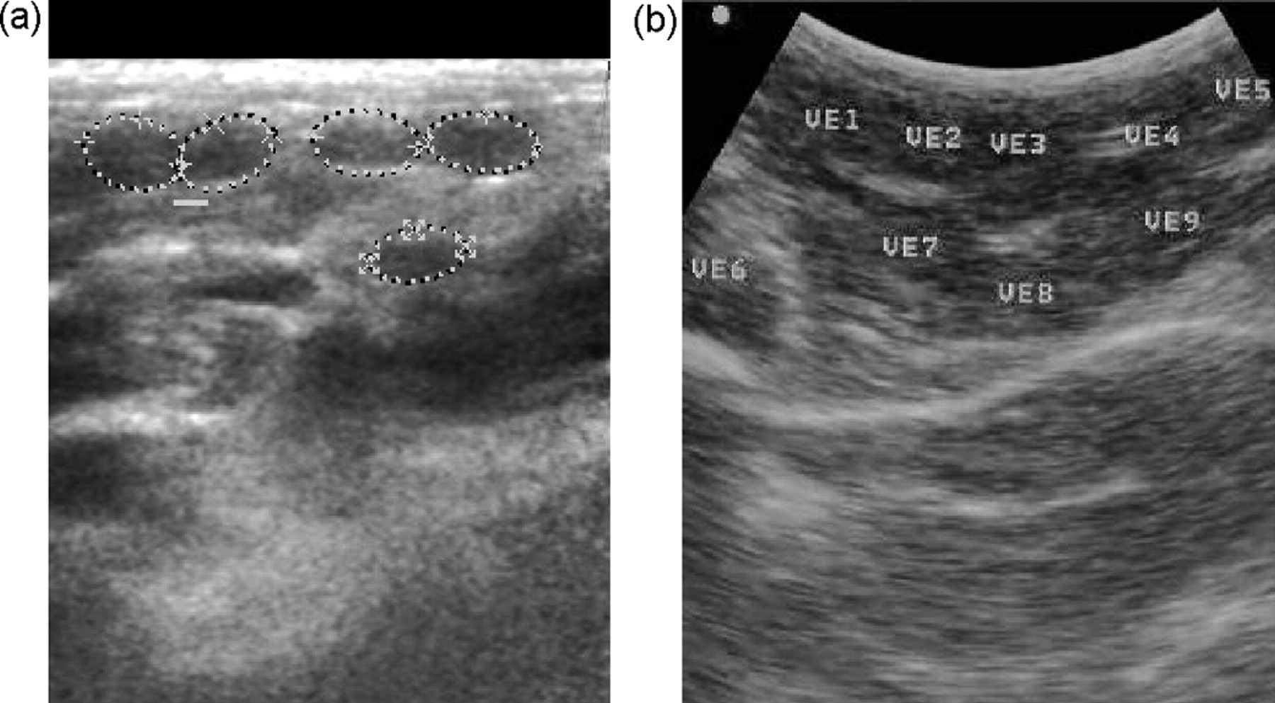
Determination of litter size in mice at Day 8.5 post-coitum. Ultrasonographic images obtained with (a) 10 MHz linear and (b) sectorial probes
Discussion
This study shows the suitability of abdominal ultrasonography for early pregnancy diagnosis in mice, with a precocity of 4.5 days after vaginal plug and an efficiency of 100% at Day 5.5 post-coitum when using a probe with 10 MHz frequency. The precocity and reliability were limited when using a transducer with 7.5 MHz, whereas the quality of the images improved when using the linear probe. The accuracy of ultrasonography is thus higher than that by abdominal palpation (estimated to be around 12 days post-coitum depending on litter size and ability of the operator). 1,3 The results from the current trial report, to our knowledge, the highest precocity for pregnancy diagnosis in mice just after implantation. The previous ultrasonographic studies by Russo et al. 9 and Brown et al. 8 started at Days 7.5 and 9.5, respectively; however, both reported measurement from their first day of observation, so an earlier detection could not be discarded. Efficiency of ultrasonographic examination for the determination of the exact number of conceptuses was very low, but adequate to estimate the range of the litter. In any case, it was higher than with palpation and adequate for management of experimental groups.
The examination for pregnancy diagnosis and determination of litter size was easy to perform and did not require the use of anaesthesia or abdominal clipping, which favoured the animal welfare. Moreover, the use of anaesthesia may alter further the development of pregnancy. 10 The time elapsed for the examination avoided the induction of possible changes of ultrasound on pre- and postnatal growth and development. These effects have been reported with exposures higher than 30 min and with frequencies up to 3.5 MHz. 11 Thus, the use of ultrasonography for pregnancy diagnosis meets the recommendations for refinement in animal experimentation (3Rs model of Russell and Burch) 4 as an alternative to abdominal palpation, laparotomy and other techniques needing anaesthesia. The second R (reduction) is also fulfilled by the use of ultrasound, as this technique will avoid the unwarranted use and killing of non-pregnant females showing vaginal plugs after infertile coitus. This is particularly important when working with mice of modified genetic backgrounds, some of them bearing altered fertilization and gestation processes. The use of ultrasound diagnosis would be beneficial not only by reducing the number of experimental animals (ethical commitment), but also assuring an adequate number of animals for the consecution of the experimental objectives (competence commitment), as non-pregnant animals can be recycled or replaced for others, obtaining more homogeneous groups.
In conclusion, ultrasonography can be accurately used as an alternative non-invasive technique for pregnancy diagnosis and determination of litter size in the mouse from very early stages of gestation, replacing procedures currently used and increasing efficiency in animal management and research.
Footnotes
ACKNOWLEDGEMENTS
The authors gratefully acknowledge the expertise of Santiago Rodriguez and the staff of the CNIC Animal Unit for skilled technical assistance. The help, providing different ultrasound machines and probes, from Ana Vidaurrazaga (Aloka España, Madrid, Spain) and Isaac Cisneros (RX Cisneros Electromedicina, Madrid, Spain) is deeply recognized. CNIC is supported by the Spanish Ministry of Health and Consumer Affairs and the Pro-CNIC Foundation; there were no other outside funding.