
Abstract
Blood examination is a key element in studies of laboratory animals. In rodents, retrobulbar venous plexus puncture is a commonly used method for obtaining a blood sample. Although this technique yields large volumes of blood, the disadvantage is that it can lead to severe tissue damage. The aim of the present study was to develop the puncture of
The examination of blood and its parameters in laboratory animals has elementary importance for veterinary practice as well as for research and drug development. Choosing the ideal blood collection technique is crucial for an experimental schedule. Different techniques are available to achieve the aim of a study, but the most harmless method has to be used in order to minimize stress to the animals. Of the various techniques for collecting minimal to large volumes of blood from mice and other rodents that have been published,
1–6
one of the most commonly utilized for mice or rats, and to a lesser extent for guineapigs and hamsters, is the retrobulbar venous plexus puncture.
7
Although the puncture of the retrobulbar venous plexus is known as a reliable technique for extracting large volumes of blood; this method is controversial due to the resulting tissue damage. Histopathological alterations of the Harderian gland and other tissues surrounding the eyes were described in rats by several authors.
8–12
Based on the studies of Van Herck
In rats, an alternative method to retrobulbar bleeding was developed in 1998, the puncture of the sublingual vein. This technique permits large volume blood sampling without a severe impact on the welfare of the animals used. 14 This method is established and recommended in rats. 1
In the first part of our study, the feasibility of the sublingual method in mice, hamsters and guineapigs was investigated. In the second part of the study, the sublingual blood collection technique was compared with the retrobulbar method using mice only. This study was conducted under animal experimentation licence number 5093 (Kanton Baselland) according to Swiss animal welfare legislation.
Materials and methods
Sourcing of animals and husbandry
For the development of the technique of sublingual blood collection the following animals were used: 20 male and female mice, approximately eight weeks old [Crl: CD-1 (CR) and C57BL/6J from Charles River Laboratories, France]; 10 male and female Syrian golden hamsters (HsdHan:AURA from Charles River Laboratories, France), aged five to eight weeks at delivery; two male albino shorthair guineapigs (Crl:[Ha] from Harlan Netherlands BV, The Netherlands). Comparison of sublingual vs. retrobulbar blood collection techniques was conducted using 60 male mice (Crl: CD-1 [CR] from Charles River Laboratories, France). Upon arrival, the mice were randomly grouped. The animals were given 16 days for acclimatization before starting the experiment. On day 1 of the study, these animals were 11 weeks old and weighed 32.0–39.3 g. Thirty mice were used for the sublingual technique, and 30 mice were used for the retrobulbar blood sampling technique.
The male mice were individually housed, and the female mice were group housed (
Blood collection techniques
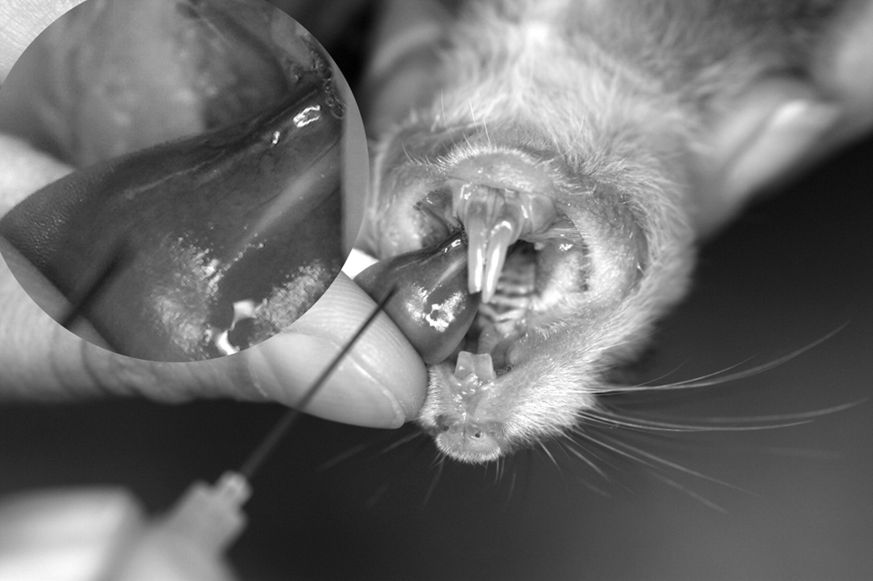
For blood collection, animals were anaesthetized with 3% (mouse) to 4% (hamster and guineapig) isoflurane anaesthesia (Forene™, Abbott Laboratories SA, Switzerland, oxygen flow rate of 4 L/min). The mouse or hamster was picked up by a technician, and then, the neck skin was grasped in order to assure a partial congestion of the jugular and lingual veins. The animal was brought into a supine position. A second person extended the tongue by picking it up between the thumb and a cotton bud (Figures 1 and 2). The thick caudal part of the left
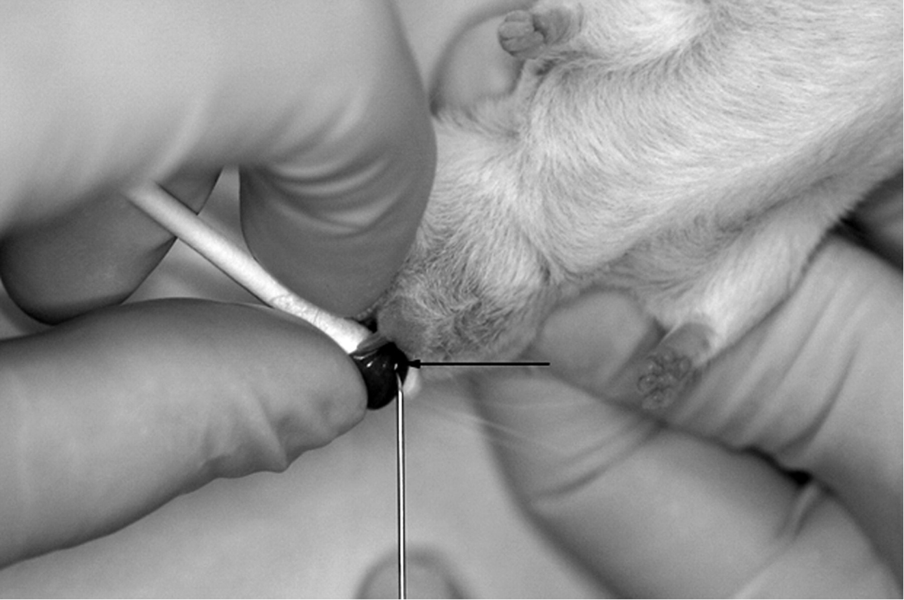
Handling of a mouse that is about to be punctured using the sublingual technique. The second technician views the sublingual surface of the tongue where two veins are running from the basis to the apex of the tongue. In contrast to hamsters or rats that possess large sublingual veins, only the caudal part of the sublingual vein in mice is thick enough for a successful puncture (indicated by arrow)
Left
The technique was also performed by one person using the same sequence as described above, except the neck skin was grasped after puncturing the sublingual vein.
Visual inspection for tongue damage was performed in anaesthetized mice and hamsters after 2 h and at three days post-punctionem.
The mice and hamsters were reused on up to three occasions and one occasion, respectively, for technique development after a recovery period of at least two weeks. After completion of the study, the mice were used for terminal bleedings and euthanized with overdosed carbon dioxide. After a veterinary health check, the hamsters were handed over as pets to private individuals. After an experimental blood collection, both guineapigs were exposed to isoflurane-overdosed carbon dioxide followed by exsanguination. A histological investigation of the tongue of both guineapigs was conducted.
Experimental design to compare sublingual and retrobulbar collection techniques in mice
The daily monitoring of clinical signs took place between 07:30 h and 10:00 h in the morning from day 1 to day 13. On study day 7, 300 μL of blood was collected from each animal (either from the sublingual vein or from the retrobulbar plexus). Sublingual blood collection was conducted as described above. The retrobulbar blood collection was carried out in the isoflurane-anaesthetized mouse using a 20 μL non-heparin glass microhaematocrit capillary. The capillary was inserted at a 45° angle into the medial corner of the right eye. Adequate congestion of the head veins was created by grasping the neck skin, at which point blood dropped immediately from the capillary end into a micro collection tube. Releasing the neck skin and removing the capillary stopped the bleeding immediately.
Necropsy of all mice was done six days after blood collection (day 13 of the study). Overdosed carbon dioxide was used for euthanasia. The tongues of the sublingually punctured mice and the orbital regions of both eyes of the animals punctured using the retrobulbar method were histopathologically processed. Transversal serial sections were prepared. The non-punctured left eyes were used as control tissue. To determine the extent of injury, the affected tissues were graded using a scale from 1 to 5 according to the percentage of affected tissue area visible on the slide. The severity grade corresponded to the percentage of affected tissue as follows: 1 = 12.5% of affected tissue, 2 = 25% of affected tissue, 3 = 50% of affected tissue, 4 = 75% of affected tissue, 5 = 100% of affected tissue. In sublingually punctured mice the tongue, and in retrobulbar punctured mice the Harderian gland, eye muscles and optic nerve were evaluated separately.
Results
Blood collection techniques
Using sublingual blood collection it was possible to routinely draw blood samples of 300 μL from mice with an optimized technique, as well as volumes up to 1.2 mL in individual animals during terminal bleeding. Only the puncture of the thicker caudal part of the vein, an area of 2–3 mm, yielded a quick and satisfactory blood flow. In addition, the punctures closer to the apex of the tongue revealed only small collectable amounts and were combined with the risk of haemorrhage and subsequent swellings (observed in a few individuals 2 h after blood collection). Haemorrhages and swellings required no medical intervention and did not affect the animals in their locomotory activity or feeding behaviour. Finally, the technique precipitated blood aspiration in some animals and the nose of the affected mice was cleaned with a cotton swab and blood collection was terminated. In order to prevent the aspiration of blood with subsequent dyspnoea, mice had to be held in a horizontal position during blood collection. The prevalence of these complications was not documented, because this was not the primary purpose of the investigations.
This technique can be performed successfully by one or two persons. However, the puncture is more difficult for one person working alone, because the veins are less visible without prior vein congestion. It is, nevertheless, an effective and secure method, when carried out by experienced individuals.
Sublingual blood sampling was also found to be an appropriate method for hamsters and a volume of 750 μL of blood per animal can be collected quickly. Due to the larger size of the mouth area and sublingual vein diameter, the risks of respiratory tract obstruction by blood does not exist to the same extent as in mice, but attention has to be paid to make sure that the slippery and compact tongue of the hamster is immobilized properly. No complications arose during the blood collection procedure, and no continuous bleeding was noted.
No sublingual vein was visible in the tongues of both guineapigs. The experimental puncture of the expected location of the vessel was not successful. Histological examination confirmed that no large blood vessel can be identified macroscopically.
Comparison of sublingual vs. retrobulbar blood collection techniques in mice
Both blood sampling techniques lasted about 20–30 s in mice and were carried out without complications, with the exception of one animal that did not recover from anaesthesia after sublingual blood collection. Histopathological examination of this mouse revealed a focal haemorrhage on the ventral side of the tongue that was considered to be insufficient to induce death. The death of this animal was most likely related to the anaesthesia and not to the blood sampling method. Apart from this death, no other in-life complications were noted.
Injuries were induced in 15 out of 30 sublingually punctured mice, whereas 28 out of 30 retrobulbar punctured mice showed tissue damage (Table 1). The evaluated mean severity grade of the lesions in tongues of mice after sublingual blood collection was determined to be 1.1. The puncture of the sublingual vein caused minimal focal injury, in the form of regenerative processes after destruction of tongue muscle fibrils, and focal accumulation of erythrocytes in the submucosa (Figures 3 and 4). The muscle cells of the affected areas were brightened and surrounded by phagocytes (mostly macrophages) and lymphocytes.
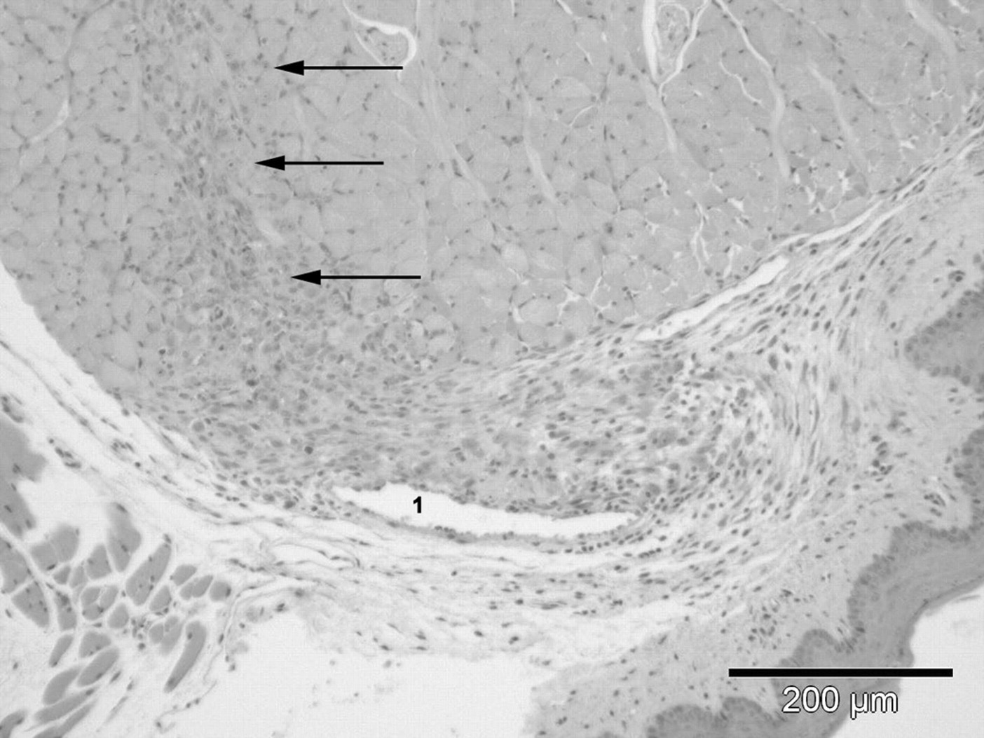
Transversal section of a tongue six days after sublingual blood collection. The sublingual vein is found in the middle of the slide (1). On the left side above the vein, an area of muscle destruction with debris and infiltration of phagocytes is visible and indicated by arrows (severity grade 1 = minimal lesion; staining: haematoxylin-eosin, 10× magnified)
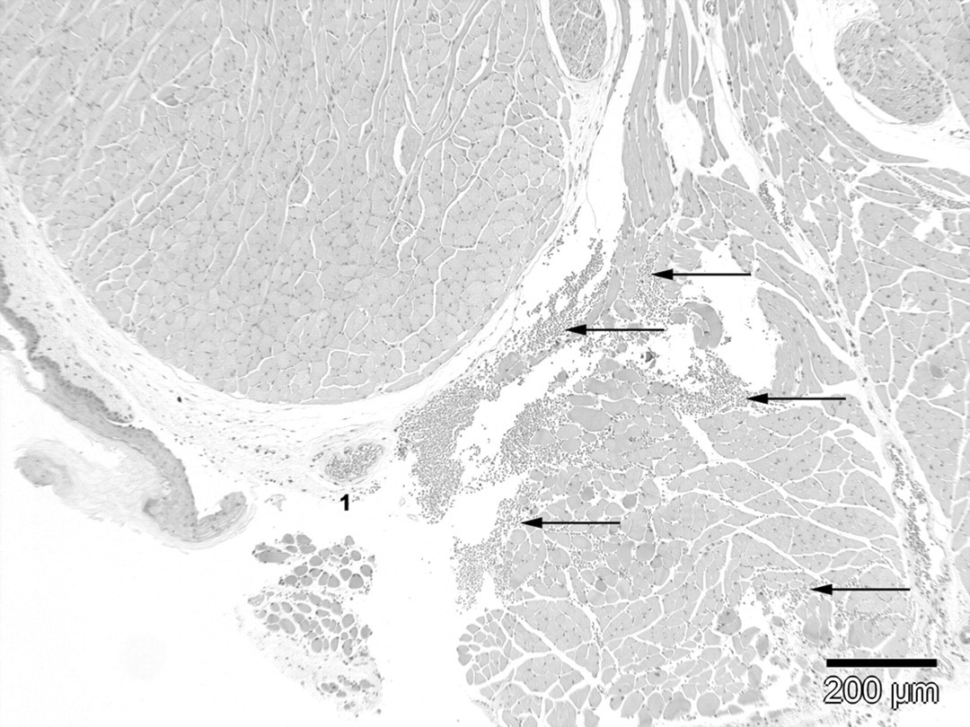
Transverse section of haemorrhages in a tongue six days after sublingual blood collection. The sublingual vein with erythrocytes in its lumen is found in the middle of the figure (1). To the right of the vein, erythrocytes infiltrated the intermuscular space and the connective tissue indicated by arrows (severity grade 1 = minimal lesion; staining: haematoxylin-eosin, 10× magnified)
Histological results of tongue and eye region tissue after sublingual and retrobulbar blood collection
Although 30 mice in the retrobulbar group were examined, in a few slides some tissues could not be shown. For this reason the examination of tissue in these slides was not possible and the number of affected animals did not correspond to the number of animals used. The key used to evaluate the grades of severity from 1 to 5 according to the percentage of affected area per tissue visible in the slide was defined in accordance with standard common practice at Novartis Toxicology, Switzerland (grade 1 = 12.5% of tissue area affected; grade 2 = 25% of tissue area affected; grade 3 = 50% of tissue area affected; grade 4 = 75% of tissue area affected; grade 5 = 100% of tissue area affected). Concerning the individual severity grades per tissue, a summarized severity grade of all lesions in an animal was determined
The orbital puncture induced unilateral lesions which involved mostly adnexa structures of the eyes: Harderian glands and eye muscles. Minimal to marked, focal to diffuse necrosis, and minimal to slight atrophic changes were noted in the Harderian gland (Figures 5 and 6). The lesions were consistently noted in the part of the Harderian gland located close to the medial corner of the eye. More diffuse lesions involving the whole gland were noted only in individual animals. Minimal to slight muscular lesions including signs of necrosis, regeneration and haemorrhage were noted in the eye muscles. In addition, a focal subacute inflammation of traumatic origin was noted in the eye of one single mouse and a focal traumatic lesion was noted in the optic nerve of one animal (Figure 5). The mean severity grade of tissue damages after the retrobulbar puncture was 1.5 (Table 1).
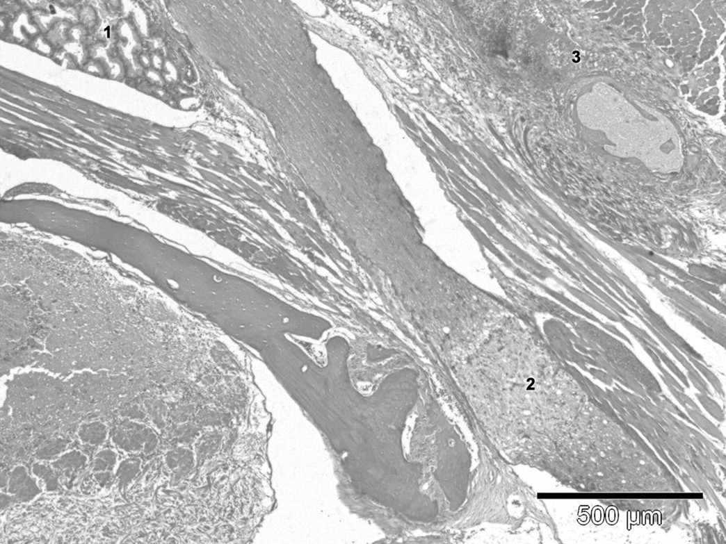
Eye region of one retrobulbar punctured mouse that was affected by lesions in bulbus muscles and the optic nerve (six days after blood collection). In the upper left corner, parts of the Harderian gland with typical tubular structure are shown (1). Diagonally from the upper left middle to the lower right corner the optic nerve is visible. On the lower part of the nerve, the structure of the
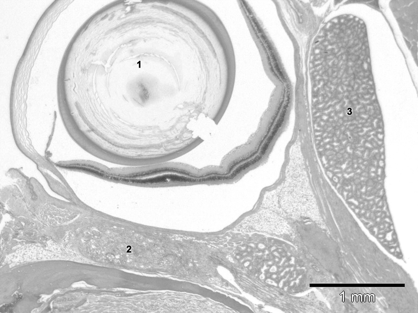
Eye region of one retrobulbar punctured mouse that was affected by lesions in the Harderian gland (six days after blood collection). In the upper left quadrant, the bulbus of the eye with cornea, lens, iris and retina (1). The space between the retina and orbita is an artefact. Below the bulbus, parts of the Harderian gland with partial loss of tubular structure (focal to diffuse atrophy and necrosis, severity grade 2 = slight) and phagocyte infiltration are shown (2). Unaffected Harderian gland area with intact
Discussion
One purpose of this study was to apply the technique of sublingual blood collection (successfully used in rats) to mice, hamsters and guineapigs. Sublingual bleeding was developed for use in mice and hamsters, but was not possible in guineapigs. In guineapigs sublingual veins are not peripherally located. Therefore, sublingual blood collection is not suitable in this species. Subsequently, the effects of blood sampling in mice using the retrobulbar and the sublingual methods, respectively, were compared. Blood collection from the sublingual vein in mice causes less tissue damages than the retrobulbar method. Therefore, it can be recommended as an alternative method to retrobulbar bleeding.
Blood sampling from the sublingual vein is suitable in mice, but differs from the way the method is applied to rats or hamsters. Use of the sublingual method of blood sampling for mice requires more practice. Even though the technique is simple, it seems to be easier to learn the procedure using a larger species (like rats or hamsters). In mice, only the caudal part of the sublingual vein has a sufficient diameter to permit successful puncture. If the vein is punctured closer to the tongue's apex, haematoma and swelling may result as a consequence of insufficient blood flow. During the collection, the mouse has to be held strictly horizontal; otherwise it could aspirate the blood resulting in dyspnoea or even gasping. Furthermore, the grasp of the animal's neck skin in order to ensure an adequate blood flow, should not be too tight. If sublingual blood collection in mice is carried out less than optimally, an unsatisfactory blood collection will probably result. However, it will not result in severe tissue damage, which is known to be the case with retrobulbar blood sampling under comparable conditions.
This technique permitted the routine collection of 300 μL of blood per mouse and 750 μL of blood per hamster. Terminal collection of larger amounts in mice up to 1.2 mL was possible, but was not further evaluated systematically. Like the retrobulbar technique, sublingual bleeding can also be done by one person. This procedure does not pose a challenge for an experienced technician. Nevertheless, we propose to exercise the method with two persons to facilitate the complete procedure.
Blood collection from the sublingual vein or retrobulbar venous plexus was very quick and lasted only 20–30 s. This quick and effective collection is very important, because mice recover quickly from isoflurane anaesthesia.
Typically, rats have been used for investigating the retrobulbar technique, its risks and influences on blood parameters. The anatomy of the eye and adnexa structures of mice, guineapigs and hamsters is comparable with the rat's anatomy. 16 Therefore, references to rat studies seem to be valid and necessary if we discuss the results obtained in other rodents.
In rats, a recovery period of two weeks after retrobulbar bleeding is recommended by Diehl
For hamsters, sublingual bleeding is easier than the procedure for mice. The oral cavity of the hamster is wider than that of the mouse, and as a corollary the sublingual veins have a larger diameter, a fact that facilitates the puncture. Because of the simple procedure and reduced risk of tissue damage, this method can also be recommended as an alternative method to retrobulbar bleeding in hamsters.
Retrobulbar bleeding may cause severe tissue damage in rats, 1,10,17 however such studies are not available in mice. The rate of incidence of damaged tissue due to the sublingual and the retrobulbar puncture methods, respectively, can be seen in the pathology examination of the tongue as well as the eye region. In the sublingual group, 15 out of 30 mice (50%) had lesions of the tongue with a mean severity of 1.1. In contrast, 28 out of 30 animals in the retrobulbar group (93.3%) showed tissue damage to the eye's adnexa structures (including optic nerve lesion in one animal) with a mean severity of 1.5. The clinical comparison of physiological damages between the eye and the tongue seems to be justifiable, even though it is uncertain to what extent mice rely on their sense of sight.
From the point of view of pathology, the method of sublingual blood collection is suitable in mice. This evaluation is based on the lower grade of severity and the smaller number of affected animals in the sublingual group compared with the retrobulbar group. This is analogous to the results obtained in rats. 14,18 Based on these pathology results, we assume that pain and discomfort after sublingual blood collection are most likely reduced compared with retrobulbar bleeding.
In conclusion, puncture of the sublingual vein can be recommended as a suitable technique for mice and hamsters, but not for guineapigs. The technique is simple, fast and effective. The method permits the collection of large volumes of blood: 300 μL (in mice) or 750 μL (in hamsters) were collected either by one or two persons using this method. The major advantage of sublingual puncture is the fact that it causes less tissue damage than retrobulbar bleeding. Therefore, it can be expected that this method produces fewer side-effects and has less effect on the animals' wellbeing.