
Abstract

Case report
An elderly man presented to the A&E Department of his local district hospital complaining of worsening dyspnoea and cough. His past medical history included carcinoma of the bronchus, diagnosed several years earlier and treated with chemotherapy and radiotherapy. On examination, there was tracheal deviation towards the left and his left chest was dull to percussion, with decreased air entry in the mid and lower zones. A plain chest radiograph (Figure 1) showed a whiteout of the left lung field and his breathlessness was attributed to a large pleural effusion.
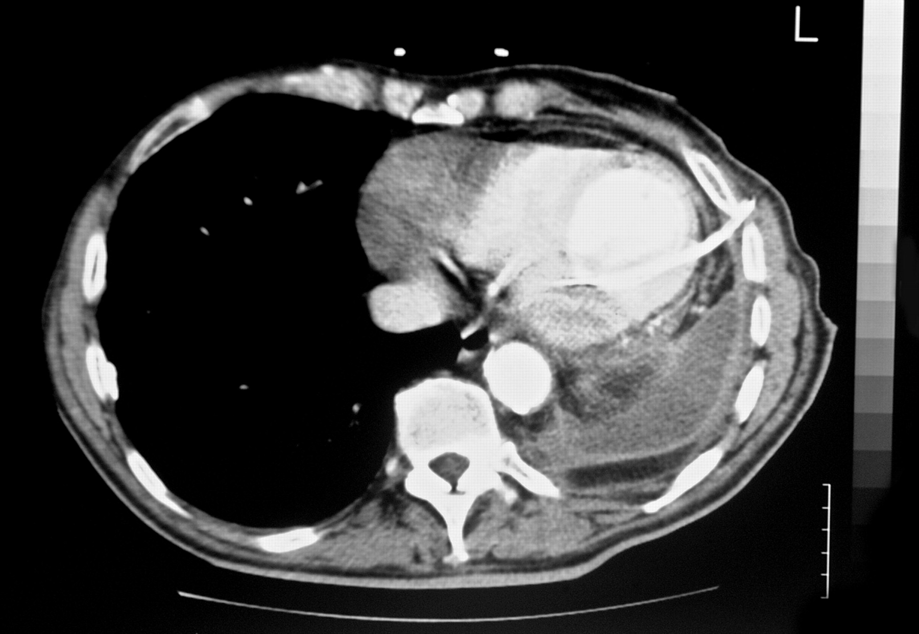
A trainee emergency physician inserted a percutaneous 12 French pigtail drain into the left chest. There was immediate and massive pulsatile haemorrhage from the drain which was quickly clamped. There was no further visible blood loss and the patient remained haemodynamically stable. Suspecting that a major vascular structure had been punctured, the drain was secured in situ. Emergency computed tomography (CT) of the thorax showed the drain within the left ventricle (Figure 2) and the patient was expeditiously transferred to our regional cardiothoracic surgical centre.
On arrival, the patient was anxious, tachycardic and hypertensive. The drain was sutured to the skin, entering the left chest in the anterior axillary line via the sixth intercostal space. At operation, an anterolateral minithoracotomy was performed through the fourth interspace and the drain was found to enter the left ventricle through the apex; it was easily removed and the puncture site repaired under direct vision with a pledgeted 3/0 polypropylene suture. The patient made an uneventful recovery and was transferred back to his local hospital for further management.
Discussion
Hemithorax opacification on chest X-ray has four potential causes: collapse
(atelectasis) of an entire lung; pleural effusion; consolidation; and
post-pneumonectomy. In atelectasis, there is volume loss of the affected lung resulting
in mediastinal shift towards the opacification. On the other hand, a
large pleural effusion exerts a mass effect, pushing the trachea away
from the affected side. In consolidation due to pneumonia, there is typically no
mediastinal shift and an air bronchogram may be seen against the consolidated lung.
Finally, when an entire lung is surgically removed, there is volume loss with subsequent
fluid accumulation and fibrotic opacification of the pleural space; a resected rib may
be absent on the radiograph although this diagnosis should be clinically apparent. The
British Thoracic Society (BTS) has published guidelines on the insertion of chest drains
and advise that ‘it is important to differentiate between the presence of collapse and a
pleural effusion when the chest radiograph shows a unilateral whiteout’.
1
The left lung field whiteout with the trachea deviated towards
the pathology in this case is classical for collapse and therefore chest drainage is not
indicated. Plain chest radiograph of left lung field whiteout with ipsilateral tracheal
deviation CT demonstrating the percutaneous drain in the left ventricle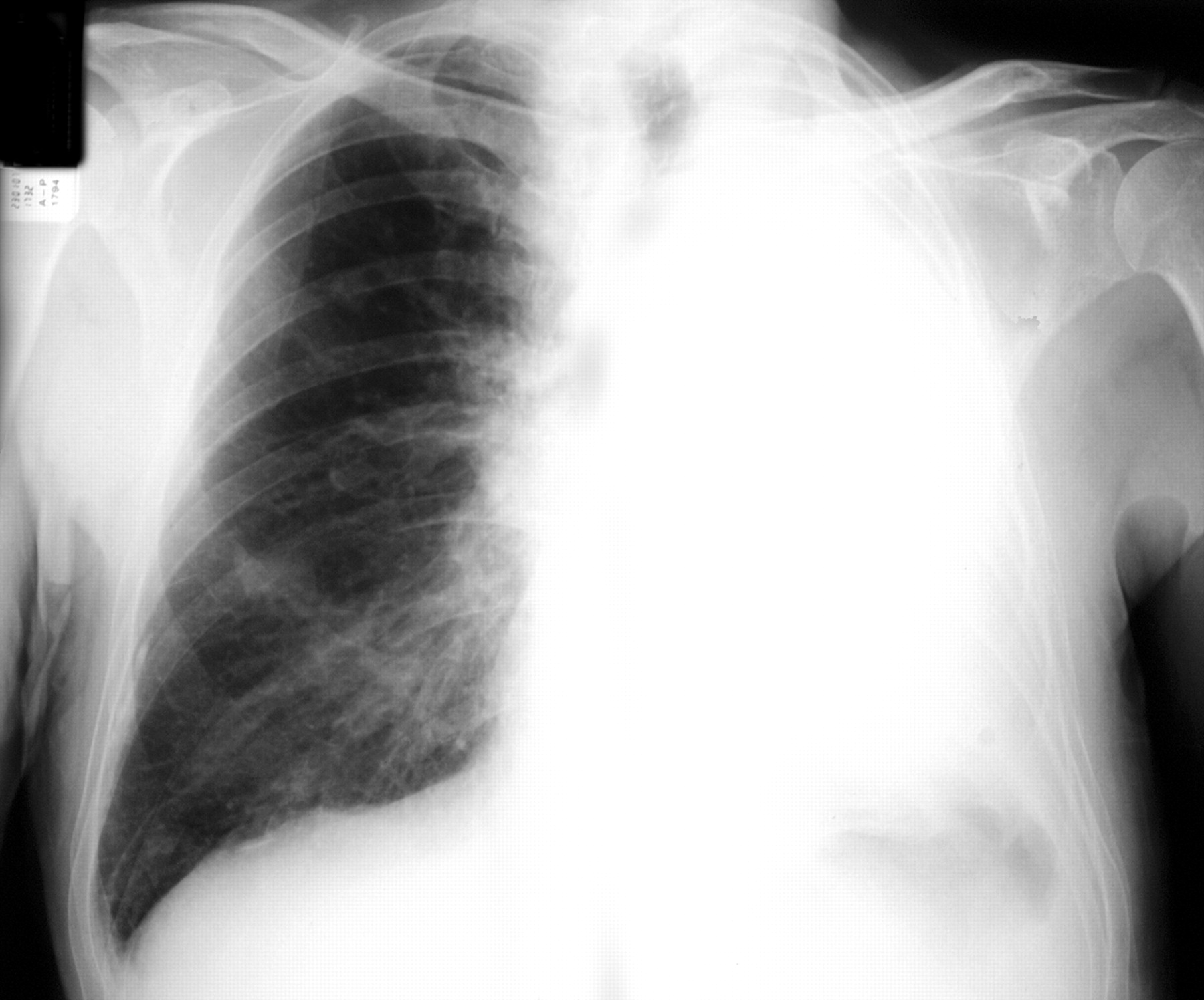
Chest drain insertion is a procedure commonly performed by many physicians and surgeons. Complications occur in up to 20% of patients including failure to enter the pleural space, early tube dysfunction and laceration of the lung. The BTS describe a truncated ‘safe triangle’ through which tube placement is advocated to minimize risk of injury to underlying structures. This area is formed by ‘the anterior border of latissimus dorsi, the lateral border of the pectoralis major muscle, a line superior to the horizontal level of the nipple, and an apex below the axilla’. Complications most commonly occur when the drain is placed outside of this zone, as in our case. In a recent survey of junior doctors in a UK teaching hospital, 45% opted to place a chest tube outside of the safe triangle, most commonly at a site deemed too low. 2 In addition, thoracic pathology including kyphoscoliosis, lung volume loss or a complex pleural space may lead to displacement of the mediastinum, tethering of the lung or elevation of the hemidiaphragm. In such cases, there is greater risk of injury to underlying structures and the drain should be placed under image guidance such as real-time ultrasound. 1 Similarly, when using the Seldinger technique, the BTS state that a drain should not be inserted without imaging ‘if free air or fluid cannot be aspirated with a needle’. In our case, the subsequent CT demonstrated lung collapse with displacement of the heart towards the chest wall, increasing the likelihood of cardiac puncture with the technique employed.
Iatrogenic cardiac injuries due to chest drain insertion are rare but often fatal. The National Patient Safety Agency recently found at least 12 deaths and 15 cases of severe harm directly attributable to chest drains over three years; puncture of the heart, lung and liver were reported, most commonly with the Seldinger technique. 3 In a quoted example: ‘Chest drain penetrated the heart and the patient had a cardiac arrest when it was removed’. On searching Medline, we identified eight cases with survival following cardiac perforation with a chest drain and all followed a similar course of action once suspected: immediate clamping of the drain, non-removal and transfer to a cardiothoracic centre for further management. 4, 5, 6, 7, 8, 9, 10, 11 We believe that these steps were also the key to salvage from the perilous situation in our case.
A cornerstone of ethical medical practice is primum non nocere: first, do no harm. The misdiagnosis of lung collapse as effusion despite clinical and radiological hallmarks exposed the patient to unnecessary intervention and risk. Directly Observed Procedural Skills (DOPS) is a validated tool for the assessment of competence in practical procedures. 12 Among others, it requires an understanding of the indications, anatomy and technique of a procedure, interpretation of diagnostic information, and awareness and management of complications. The cognitive aspects of determining the appropriateness of a procedure are as important as the technical skills. DOPS is a component of workplace-based assessment and forms an integral part of a learning portfolio as evidence of competence and performance.
In summary, differentiating between collapse and a pleural effusion on a unilateral whiteout chest radiograph is essential. Cardiac injury due to chest drain insertion is rare but often fatal; if suspected, the drain must not be removed and an emergency referral made to a cardiothoracic surgeon. Life-threatening complications may be minimized through education, adherence to BTS guidelines and competency-based assessment.
Footnotes
DECLARATIONS
Footnotes
Acknowledgements
Contributors from the referring hospital have approved the contents and accuracy of the manuscript but declined authorship or acknowledgement. We thank Nicole Breitenfeldt, Addenbrooke's Hospital, Cambridge for German–English translation