
Abstract

Antenatal screening for Down's syndrome involving the measurement of fetoplacental markers in maternal serum to determine the mother's risk of having an affected child is common practice in many countries, and is also utilized to identify pregnancies at risk for fetal open neural tube defects and trisomy 18. Second-trimester maternal serum screening has also been reported to detect other forms of chromosomal abnormalities, such as triploidy.1,2 Here, we report five cases of triploid pregnancies, three identified through second-trimester maternal serum screening, and two detected incidentally by early ultrasound.
Between January 2000 and December 2007, 78,947 women with singleton pregnancies underwent triple screening tests at 15 to 20 weeks of gestation at the Prenatal Diagnostic Center at Guangzhou Maternal & Neonatal Hospital, Guangzhou, Guangdong, People's Republic of China. Biochemical analysis of the markers free β-human chorionic gonadotrophin (hCG), alphafetoprotein (AFP) and unconjugated oestriol (uE3) was performed using a commercial time-resolved fluoroimmunoassay (AutoDELFIA, Perkin Elmer Life Sciences, USA) method. Down's syndrome and trisomy 18 risks were calculated using computer software (2T-Risks, Perkin Elmer Life Sciences, USA).
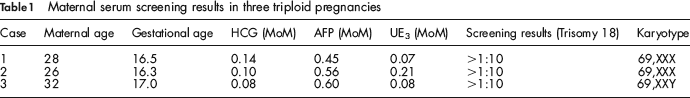
Three cases of fetal tripoidy were identified, with a prevalence of 0.38 per 10,000 fetuses. Table 1 summarizes the maternal serum screening results for the three pregnancies. All cases had very low hCG and low AFP and uE3, and had been detected by the screening tests for a high risk for trisomy 18. In all three cases, follow-up ultrasound showed viable fetuses with evidence of fetal growth restriction and fetal anomalies.
Maternal serum screening results in three triploid pregnancies
During the study period, combined first-trimester screening using nuchal translucency (NT) and maternal serum markers was not offered at our centre; however, an early ultrasound scan including NT measurement, was offered to mothers presenting at 11 to 14 weeks of gestation. 3 For pregnancies with increased NT thickness (>3.0 mm), chorionic villus sampling (CVS) or amniocentesis was suggested. Among the 3598 patients who underwent the routine 11 to 14-week scan, two cases of triploidy were detected with NT measurements of 4.4 mm in one case (69, XXX) and 5.1 mm in another case (69, XXY).
In order to determine the origin of the extra haploid chromosome, DNA was isolated from the CVS samples or amniotic fluid cells and the peripheral blood of the parents. For quantitative fluorescent polymerase chain reaction (QF-PCR) amplification, fifteen highly polymorphic markers (CSF1PO, D7S820, D8S1179, D21S11, D2S1338, D3S1358, D13S317, D16S539, TH01, D18S51, D19S433, TPOX, vWA, D5S818 and FGA) were used. These analyses revealed that the extra set of chromosome was maternal in origin in the three second-trimester cases and paternal in origin in the two first-trimester cases.
Triploidy is a rare lethal chromosome abnormality caused by the presence of an entire extra set of chromosomes. Triploid pregnancies appear to present as two distinct profiles of second trimester maternal serum analytes: (1) those screened positive for both Down's syndrome and an open neural tube defect with elevated AFP, grossly elevated hCG, and low/normal uE3 or (2) those screened positive for trisomy 18 with very low hCG, and low AFP and uE3. 4 In a report, which was the largest ever study of its type, Benn et al. summarized the results of 74 triploid pregnancies, and found that the relative frequencies between the two distinct patterns were equal. 1 However, only the latter serum marker pattern was seen in our triploid cases. This might be due to our small sample. Another explanation might be that triploid with high level of hCG is more likely to be diandric. 5 Diandric triploid in the first trimester period tends to have large NT measurements, which can be detected by early ultrasound screening. 5 This is also the case in our two early triploid pregnancies, both of which had large NT measurements.
By using molecular markers, we could reveal that all of the three second-trimester cases were digynic triploidy, which confirmed the previous report that digynic triploidy was characterized by very low hCG. 6 Our series is rather small and more data may be needed to draw definite conclusions. Nonetheless, our findings are the first cases of triploidy identified by antenatal screening programmes in the native Chinese population, and further confirmed the results of other studies that triploidy, like trisomies, can be incidentally detected as part of a screening programme for Down's syndrome. Rapid antenatal diagnosis can be obtained by interphase fluorescence in situ hybridization (FISH).