
Abstract
Introduction:
Tenosynovial giant cell tumours (TSGCT) are rare mesenchymal tumours that develop in joints, bursae and tendon sheaths. The aim of this study was to describe the epidemiological, clinical and histopathological aspects of this pathology in Togo.
Methodology:
This was a retrospective descriptive and analytical study of histologically confirmed cases of tenosynovial giant cell tumour from 1 January 2014 to 31 December 2023 in the Pathology Department of the Sylvanus Olympio Teaching Hospital in Lomé, Togo.
Results:
We identified 12 cases of tenosynovial giant cell tumours. We found 8 male subjects and 4 female subjects. The mean age of our patients was 24.2 ± 12.8 years. The tumour was located in the fingers in 9 cases (75%) and in the toes in 3 cases (25%). The mean time to diagnosis was 26.3 + 20.8 months. Pain and a palpable mass were the main symptoms. The nodular type was found in 7 cases (58.33%) and the diffuse type in 5 cases (41.67%).
Conclusion:
Tenosynovial giant cell tumour is a benign proliferative disorder of the synovium, the aetiopathogenesis of which remains undetermined.
Introduction
Tenosynovial giant cell tumours are rare mesenchymal tumours, affecting joints, tendon sheaths and bursae. 1 There is a nodular type and a diffuse type; the nodular type has a mainly indolent course and the diffuse type is locally aggressive.1 -3 It is mainly caused by a mutation in the stromal cells of the synovial membrane, leading to overexpression of colony-stimulating factor 1, which recruits CSF1R-expressing cells of the mononuclear phagocyte lineage into the tumour mass.2,4 Sarcomatous transformation with metastatic spread is extremely rare. 5 Most patients with tenosynovial giant cell tumours are young and, although generally not life-threatening, the disease and its treatment can have an impact on quality of life.3,6,7
In national studies, incidence rates vary from 30 to 34/1 000 000 for nodular tenosynovial giant cell tumours affecting the fingers and 11/1 000 000 for those located in the extremities. Incidence rates for the diffuse type range from 5 to 8.4/1 000 000.6,8,9 Hospital studies have reported a higher proportion of the diffuse type (70%-90%).10 -12
In our Togolese context, no study of tenosynovial giant cell tumours has been reported. We therefore initiated this study with the aim of describing the epidemiological, clinical and histopathological aspects of tenosynovial giant cell tumours in our context.
Methodology
We carried out a retrospective, cross-sectional, descriptive and analytical study of cases of tenosynovial giant cell tumours diagnosed at the Pathology department of the Sylvanus Olympio Teaching Hospital in Lomé from 1 January 2014 to 31 December 2023, that is, over a period of 10 years.
Data were extracted from laboratory records. Study material consisted of biopsies and surgical specimens fixed in 10% buffered formalin and processed using standard histology techniques. Variables included age, sex, site of lesion, clinical signs, time to consultation and histological type. The results are presented in the form of proportions for qualitative variables and means and standard deviations for quantitative variables. The Student’s t test was used to compare means, and the chi-2 test or Fisher’s exact test to compare percentages. The significance threshold was set at .05.
The reporting of this study conforms to the Strengthening the Reporting of Observational Studies in Epidemiology (STROBE) Statement: guidelines for reporting observational studies. 13
Results
Epidemiological Data
We identified 12 cases of tenosynovial giant cell tumours over a period of 10 years. There were 8 males and 4 females. The mean age of our patients was 24.2 ± 12.8 years, with extremes of 12 and 58 years. Five of our patients were aged between 10 and 20 years and between 20 and 30 years respectively, representing 41.7% of all cases in each age group. The lesion was located on the fingers in 9 cases (75%) and on the toes in 3 cases (25%; Table 1).
Summary of sociodemographic and clinicopathological characteristics of patients.

Clinical Data
The presence of an isolated mass was the clinical sign found in 7 cases (58.33%), and associated with pain in 5 cases (41.67%). The average consultation time was 26.3 + 20.8 months, with a range of 2 to 72 months (Table 1).
Histopathological Data
Histological diagnosis was made on 8 biopsies (66.67%) and on 4 surgical specimens (33.33%). The surgical specimens included 3 lumpectomy specimens and 1 finger amputation specimen (Table 1). Histologically, the tumour proliferation was made up of small monotonous cells with rounded, vesicular nuclei; mixed in variable proportions with numerous multinucleated giant cells, on a background of fibrous hyperplasia, richly vascularized (Figures 1 and 2). The nodular type was found in 7 cases (58.33%) and the diffuse type in 5 cases (41.67%).

Proliferation of giant cells, mixed with a few mononuclear cells (HE 100×).

A few giant cells at high magnification, within abundant fibrous tissue that is sometimes hyalinized (HE 400×).
A statistically significant relationship was found between clinical symptomatology and the histological nature of the lesion (P-value = .001263; Table 2).
Histological type/symptoms.

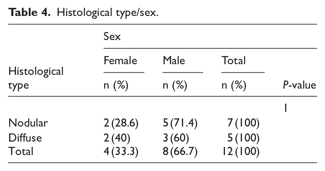
There was no statistically significant relationship between histological nature and age (P-value = 1), sex (P-value = 1), or time to consultation (P-value = 1; Tables 3–5).
Histological type/age.

Histological type/sex.
Histological type/diagnosis delay.

Therapeutic and Prognostic Data
Only one of our patients underwent amputation. The remaining patients underwent per- or post-diagnostic lumpectomy. No iatrogenic complications, in particular nerve damage, were observed.
After 1 year of follow-up, 3 patients were lost to follow-up. We noted a recurrence in 2 patients at 3 and 8 months respectively.
Discussion
Epidemiological Data
Tenosynovial giant cell tumours are mesenchymal tumours arising in the bursae, joints and tendon sheaths.1,14,15 It is a rare soft tissue tumour that often affects adults aged between 30 and 50 years, with an overall incidence of 1 in 50 000.16 -18 We found an annual incidence of 1.2 cases of tenosynovial giant cell tumours. The average age of our patients was 24.2 ± 12.8 years. A predominance of females has been reported in the literature.10,19 In our series, there was a predominance of males. Çevik et al 15 found the same male predominance. The lesion was located on the fingers in 75% of cases. Approximately 75% of these tumours are located in the fingers, particularly on the palmar surfaces of the fingers and are the second most common tumours of the hand.20,21
Radio-Clinical Data
Tenosynovial giant cell tumours have a highly variable clinical presentation and are associated with pain, swelling, limitation of movement, haemorrhagic joint effusions and progressive cartilage destruction; all of which can have a considerable impact on activities of daily living (eg, walking, work, sports).17,22 The average time to diagnosis is 6 months for the majority of patients. 16 The presence of an isolated mass was the clinical sign found in 58.33% of cases, with average time to diagnosis of 26.3 months ±20.8. Xie et al found an average diagnosis time of 18 months. 10 Because the bones are shallow, individuals with tenosynovial giant cell tumours of the hand most often present symptoms rapidly. 16 The relatively long delay in diagnosis in our context can be explained by the absence of pain in the majority of our patients (58.33%).
Standard radiography is indispensable, and is contributive as there are bone abnormalities in 33% of diffuse forms in the knee. Bone lesions, in the form of cystic erosion, are more frequent and suggestive in the hand and hip. Cysts in a non-weight-bearing region, often symmetrically on either side of the joint line or at the capsular insertion lines, without calcification, are often suggestive of articular TSGCT. 23
The most contributive examination is MRI, indispensable to diagnosis and surgical planning. Localized forms typically show as a well-delineated lesion, off-centre from or else more-or-less completely enclosing the tendon in question. Diffuse forms are usually articular, showing as a soft-tissue mass with homogeneous uptake, with associated joint effusion. Fossa and extra-articular extension is often associated and should be screened for on preoperative work-up. The localized or diffuse lesion signal indicates the form of the TSGCT: variable tissue hemosiderin loading accounts for weak or intermediate signal on T1 and spin-echo T2-weighted sequences. The signal is enhanced on gadolinium injection. Gradient-echo sequences are very useful for detecting hemosiderin deposits, showing in low signal, even after injection.23 -25
Ultrasonography is now widely used; it does not replace MRI, but can be indicative, showing a mass of variable aspect but with suggestive location. TSGCT appear hypervascularized on Doppler ultrasound, 26 which optimally guides synovial biopsy.
As regards differential diagnosis, in the limbs, most lesions show different MRI characteristics: ganglion cysts, haemangioma, synovial sarcoma of the foot and nerve-sheath tumour show high T2 signal. Like TSGCT, other lesions may show intermediate T2 signal: in the foot, fibromatosis, xanthoma, Morton neuroma and certain desmoid sarcomas or tumours; location, symptomatology and clinical findings guide diagnosis, which must, however, be confirmed on specialized radiological/clinical assessment. 23
In periarticular, peripheral or proximal articular locations, synovial sarcoma, which is rarely intra-articular, is a difficult differential diagnosis, with high stakes. Location may be in the sheaths or bursae, like TSGCT; in 20% to 30% of cases, there are calcifications, which are also, although rarely, found in TSGCT. MRI typically associates a threefold signal: high in fluid, iso or intermediate in lipid and low in fibrous tissue; but, although typical, this aspect is not systematic. Biopsy should be performed in case of the slightest doubt, especially in nodular forms. 23
In intra-articular locations, erosion and images of cysts in non-weight-bearing regions are suggestive, especially when associated with the above typical MRI aspect. Recurrent hemorrhagic synovitis in incipient haemophilia is the most difficult differential diagnosis; TSGCT with low hemosiderin load may mimic other pathologies: incipient rheumatic pathology, or synovial chondromatosis. 23
Pathological Data
There are many names given to tenosynovial giant cell tumour, reflecting its clinicopathological heterogeneity.20,27 Terms used to describe the tumour include tenosynovial giant cell tumour, nodular tenosynovitis, pigmented villonodular synovitis and giant cell tumour of tendon sheath.1,20 It is now known as tenosynovial giant cell tumour according to the World Health Organisation’s 2020 classification of bone and soft tissue tumours. 28
Nodular forms are more frequent in females (2:1), whereas female predominance is slight in diffuse forms. 23 In our series, 58.33% were of the nodular type and 41.67% of the diffuse type, with a strong statistically significant relationship between clinical symptoms and the histological nature of the lesion (P-value = .001263). The local aggressiveness of the diffuse type would motivate these patients to consult a lot more.
The aetiopathogenesis of tenosynovial giant cell tumours remains unclear. Multiple factors have been proposed as possible aetiologies, including inflammation, trauma, toxic causes, allergy, clonal chromosomal abnormalities and aneuploidy. 20 Initially, this pathology was considered to be an inflammatory disease; however, the existence of aneuploidy in some patients and the demonstration of clonal chromosomal abnormalities strongly suggest a tumour origin.20,29 Chromosomal translocation occurs when a section or portion of a particular chromosome separates and rearranges itself, resulting in a shift of genetic material, with modification of the chromosomes as a whole. Certain areas of chromosomes 1 and 2 are involved in the translocation that occurs in these tumours. Colony stimulating factor 1, or CSF1, is a type of protein overproduced by the cells during this translocation. Tenosynovial giant cell tumour cells use CSF1 to recruit white blood cells, incorporating them into the tumour; this explains the inflammatory changes induced by these tumours.16,20 The permissive and immunosuppressive properties of the histiocytes associated with giant cell tenosynovial tumours have prompted interest in the therapeutic targeting of these cells. The colony-stimulating factor 1 (CSF1)/colony-stimulating factor 1 receptor (CSF1R) axis has therefore been favoured, and various approaches targeting either the ligands or the receptor are currently in clinical development. 29
Immunohistochemistry is not necessary for positive diagnosis, but is most often used to assess tumour progression. 30 Ki-67 is a reliable marker for malignant tumour proliferation, and the results of detection are accurate. In diffused-type, large-volume and recurrent TSGCT patients, increased levels of Ki-67 expression were documented in earlier research.31,32 Wang et al 33 provides the first analysis of Ki-67 in diffuse-type TSGCTs, demonstrating significantly higher Ki-67 indices in recurrent versus non-recurrent TSGCTs. However, the combined expression of the colony stimulating factor 1 receptor (CSF1R) mutation and Ki-67 is lacking evidence in diffuse-type TSGCT, representing an area requiring further study. 30
Therapeutic and Prognostic Data
The majority of our patients (91.66%) underwent per- or post-diagnostic lumpectomy, with recurrence in 2 patients at 3 and 8 months respectively.
After complete resection, the recurrence rate for diffuse-type TSGCT is between 20% and 50%.23,34
Patients affected by TSGCT should be managed within expert centres or reference networks, by a dedicated, experienced sarcoma multidisciplinary treatment team, including a pathologist, radiologist, orthopaedic surgeon, pain specialist, surgical, radiation and medical oncologists; with new guidelines Other specialists, such as neurosurgeons and physical therapists, should be involved as required. 1 The surgical treatment should be emphasized for the treatment of this disease, sometimes target therapy might be beneficial for those with CSF-1R overexpression.
CSFR1 inhibitors are effective and beneficial for quality of life and symptoms, but are not widely available in most countries. The degree of uncertainty in choosing the most appropriate treatment and the lack of guidelines on the clinical management of tenosynovial giant cell tumours mean that the uptake of new treatment lines is inconsistent, with sub-optimal outcomes for patients. 9 Spierenburg et al recommend open surgical excision as the reference treatment. Systemic treatment is indicated for unresectable or refractory cases. However, there are regular side-effects and relapse may occur when treatment is stopped. 35
Limitations
Some information was not found, in particular the profession, socio-economic situation and the fate of certain patients.
Conclusion
Tenosynovial giant cell tumours are rare mesenchymal tumours arising in the joints, bursae and tendon sheaths. It mainly affects the fingers, with a predominance of females. We found a relatively low incidence, with a predominance of males. The average delay in diagnosis was relatively long in our series, reflecting the lack of awareness of this condition in our context. This highlights the need for greater awareness of this tumour among the general population, and among healthcare staff in particular.
Supplemental Material
sj-docx-1-pat-10.1177_30502098251341137 – Supplemental material for Tenosynovial Giant Cell Tumours: A Clinicopathologic Study of a Case-Series
Supplemental material, sj-docx-1-pat-10.1177_30502098251341137 for Tenosynovial Giant Cell Tumours: A Clinicopathologic Study of a Case-Series by Toukilnan Djiwa, Mayi Bombone, Bagassam Sama, Panakinao Simgban, T. H. Towoezim and Tchin Darré in Sage Open Pathology
Footnotes
Acknowledgements
Not applicable.
Ethics Considerations
This study was approved by the ‘Comité de Bioéthique pour la Recherche en Santé (CBRS)’ (Bioethics Committee for Health Research) from the Togo Ministry of Health, Ref No. 0101/2016/MS/CAB/DGS/DPLET/CBRS). The study has been carried out in accordance with relevant guidelines and regulations.
Consent to Participate
All patients and relatives of patients had received information on the purpose and procedures of this study and provided written and informed consent.
Consent for Publication
All patients and relatives of patients had received information on the purpose and procedures of this study and provided written and informed consent.
Authors Contributions
T. Djiwa was responsible for the conception of the study; participated in the study design; undertook the field study; conducted the data collection, analysis and interpretation; and wrote the manuscript. M.B., B.S., P.S., T.TH. and T.D. were involved in the data collection, analysis, and interpretation. They wrote and finalized the manuscript. T.D. is responsible for the overall scientific management of the study, for analysis and interpretation and for the preparation of the final manuscript. All authors have read and approved the final manuscript for submission for publication.
Funding
The author(s) received no financial support for the research, authorship, and/or publication of this article.
Declaration of Conflicting Interests
The author(s) declared no potential conflicts of interest with respect to the research, authorship, and/or publication of this article.
Data Availability Statement
Extracted data are with the corresponding author and available under reasonable request.
Supplemental Material
Supplemental material for this article is available online.
References
Supplementary Material
Please find the following supplemental material available below.
For Open Access articles published under a Creative Commons License, all supplemental material carries the same license as the article it is associated with.
For non-Open Access articles published, all supplemental material carries a non-exclusive license, and permission requests for re-use of supplemental material or any part of supplemental material shall be sent directly to the copyright owner as specified in the copyright notice associated with the article.