
Abstract
With the growing number of bioprosthetic aortic valve replacements (AVR), whether by surgery or transcatheter (TAVR), 1 diagnosing bioprosthetic valve dysfunction (BVD) has become increasingly important. Echocardiography is the only widely available, non-invasive and non-irradiating modality needed to confirm BVD and failure (BVF) following TAVR by 1) imaging the valve morphology to determine the mechanism and etiology of dysfunction, avoiding the risk of complications associated with transvalvular catheters, particularly in the setting of endocarditis or thrombosis, and 2) assessing the severity of hemodynamic valve deterioration longitudinally. Discordance between echocardiography and catheterization immediately after TAVR is related to Doppler physics and resolves when significant dysfunction occurs. 2
Accessibility, safety, ease of use and cost-effectiveness
There are several advantages in using echocardiography rather than catheterization for BVD diagnosis and follow-up. First, echocardiography is known to be safe and reproducible, enabling regular, accurate follow-up. As recommended in latest guidelines, transthoracic echocardiography (TTE) should be performed early following new valve implantation to establish the baseline morphologic appearance and hemodynamic function. Guidelines recommend routine TTE follow-up every year following bioprosthetic AVR 3 or whenever there is a change in clinical signs or symptoms. This allows for early detection of structural valve deterioration (SVD) and determine optimal timing for reintervention.3,4 In addition, TTE can be performed at rest as well as during exercise,5,6 without change in medications or addition of sedation, thus better reflecting the hemodynamic status of normal daily activities. Second, echocardiography is also widely accessible as it can be performed by different healthcare professionals (eg, cardiologists, sonographers, dedicated nurses) in any location. As healthcare disparities increase,7–9 echocardiography, thanks to its portability, affordability and accessibility, is a key tool for improving access to care. 9 Thus, conducting studies evaluating the cost-effectiveness of echocardiography versus invasive catheterization is akin to conducting a randomized trial on the safety of a parachute. 10 Finally, gradients by echocardiography and catheterization are highly correlated in advanced SVD, 2 but more importantly, echocardiography offers multiple confirmatory parameters to improve diagnostic accuracy such as morphologic changes to valve leaflets or stent frame, left ventricular dimensions and function, and presence of pulmonary hypertension. If the results are similar between the two methods, it doesn't seem justified to expose patients to the possible complications of an invasive procedure such as systemic embolism, vascular complications, and rarely death, myocardial infarction and stroke.
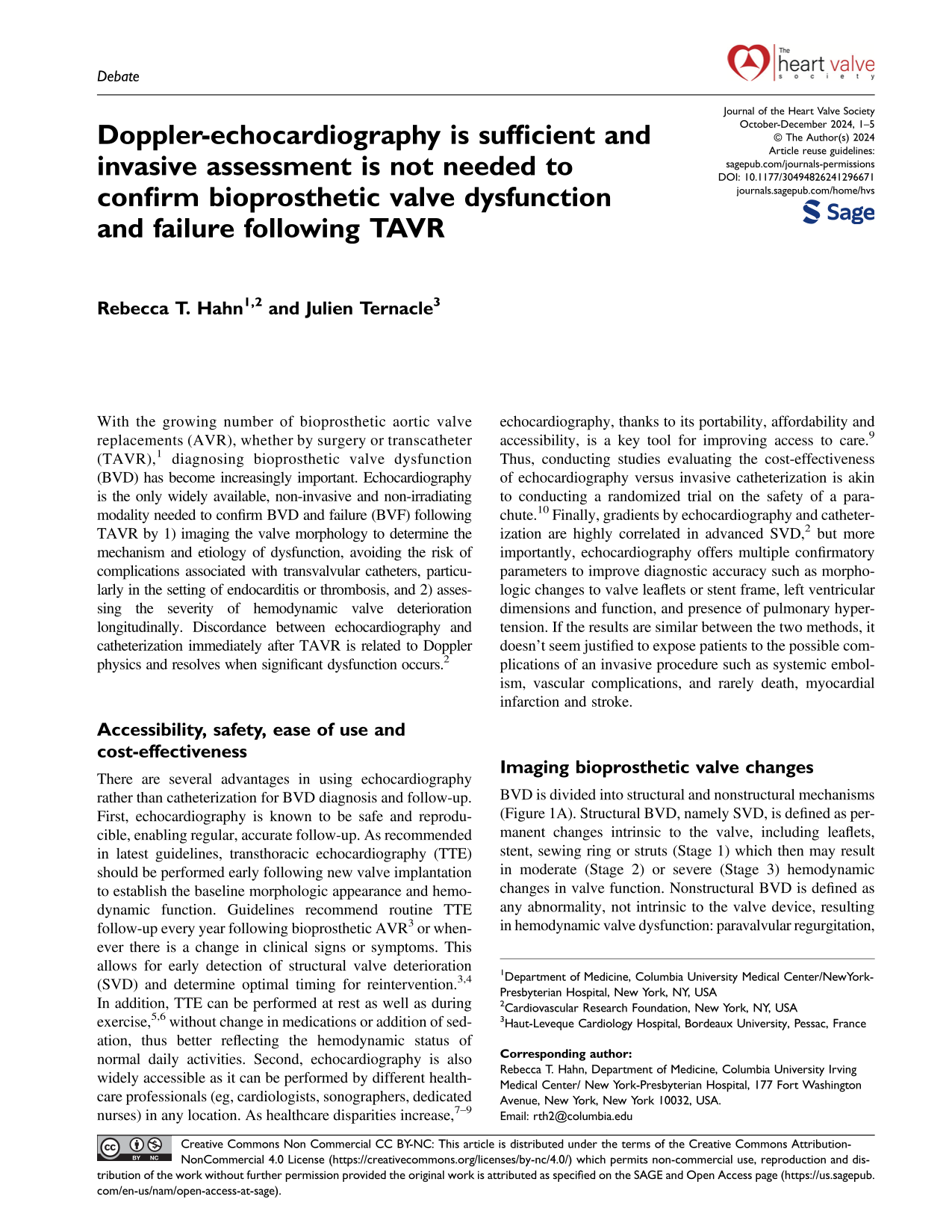
Imaging bioprosthetic valve changes
BVD is divided into structural and nonstructural mechanisms (Figure 1A). Structural BVD, namely SVD, is defined as permanent changes intrinsic to the valve, including leaflets, stent, sewing ring or struts (Stage 1) which then may result in moderate (Stage 2) or severe (Stage 3) hemodynamic changes in valve function. Nonstructural BVD is defined as any abnormality, not intrinsic to the valve device, resulting in hemodynamic valve dysfunction: paravalvular regurgitation, sub-valvular pannus overgrowth, inappropriate positioning (including procedural malposition and post-deployment migration) or sizing (under- or over- sizing), and prosthesis-patient mismatch (PPM). Other mechanisms of abnormal valve structure and function include thrombosis and endocarditis (Figure 1A). Differentiating these etiologies of BVD is essential to define optimal management, which cannot be achieved by measuring valve hemodynamics alone, but by imaging the valve. Therefore, echocardiography is the imaging modality of choice by the current guidelines to identify Red Flags requiring further investigations (ie, multimodality imaging) to confirm BVD diagnosis and its mechanism (Figure 1A). 11

Role of echocardiography to confirm bioprosthetic valve dysfunction and its correlation with invasive assessment.
Staging severity of hemodynamic valve deterioration
Echocardiography provides a comprehensive evaluation of bioprosthetic hemodynamic performances by assessing several parameters, including mean gradient, maximal peak velocity, effective orifice area (EOA), Doppler velocity index (DVI) and severity of intra- and/or peri-prosthetic aortic regurgitation,12,13 whereas catheterization provides only trans-prosthetic gradients. Although aortic regurgitation may also be quantified by aortography, this requires contrast injection with its associated complications (eg, contrast-induced nephropathy) and cannot determine whether the regurgitation is intra- or peri-prosthetic. However, all the hemodynamic parameters necessary to confirm the diagnosis of BVD and determine its severity according to VARC-3 (Valve Academic Research Consortium) 14 and HVC (Heart Valve Collaboratory) criteria 11 are provided by echocardiography (Figure 1A). This step of BVD severity classification is essential for therapeutic decision and subsequent follow-up. Once again, echocardiography is the imaging modality of choice for assessing the severity of BVD.
Why may echocardiography and catheterization be discordant?
Discrepancies in gradient measurements may occur between catheterization and echocardiography immediately following TAVR, 2 which may be explained by: 1) Principles of fluid mechanics and pitfalls of the modified Bernoulli equation, 2) Variability of the contraction coefficient depending on the orifice shape and, 3) pressure recovery distal to the vena contracta in a tubular structure.
Principles of fluid mechanics and pitfalls of the modified Bernoulli equation: Because of the Conservation of Momentum Principle, the average velocity at any position within the tube is constant. For laminar flow, the fluid adjacent to the vessel wall is motionless and blood flow in adjacent regions is reduced, the central flow accelerates to maintain the average. When velocities are measured by continuous wave Doppler, the highest velocities are recorded thus overestimating the “average” velocity. Thus, continuous wave Doppler may overestimate velocities across the valve orifice in a normal bioprosthesis. Conversely, turbulent flow has a well-defined average velocity profile across the orifice with a good correlation between Doppler and invasive measurements. 15 For pulsatile flow, the pressure gradient between two locations along a streamline is expressed by the Bernoulli equation, with the fundamental assumptions that proximal velocity is significantly smaller than distal velocity. However, proximal and distal velocities are of same magnitude in bioprosthetic valve, requiring subtraction between proximal and distal velocities to calculate the true pressure gradient. Thus, pressure gradient reported immediately after TAVR is likely an overestimate of the true transvalvular gradient.
Variability of the contraction coefficient depending on the shape of the orifice: The vena contracta is the point at which a jet has its minimal area. If fluid is led smoothly into an orifice, there is no contraction of the jet area before it expands radially beyond the orifice. If the jet passes through a sharp edged orifice, it continues to constrict beyond the orifice before then expanding radially. This contraction coefficient, defined by minimal jet area divided by orifice area, ranges from 0.6 (abruptly narrowed orifice) to 1.0 (gradually narrowed orifice), and is affected by the geometry of the orifice, as well as flow rate. Doppler echocardiography measures the highest velocities at veina contracta level, while invasive measurements are more downstream.
Pressure recovery distal to the vena contracta: Downstream from the vena contracta, pressure begins to recover but at a lower level than the original static level because of irrecoverable loss due to viscous effects. Invasive gradient measured at the recovered pressure site will be clearly lower than Doppler measurement at the vena contracta. Pressure recovery can cause major discrepancies in the setting of small ascending aorta diameter (<30 mm), larger orifice size and non-eccentric jets.16–18 As these last two situations are present after TAVR, it is likely that this Echo-Cath discordance is explained, at least in part, by pressure recovery, which cannot be directly measured non-invasively by echocardiography.
Does this discordance have an impact?
For all these reasons, pressure gradients immediately after TAVR can be expected to be higher on echocardiography than on catheterization (Figure 1B). However, the correlation between the methods improves as the bioprosthesis becomes stenotic because there is now turbulent flow with lower proximal versus distal velocity, constant (ie, does not vary with flow) contraction coefficient with stenotic orifices, and less pressure recovery. Thus, discrepancies between the two measures will be minimized as BVD severity increases without the need for invasive confirmation (Figure 1B). The clinical impact of Echo-Cath discordance is unknown as the large majority of studies evaluating the association between post-TAVR transprosthetic gradient and outcomes have used echocardiography. In a large retrospective analysis, the impact of immediate post-TAVR transaortic echocardiographic and invasive gradients on mortality showed opposite results for echocardiographic and invasive measurements in low-gradient patients. 19 Low echo gradients associated with higher mortality, explained by an association of low gradients with lower ejection fraction and stroke volume index. In contradistinction to this clinically logical finding with echocardiographic gradients, low invasive gradients showed no association with ejection fraction or stroke volume index and were associated with lower mortality. Therefore, catheterization evaluation does not appear to offer any advantages over ultrasound, while it carries some risks. 20
Conclusion
Echocardiography is the only exam needed to confirm the diagnosis of BVD, identify its mechanism and etiology, and quantify its severity, while catheterization may be associated with complications without offering any advantage over echocardiography. The additional use of noninvasive multimodality imaging is necessary in selected situations.
Footnotes
Funding
The authors received no financial support for the research, authorship, and/or publication of this article.
Declaration of conflicting interests
The authors declared no potential conflicts of interest with respect to the research, authorship, and/or publication of this article.