
Abstract
Background:
Massive, irreparable rotator cuff tears (RCTs) remain a challenging clinical problem with numerous described treatment options. Bursal acromial reconstruction (BAR) represents a promising and evolving technique for a subset of patients with irreparable RCTs.
Indications:
BAR is indicated for patients with massive, irreparable RCTs with a primary complaint of pain, well-compensated shoulder function, and minimal radiographic degenerative changes of the glenohumeral joint as an alternative to reverse total shoulder arthroplasty or superior capsular reconstruction.
Technique Description:
Positioning per surgeon preference and diagnostic arthroscopy is performed. Subacromial decompression with a minimal and gentle acromioplasty is performed, followed by assessment of RCT repairability. If the tear is deemed irreparable, acromial measurements in the medial-lateral and anterior-posterior dimensions are obtained. Two pieces of acellular dermal allograft are cut to the acromial dimensions and affixed together using fibrin glue. The reactive side (facing the acromion), medial, and anterior sides of the graft are labeled. Two suture tapes are passed through the corners of the graft and self-locked and run diagonally in a cruciate configuration using an antegrade suture passer. Medial and lateral #2 fiberwire sutures are placed in a luggage-tag configuration. Neviaser (posterior), middle, and anterior acromioclavicular joint portals are created for medial sided suture passage. Medial graft sutures are shuttled through the respective medial portals and the graft is pulled into the subacromial space. The lateral sutures are then removed from percutaneous posterolateral, middle lateral, and anterolateral portals along the acromial edge. Medial sutures are retrieved using a suture grasper subcutaneously on top of the acromion through the percutaneous lateral portals. The sutures are tied through the lateral portals, starting with the medial-lateral sutures, and the knots are buried. Postoperatively, patients are progressed through passive, active-assisted, and active range of motion between weeks 2 and 6 and strengthening is progressed at 6 weeks.
Results:
Clinical results are lacking in the literature, but anecdotal results in our institution have demonstrated promising early outcomes.
Discussion/Conclusion:
BAR represents a promising alternative in the array of surgical options for treatment of irreparable RCTs.
This is a visual representation of the abstract.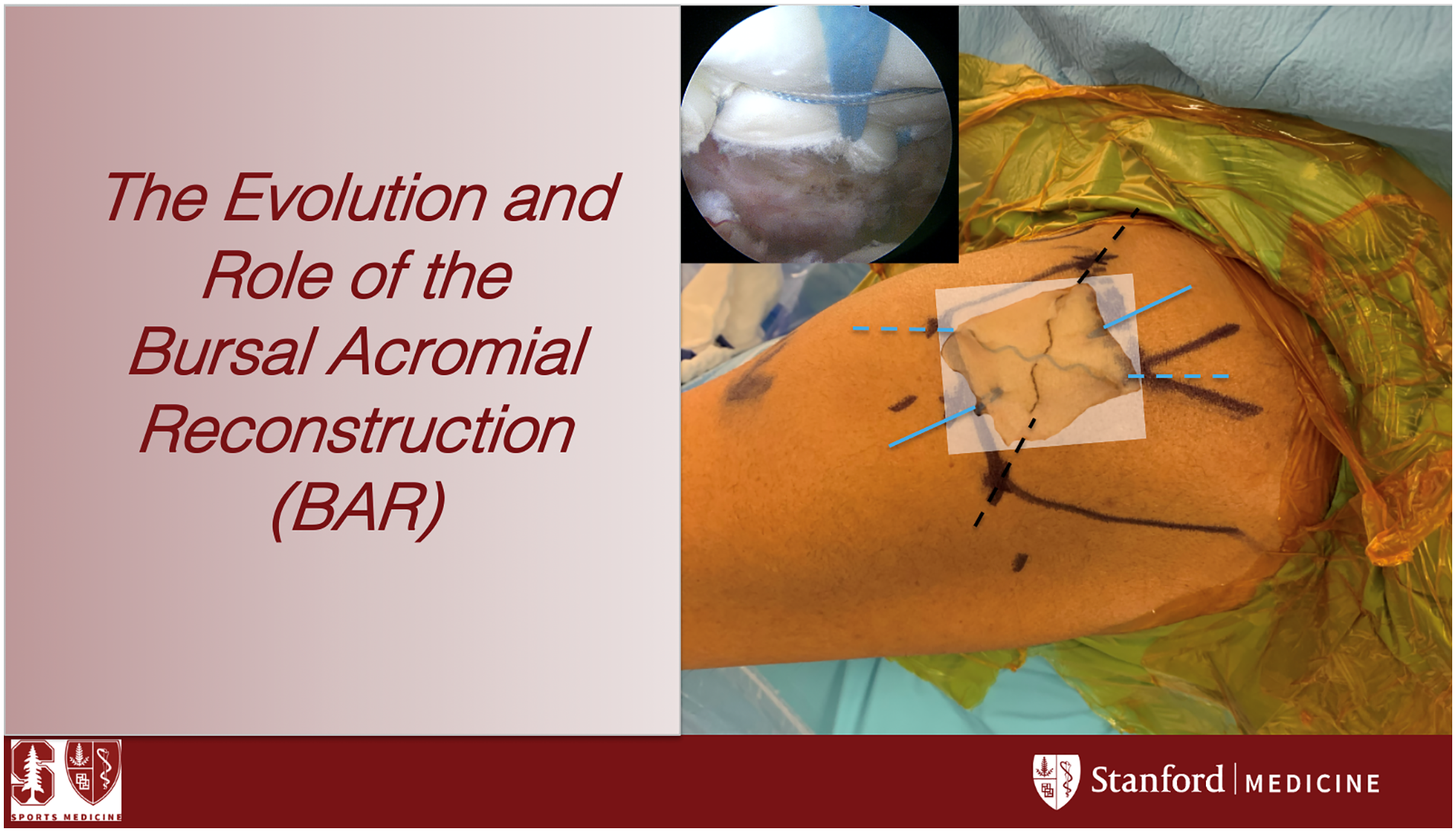
Video Transcript
In this video, we will discuss the evolution and role of the bursal acromial reconstruction (BAR). Rotator cuff pathology is the leading cause of shoulder-related disability, and approximately 250,000 rotator cuff repairs are performed annually in the United States. Massive tears make up nearly 20% of all rotator cuff tears and, although a high percentage of these tears may be deemed “repairable,” they are complicated by a high reported rate of structural failure. Numerous treatment options for irreparable rotator cuff tears are described in the literature, without a clear advantage to 1 procedure for all patients.
The BAR is a newly proposed technique for treating rotator cuff tears that consists of securing acellular dermal matrix allograft to the undersurface of the acromion, thus theoretically reducing acromial contact pressure and pain. Two recent studies have described good results for both pain and function in patients with failed superior capsule reconstruction (SCR) in which the graft covers the tuberosity, acting as an interposition between the acromion and humeral head. These studies beg the question—is it actually the interposition phenomenon that allows greater pain relief and biomechanical restoration?
To that end, multiple authors have described techniques for creating an interposition allograft between the humeral head and acromion. Makovicka and colleagues described a variation of the BAR that they referred to as a subacromial allograft spacer, intended as an adjunct for SCR. Their technique included graft fixation through acromial drill tunnels, posing a potential risk of acromial fracture. Ravenscroft has elegantly demonstrated the BAR without acromial tunnels using a cruciate suture technique in which sutures are passed subcutaneously over the acromion, thus eliminating the risk of acromial fracture while ensuring excellent graft fixation. We will present this technique with the authors’ modifications.
Our proposed indications are presented here. The authors believe BAR could be used across functional demand levels depending on the patient’s goals, as it provides a minimally invasive option that allows faster recovery and pain relief. Indicated patients primarily complain of pain and have well-compensated shoulder function despite their massive rotator cuff tear, as opposed to SCR and reverse total shoulder arthroplasty, which are indicated in patients with decompensated shoulder function. In addition, the BAR is indicated for patients desiring a less invasive option with a faster return to activities. This technique is not proposed as a single solution for all patients with irreparable rotator cuff tears, but rather as an additional strategy for surgeons treating these patients.
Next, the authors provide a brief case presentation and the surgical technique video. In this case, a 65-year-old right-hand–dominant man presented with left shoulder pain after a failed rotator cuff repair 10 months prior. He is very active and is an avid surfer. Preoperative shoulder outcome scores are provided. In addition to positive impingement signs, the patient’s physical examination demonstrated preserved active range of motion, but weakness to both Jobe and external rotation testing. Plain radiographs demonstrate minimal degenerative changes of the glenohumeral joint and an adequately preserved acromiohumeral interval of 8 mm. Representative coronal T2 and sagittal T1 images demonstrate a massive rotator cuff tear with retraction nearly to the level of the glenoid and grade 3 fatty infiltration of the supraspinatus with a tangent sign. After discussing multiple treatment options, the patient elected to proceed with BAR, because it promised improved pain relief and a faster return to surfing than other options. In addition, although his examination demonstrated significant weakness, this did not limit him from surfing and his daily activities.
The steps of the surgery are provided here and will be demonstrated in the course of the video. Here, the patient is in the beach chair position and anatomic landmarks have been drawn. In addition to the standard posterior, anterior, and lateral portals, medial portals, including a Neviaser portal, central and anterior AC joint portals are created for medial suture passage. Lateral portals at the acromial edge will be used for suture passage and tying laterally. This figure demonstrates the graft placement on the undersurface of the acromion. As demonstrated, the portals will allow for graft fixation through medial-lateral and cruciate sutures.
Subacromial decompression and gentle acromioplasty, meant to create a level surface, are performed. The rotator cuff is then evaluated for repairability and, if the tear is deemed irreparable, the surgeon can proceed with the BAR technique. The acromial dimensions are then measured using an arthroscopic measuring device to determine the anterior to posterior length and the medial to lateral length. After arthroscopic measurements are obtained, 2 pieces of human dermal allograft tissue are measured and cut accordingly. It is also our preference to ensure that the reactive side of the dermal allograft is positioned to face the acromion for optimal in-growth. The 2 pieces are then layered and secured together using a fibrin glue to increase the thickness of the applied graft. The graft is also marked to ensure proper orientation. The midpoint of a free suture tape is then passed in the corner of the graft, and the tails are passed through a loop to produce a locking stitch.
One limb of suture tape is then passed through the opposite corner of the graft and locked again. Slack is removed from the mid-portion of the suture. This is then repeated in the remaining corners to produce the cruciate suture configuration. Last, 2 looped 0-FiberWire (Arthrex) sutures are then passed in a luggage-tag configuration at the midpoint of the medial and lateral margins of the graft. The final suture configuration is demonstrated here and allows for compression of the graft against the acromial surface.
A portal is then created through the acromioclavicular joint and enlarged using a windshield wiper technique. In addition to a central AC joint portal, an anterior AC joint portal and a posterior AC joint portal (or Neviaser portal) are then created for medial suture passage. The graft is then folded and delivered using a soft tissue grasper through a 10-mm lateral cannula. Tension on the medial sutures is also used for delivery of the graft. Once the graft has been delivered, percutaneous lateral, anterolateral, and posterolateral portals are created for lateral suture retrieval. Next, the medial sutures are retrieved using a suture grasper subcutaneously, superficial to the acromion, from the lateral, posterolateral, and anterolateral portals. The sutures are then tensioned and tied, beginning with the medial and lateral sutures, followed by the cruciate suture tapes. Knots are secured over the lateral aspect of the acromion and are subsequently cut with an arthroscopic knot cutter. During the process of tensioning, tying, and burying the suture knots, the graft is visualized in the subacromial space to ensure adequate position and tension on the acromial undersurface.
Postoperative rehabilitation is provided here, which includes range of motion beginning at 2 weeks and strengthening at 6 weeks. This protocol can be modified for associated procedures performed at the time of BAR.
The proposed advantages of BAR include faster return to activities than many other treatment options. In addition, BAR is less costly and less complex than many alternatives, including SCR. Avoiding acromial tunnels minimizes the risk of acromial fracture seen in other techniques. Although there is the potential for improved biomechanics due to graft interposition, this is yet to be verified biomechanically. Disadvantages of the procedure include that this is a relatively new technique with little clinical data at present. Furthermore, the durability of the graft has been questioned. And although the cost is decreased relative to some alternatives, it still remains more costly than a simple arthroscopic debridement.
Further biomechanical and clinical research is needed to understand the role of BAR in patients with irreparable rotator cuff tears. In addition, there is a general need for comparative studies regarding the numerous treatment options for irreparable rotator cuff tears.
Footnotes
Submitted February 12, 2021; accepted February 18, 2021.
The authors declared that they have no conflicts of interest in the authorship and publication of this contribution. AOSSM checks author disclosures against the Open Payments Database (OPD). AOSSM has not conducted an independent investigation on the OPD and disclaims any liability or responsibility relating thereto.