
Abstract
We report a unique echocardiographic sign, pathognomonic of a rare but serious complication of acute coronary syndrome.
Keywords
Case Description
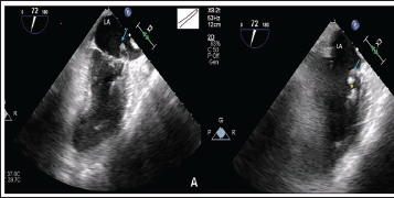
A 58-year-old woman presented to us with complaints of left-sided chest pain of a compressive type, radiating to the left arm and back for 3 days. On admission, she was in cardiogenic shock. 1 Electrocardiogram showed sinus tachycardia with extensive ST-segment elevations in precordial and inferior leads. Transthoracic echocardiography revealed moderate left ventricular dysfunction with akinesia of the posterior, lateral, and apical segments, with mitral regurgitation (MR). She was electively intubated, and an intra-aortic balloon pump was inserted. Coronary angiogram showed an occluded distal left anterior descending artery and distal left circumflex artery, with critical (90%) stenosis in the obtuse marginal artery. Transesophageal echocardiography showed an interesting feature of an “inverted V” sign or “hairpin” sign: a transected tertiary head of the anterolateral papillary muscle, which, along with two chordal structures, was prolapsing into the left atrium in systole. In diastole, this transected tertiary head of the papillary muscle was seen to rest back on the anterolateral papillary muscle, giving an appearance of an intact papillary muscle chordal apparatus. This striking appearance can be considered pathognomonic of this pathology (Figure 1). Severe MR resulted from this transection. 2 She underwent emergency coronary artery bypass grafting and mitral valve replacement with a 25 mm St. Jude mechanical heart valve.
(A) Echocardiography in Systole Showing Transected Tertiary Head of Anterolateral Papillary Muscle Along with Two Chordal Structures Prolapsing into the Left Atrium Suggestive of Hair Pin Sign or Inverted V-sign (Blue Arrow). (B) Echocardiography in Mid-diastole Showing Transected Tertiary Head of Anterolateral Papillary Muscle Along with Two Chordal Structures (Blue Arrow), and a Transection Gap (Yellow Star) with Papillary Muscle Body. (C) Echocardiography in End-diastole Showing the Transected Tertiary Head of the Papillary Muscle-chordal Apparatus was Seen Resting Back on the Anterolateral Papillary Muscle Giving an Appearance of Intact Papillary Muscle Chordal Apparatus (Blue Arrow).
Footnotes
Declaration of Conflicting Interests
The authors declared no potential conflicts of interest with respect to the research, authorship, and/or publication of this article.
Ethical Approval
Our institution does not require ethics approval for reporting individual cases or case series.
Funding
The authors received no financial support for the research, authorship, and/or publication of this article.
Informed Consent
The participant has provided informed consent for the submission of the article to the journal.