
Abstract
Background.
The management and control of malaria has become gradually challenging due to the spread of drug-resistant parasites, lack of effective vaccine, and the resistance of vector to insecticides. Consequently, novel agents are urgently needed from different sources including from medicinal plants. In Ethiopia and Uganda, Myrica salicifolia root is traditionally claimed for the treatment of malaria. The aim of this study was to evaluate the in vivo antimalarial activity of root crude extract of M salicifolia.
Methods.
The parasite, Plasmodium berghei was used in this study since it is an appropriate parasite that is most commonly used because of its higher accessibility. A 4-day suppressive test was employed to evaluate the antimalarial effect of crude extract against early infection. The curative and prophylactic effect of the crude extract was further tested by Rane’s test and residual infection procedure. Parasitemia, survival time, packed cell volume, body weight, and rectal temperature of mice were used as evaluation parameters. Windows SPSS version 24 was used to analyze the data and analysis of variance followed by Tukey’s honestly significant difference to compare results between groups.
Results.
The root crude extract of M salicifolia significantly (P < .05-.0001) suppressed parasitemia. The crude extract exhibited a chemosuppression of 40.90.
Conclusion.
The development of new antimalarial agents and the finding supports the traditional claims and previous in vitro studies.
Keywords
Malaria is the most prevalent and devastating mosquito-borne infectious disease. It is a major cause of morbidity and mortality throughout tropical and subtropical regions of the world, where the temperature and rainfall are suitable for the development of vectors and parasites, causing huge medical, economic, and social impacts. 1
An estimated 219 million cases and 435 000 deaths due to malaria occurred worldwide in 2017. Almost 80% of all malaria cases and deaths globally were in the African region and in India. The African region bears 200 million cases (92%), followed by the Southeast Asia region (5%) and the Eastern Mediterranean region (2%). 2 The burden of disease can be exceptionally high among the most vulnerable groups, such as children <5 years old, pregnant women, and migrant laborers traveling to endemic areas, especially when worsening nutritional conditions impair their capacity to fight the disease. 3 Plasmodium falciparum is the most prevalent malaria parasite in the African region, accounting for 99.7% of estimated malaria cases in 2017, as well as in the regions of Southeast Asia (62.8%), the Eastern Mediterranean (69%), and the Western Pacific (71.9%). 2 Malaria is also the major public health problem in Ethiopia and it is prevalent in over 75% of the country’s area. Approximately 68% of the Ethiopian population has been estimated to live in areas of <2000 m of altitude and, thus, are considered to be at risk of malaria. According to the World Health Organization 2018 malaria report about 2 666 954 cases of malaria were reported in Ethiopia. 2,4
At present, more than 80% of the world’s population relies on ethnopharmacologic healing modalities and plants for their primary health care and wellness. 5 Due to cultural acceptability, physical accessibility, and economic affordability as compared with modern medicine, traditional medicines are used widely in Ethiopia 6 and it is estimated that about 90% of the population is dependent on traditional medicine, essentially plants. 7 Natural products continue to make an immense contribution to malaria chemotherapy providing most of the antimalarial drugs in use. For example, quinine, bitter-tasting alkaloid isolated from the Cinchona bark, is one of the earliest natural compounds that helped man in the fight against malaria, which later served as a template for the synthesis of chloroquine, primaquine, mepacrine, and mefloquine. 8 Artemisinin extracted from Artemesia annua is another example, which gave rise to the development of dihydroxy artemisinin, artemether, arteether, and artesunate. Many more natural products possessing various chemical structures, such as alkaloids, steroids, chalcones, terpenes, flavonoids, peptides, quinones, xanthones, coumarines, naphthopyrones, polyketides, phenols, lignans, chromenes, and so on have been tested as antimalarial drugs. 9 Worldwide, over 1200 plant species are reportedly used for the treatment of malaria and fever and thus are potential sources of new antimalarial treatments. 10 Traditional medicines are also often used to counteract fever in some countries. For instance, in Africa several plants are commonly used in traditional medicine for the treatment and management of fever especially due to malaria or other microbial origins and related conditions. 11
Myrica salicifolia A Rich (Myricaceae) is a shrub of 1 m in height and is found in several central and east Africa countries such as Burundi, Ethiopia, Kenya, Malawi, Rwanda, Tanzania, Uganda, and Zaire. 12 Locally, M salicifolia is known as shinet (Amharic), Kataba (Ormigna), Nihibi, (tigirigna), and Abeyi (Guaragegna). 13 Traditionally, the root extract of M salicifolia A Rich (Myricaceae) was consumed by Maasai warriors to prime them for battle. 14 The study indicates that local people along with local herbalist use M salicifolia A Rich (Myricaceae) root and bark extract with tea for aliment of different disease such as chest congestion, pneumonia, diarrhea, nervous disorders, diabetes, hypertension, and respiratory diseases. 15
To verify the traditional uses of M salicifolia, various in vitro and in vivo studies have been conducted. The studies showed that this plant has most of the claimed activities, including antibacterial activities against Gram negative bacterial strains namely Pseudomonas aeruginosa, Klebsiella oxytoca, Proteus mirabilis, Klebsiella pneumoniae, Salmonella kisarawe, Salmonella typhi, and Escherichia coli. 16 The plant also has antipyretic 14 and antiplasmodic activities, 17 is a cough suppressant, and possesses wound healing and aphrodiasic activities. 18 Studies have shown that the root extract of M salicifolia A Rich (Myricaceae) is a nonhypnotic central nervous system depressant with muscle reluctant, analgesic, hypothermic, and antipyretic properties. 18 Its widely used herbals leaf extract proved to be a central nervous system depressant effect. 19 Similar study reveals that the root of M salicifolia A Rich (Myricaceae) is used as slow acting medicine in stomach problems and head ache (symptoms associated with malaria), the barks chewed for toothache problem whereas powdered young leaves are used to treat skin infections. 17
The root part of M salicifolia has been used in the treatment of malaria orally as claimed in Ethiopia and Uganda 17,20 –22 and studies done by Kirira et al 23 in an in vitro model showed that this plants have mild antiplasmodial activity against the chloroquine-sensitive (NF 54) (IC50 [half maximal inhibitory concentration] 66.84 ± 2.88 μg/mL in aqueous extract and 51.07 ± 1.70 μg/mL in methanol extract) and against resistant (ENT 30) P falciparum strains (IC50 85.97 ± 5.48 μg/mL in aqueous extract and 55.89 ± 2.00 μg/mL in methanol extract). A MeOH extract of Nectandra salicifolia trunk bark, obtained during a diversity-based plant collection in a lower montane rainforest in Costa Rica, showed activity in an in vitro antiplasmodial assay measuring incorporation of [3H]-labeled hypoxanthine by P falciparum. 24 Yet in vivo antimalarial activity studies have not been reported, because results in vitro studies may lead to results that do not correspond to the circumstances occurring around a living organism. So, this study is designed to evaluate the in vivo antimalarial effect of the methanolic root extract of M salicifolia against Plasmodium berghei in mice.
Materials and Methods
Collection of Plants and Preparation of Extracts
Roots of M salicifolia A Rich (Myricacae) were collected from Gondar town about 15 km from central pizza at the site of Alem Ber of north Gondar zone in April 2016. The plants were identified by local herbal expert and authenticated by botanist and have a voucher number of YK001 at Department of Biology, College of Natural and Computational Science, University of Gondar. The roots were cleaned from any extraneous materials, dried at room temperature, and ground to powder for preparation of the extract in the Pharmacognosy Laboratory, College of Medicine and Health Sciences, University of Gondar. The dried plant materials were kept in polyethylene bags until maceration process begins.
The dried and ground root of the plant was mixed with methanol in separate flasks and placed in orbital shaker at room temperature for 72 hours. The extract was then filtered with cotton and filter paper. The filtrate was then oven at 40°C to yield a solid residue. The crude extract was kept in a refrigerator at −20°C until use for the experiment.
Experimental Animals and Parasites
Healthy adult Swiss albino mice of either sex (25 ± 5 g, and 6-8 weeks of age) were purchased from the Ethiopian Public Health Institute (EPHI). The animals were kept in cages and housed in a standard animal house under natural 12/12-hour light/dark cycle at room temperature, and provided with pellet diet and water ad libitum in the animal house of Department of Pharmacology, School of Pharmacy, College of Medicine and Health Sciences, University of Gondar. The mice were maintained and cared for according to the international guidelines given by Organization for Economic Cooperation and Development (OECD) and Institute for Laboratory Animal Research (ILAR) for the use and maintenance of experimental animals throughout the experiment. 25,26 Animals were allowed to acclimatize to the laboratory condition for a week before beginning the experiment.
The P berghei ANKA strain (chloroquine sensitive) was obtained from EPHI Addis Ababa and the parasite was maintained by intraperitoneal serial passage of blood from mouse to mouse.
Acute Oral Toxicity Study
Acute toxicity testing was conducted using limit test dose of 2 g/kg according to the Organization for Economic Cooperation and Development (OECD 425, 2008) guidelines. Five healthy nonpregnant female Swiss albino mice were used to carry out the acute toxicity study. The mice were withheld food, but not water, for 4 hours prior to dosing and at the end of the fasting period weight was recorded and the dose was calculated based on the measured weight. The first animal was given with a limit dose of 2000 mg/kg and no death was observed within 24 hours of dosing. Another 4 female mice were dosed and observed for toxicities like diarrhea, weight loss, and absence of tremor, lethargy, and paralysis periodically for the first four hours during the 24-hour period and later were followed for 14 days for any lethality. 27
Grouping and Dosing of Animals
Mice were randomly assigned into 3 extract-treated groups and 2 controls, 6 mice per group for each model were used. Group I was a negative control and treated with distilled water 10 mL/kg. Groups II, III, and IV were treated with 100, 200, and 400 mg/kg doses of the extract orally, respectively. The doses of the extract were 1/20th, 1/10th, and 1/5th of the LD50 (lethal dose 50%) value from the acute oral toxicity study. Group V was treated with chloroquine 25 mg/kg via the oral route using oral gavage. Volume administered was calculated based on individual mouse body weight and duration of administration was depended on the type of test performed.
Inoculation of Parasite
Mice were infected by blood sample collected from a donor mouse with a rising parasitemia of about 20% to 30%.After determination of the percentage parasitemia in the donor mouse, it was sacrificed by head blow and blood was collected via incisions of the jugular vein into a test tube containing 3.8% trisodium citrate added as anticoagulant. The collected blood was diluted by 0.9% physiological isotonic saline based on the parasitemia of the donor mice and the red blood cell (RBC) count of normal mice (4.5 × 10⁹ RBC/mL) in such a way that 1 mL blood contains 5 × 10⁷ P berghei–infected erythrocytes. Each mouse to be used in the experiment was infected intraperitoneally with 0.2 mL of infected blood containing about 1 × 10⁷ P berghei–parasitized erythrocytes. 28
Four-Day Suppressive Test
This test was performed to evaluate schizontocidal activities of the crude extract on early P berghei infection in mice using a 4-day suppressive test described by Knight and Peters. 29 The infected mice were randomly divided into 5 groups and treatment was started 3 hours after inoculation of the parasite on day 0, and continued for the next 4 days from day 0 to day 3 with 24-hour time interval between the doses. After giving the treatment for four days, thin blood film was made from the tail of each mouse on the fifth day (D4) to determine the level of parasitemia and percentage inhibition.
Rane’s Test (Curative Test)
The curative test was undertaken with the crude extract, which showed the highest parasitemia suppression in the 4-day suppressive test. Accordingly, evaluation of the curative potential against an established malaria infection was carried out according to the method described by Ryley and Peters. 30 The mice were injected intraperitoneally with standard inoculum of 1 × 10⁷ P berghei–infected erythrocytes on the first day (day 0). Seventy-two hours later, the mice were divided into 4 groups of 6 mice per group. The mice were treated once daily for 5 days from day 3 to day 7. Starting from day 3 through day 7 daily thin blood films was prepared from the tail of each mouse to monitor the level of parasitemia.
Test for Prophylactic Activity
The prophylactic activity of the crude extract, that showed the highest parasitemia suppression in the 4-day suppressive test, were tested using the residual infection procedure described by Peters. 31 Groups of mice were randomized into 5 groups of 6 mice per group for crude extract. Treatment was given daily for 4 days and all mice were infected with the parasite on the fifth day. Thin blood films were prepared from each mouse after 72 hours of infection to determine the level of parasitemia.
Determination of Parasitemia
Thin blood smears were prepared from tail snip of each mouse on the fifth day (D4) for Peters’s 4-day suppressive test, after 72 hours of infection on day 8 in prophylactic test and from day 3 after infection established to day 7 for curative test on microscopic slides. The slides were dried, fixed with absolute methanol and stained with 10% Giemsa at pH 7.2 for 10 minutes and then it was washed gently using distilled water and air dried at room temperature. Finally, the slides were examined under microscope with an oil immersion objective (×100 magnification power) by taking an average of 6 fields. The parasite count was done by an experiment blinded technician. Percentage parasitemia was calculated by counting infected RBC and total RBC from Giemsa-stained thin blood films and the average percentage suppression of parasitemia was calculated for each dose level by comparing the parasitemia in infected controls with those of treated mice with the following formulas 32 :


Determination of Mean Survival Time
Mortality was monitored daily and the number of days from the time of inoculation of the parasite up to death was recorded for each mouse in the treatment and control groups throughout the follow-up period of 30 days (D0-D29) for all the models. Mean survival time (MST) for each group was determined arithmetically by calculating the average survival time (days) of mice starting from date of infection over a period of 30 days (D0-D29). MST for each group was then calculated using the following formula 33 :

Packed Cell Volume Determination
Packed cell volume (PCV) was measured to predict the effectiveness of the test crude extract in preventing hemolysis resulting from increasing parasitemia associated with malaria. Blood was collected from tail of each mouse in heparinized microhematocrit capillary tubes. The capillary tubes were filled to 3/4th of their height with blood and sealed at one end with sealing clay. The tubes were then placed in a microhematocrit centrifuge, with the sealed end outward and centrifuged for 5 minutes at 11 000 rpm. The tubes were taken out of the centrifuge and PCV was determined using a standard microhematocrit reader. It was measured before inoculating the parasite (day 0) and day 4 in Peters’s 4-day suppressive test and D0 and D7 in the prophylactic activity test. In the case of Rane’s test, PCV was measured on the third day after infection established and on the last day of treatment on the seventh day. PCV is a measure of the proportion of RBCs to plasma in the whole blood and determined using the relation shown below. 34

Determination of Body Weight and Temperature Change
The body weight of each mouse in all groups was measured on day 0 before infection and day 4 in the 4-day suppressive test and D0 and D7 (72 hours after infection) in the prophylactic activity test while in the Rane’s test it was measured on day 3 after infection was established and on day 7, the last day of the treatment using a sensitive digital weighing balance, to observe whether the test extract of the leave prevent body weight loss that commonly reduced with increasing parasitemia in infected mice. The rectal temperature of each mouse in all groups was measured by a digital thermometer 1 hour before infection, 4 hours after infection, and then daily to see the effect of crude extract on rectal temperature in 4-day suppressive test. On the other hand, in Rane’s test, rectal temperature was measured 1 hour before infection and then daily from day 3 to day 7 and in prophylactic activity test rectal temperature was measured D0 before treatment, D4 before inoculation, and then daily till day 7.
Statistical Analysis
Results of the study were expressed as a mean plus or minus standard error of the mean (M ± SEM). Data analyzed was undertaken using Windows SPSS Version 24. One-way analysis of variance (ANOVA) followed by Tukey’s (post hoc) test to evaluate statistical significance for grouped data. The results were analyzed at 95 confidence interval and P < .05.
Results
Yield of Crude Extract of Myrica salicifolia
A total of 152.65 g of dried root crude extract was harvested at the end of the extraction process. In the preparation of crude 80% methanolic extract from the dried roots of M salicifolia, a yield of 21% was obtained.
Acute Toxicity Test of Crude Extract
The acute toxicity study indicated that the crude extract caused no mortality in limit dose of 2000 mg/kg within the first 24 hours as well as for the following 14 follow-up days. Physical and behavioral observations of the experimental mice also revealed no visible signs of overt toxicity. This indicates that LD50 of the extract is greater than 2000 mg/kg.
Phytochemical Screening
Phytochemical screening of the methanolic root extract of M salicifolia revealed that the extract contains steroids, flavonoids, terpenoids, tannins, phlobatannins, saponin, and phenolics (Table 1).
Phytochemical Screening of the Root Crude Extract of Myrica Salicifolia.

a − − − indicates negative test and + + + indicates positive test.
Effect of the Crude Extract in 4-Day Suppressive Test
As presented in Table 2, all doses of the root crude extract of M salicifolia (100, 200, and 400 mg/kg) showed statistically significant (P < .05, P < .01, and P < .001, respectively) chemosuppressive activity against P berghei infection in mice in 4-day suppressive test as compared with negative control. The highest level of inhibition (59.13%, P < .001) was obtained by 400 mg/kg dose of the crude extract, followed by 200 mg/kg of the crude extract (51%, P < .01), while the lowest suppression (40.9%, P < .05) was obtained after administration of 100 mg/kg dose of the crude extract. Similarly, chemosuppressive activity produced by the standard drug, chloroquine 25 mg/kg were significant (P < .0001) as compared with negative control, which showed 100% chemosuppression.
Effect of Myrica salicifolia Crude Extract on Percentage Parasitemia and Survival Time of Plamodium berghei–Infected Mice in the 4-Day Suppressive Test.*

Abbreviations: CQ, chloroquine diphosphate; NC, negative control.
* Values are presented as mean ± SEM (standard error of the mean). Statistical significance denoted by 1 P < .05, 2 P < .01, and 3 P < .001, 4 P < .0001, acompared with NC, bcompared with CQ 25 mg/kg.
All doses of the crude extract—100, 200, and 400 mg/kg—prolonged MST significantly (P < .01, P < .0001, and P < .0001, respectively) and with MST of 9.2 ± 0.58, 10.8 ± 0.58, and 13.8 ± 0.58 days, respectively, as compared with negative control (6.4 ± 0.40). The mean survival time of mice treated with chloroquine 25 mg/kg was significant (30.00 ± 0.00 days, P < .0001) as compared with the negative control and the entire doses of crude extract (Table 2).
Effect of Myrica salicifolia Extract on Body Weight, PCV, and Temperature Determination in 4-Day Suppression Test
In both the negative control and extract-treated experimental mice, loss of body weight between D0 and D4 was observed. All doses of the crude methanolic root extract of M salicifolia showed PCV reduction. However, Positive control treated group a slight increment of PCV on D4 as compared with D0 (Table 3).
Effect of Myrica salicifolia on the Body Weights, Packed Cell Volume, and Temperature of Plasmodium berghei–Infected Mice in the 4-Day Suppressive Test.*

Abbreviations: CQ, chloroquine diphosphate; NC, normal control; D0, day infection was initiated; D4, fifth day of infection.
* Values are presented as mean ± SD (standard deviation). Statistical significance as compared with treatment control denoted by 1 P < .05, 2 P < .01, 3 P < .001, 4 P < .0001, acompared with NC, and bcompared with CQ.
In all test groups, the methanolic root extracts did not cause significant (P > .05) prevention of loss of rectal temperature of P berghei–infected mice. The standard drug, chloroquine 25 mg/kg, showed significant (P < .01) activity in prevention against rectal temperature reduction when compared with negative control.
The crude extract did not significantly protect the loss in body weight as compared with the negative group (Table 3). There were no significant differences among all doses of the crude extract in the reduction of body weight associated with increasing parasitemia. However, standard drug (chloroquine 25 mg/kg)–treated group resulted a significant (P < .01) prevention of body weight loss as compared with negative control.
Effect of the Crude Extract in Curative Test
The crude extract showed a significant antimalarial activity in the 4-day suppressive test and was further evaluated for their effect on established parasite infection. As presented in Table 4, the crude extract of M salicifolia root on established malaria infection (Rane’s test) demonstrated a significant (P < .0001) curative action at all tested doses on the seventh day from a thin blood smear. Analysis of parasite suppressive activity indicated that crude extract (37.84%, 51.35%, and 59.46%) at doses of 100, 200, and 400 mg/kg, respectively, compared with negative control at day 7.
Effect of Myrica salicifolia Crude Extract on Percentage Parasitemia and Mean Survival Time of Plasmodium berghei–Infected Mice in Rane’s Test.*

Abbreviations: CQ, chloroquine diphosphate; NC, negative control.
* Values are presented as mean ± SEM (standard error of the mean). Statistical significance denoted by 1 P < .05, 2 P < .01, 3 P < .001, 4 P < .0001, acompared with NC, and bcompared with CQ.
Maximum inhibition (59.46%) was attained with 400 mg/kg dose of crude extract. The standard drug caused reduction in parasitemia levels right after the first dose administration on the third day and completely cleared parasitemia on the seventh day from a thin blood smear (100%).
Considering the mean survival time of mice, all doses of crude extract (100, 200, and 400 mg/kg) showed a significant (P < .5, P < .01, and P < .001, respectively) long survival date compared with the untreated group. Similarly, the MST of mice treated by chloroquine was significantly (P < .0001) longer when compared with all tested doses of the crude extract and the untreated group.
Effect of Myrica salicifolia Extract on Body Weight, PCV, and Temperature Determination in Curative Test
Analysis of the effects on PCV revealed, all doses of the crude extract prevented the PCV reduction significantly at all doses (100, 200, and 400 mg/kg) (P < .05, P < .01, and P < .001, respectively) compared with untreated group. Likewise, the standard drug also significantly prevented the PCV reduction compared with the untreated group. However, the effect of the chloroquine on prevention of PCV loss was not significantly higher than all doses of the crude extract (100, 200, and 400 mg/kg) (Table 5).
Effect of Myrica salicifolia Crude Extract on the Body Weights, Packed Cell Volume, and Temperature of P. Berghei infected Mice in Rane’s Test.*
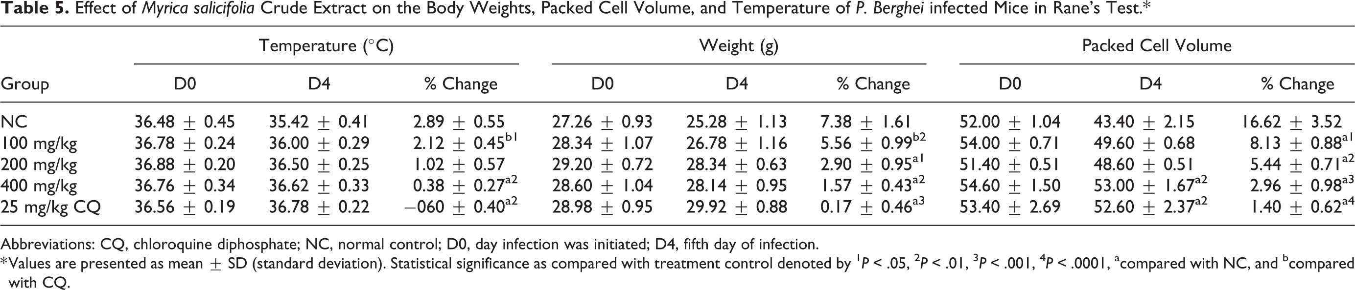
Abbreviations: CQ, chloroquine diphosphate; NC, normal control; D0, day infection was initiated; D4, fifth day of infection.
* Values are presented as mean ± SD (standard deviation). Statistical significance as compared with treatment control denoted by 1 P < .05, 2 P < .01, 3 P < .001, 4 P < .0001, acompared with NC, and bcompared with CQ.
Except the lower dose, both the middle and the higher doses of crude extract revealed significant (P < .05 and P < .01, respectively) increase of weight change between D7 and D3 compared with the untreated group. Similarly, the standard drug showed significant (P < .01and P < .001) increase of weight change between D7 and D3 compared with 100 mg/kg of the crude extract and untreated group, respectively.
As revealed from multiple comparisons, both the crude extract at the dose of 400 mg/kg and the standard drug significantly (P < .01) prevents the drop of temperature compared with the untreated group. However, the middle and the higher dose of the crude extract failed to prevent the drop of temperature as compared with the untreated group (Table 5).
Effect of the Crude Extract in Prophylactic Test
The crude extract (100, 200, and 400 mg/kg) showed dose-dependent chemoprophylactic activities against P berghei–infected mice with the following parasite suppressive activity: 37.14%, 49.52%, and 57.62%, respectively. All doses of the crude extract significantly (P < .0001) suppressed parasitemia as compared with the negative control. The standard drug showed a significant chemosuppression (P < .0001) as compared with all doses of the crude extract and the negative control.
On the other hand, 100, 200, and 400 mg/kg of the crude extract (P < .05 to P < .001) were able to significantly (P < .05, P < .0001, and P < .0001, respectively) prolong survival time as compared with the negative control. The standard drug also showed a significant (P < .0001) prolongation of mean survival time as compared with all doses of the crude extract and negative control (Table 6).
Effect of Myrica salicifolia Crude Extract on Percentage Parasitemia and Survival Time of Plasmodium berghei–Infected Mice in the Prophylactic Test.*

Abbreviations: CQ, chloroquine diphosphate; NC, negative control.
* Values are presented as mean ± SEM (standard error of the mean). Statistical significance denoted by 1 P < .05, 2 P < .01, 3 P < .001, 4 P < .0001, acompared with NC, and bcompared with CQ.
Effect of Myrica salicifolia Extract on Body weight, PCV, and temperature Determination in Prophylactic Test
Except the lower dose of crude extract, the middle and the higher dose of the crude extract prevented the weight loss significantly (P < .05 and P < .01, respectively) compared with the negative control. Similarly, the standard drug also significantly (P < .0001) prevented weight loss compared with the negative control. The temperature reduction due to inoculation of the parasite was significantly (P < .01) prevented in the mice treated at dose of 200 mg/kg extract, 400 mg/kg extract, and the standard drug compared with the negative control.
All the doses of the crude extract (100, 200, and 400 mg/kg) prevented the loss of PCV significantly (P < .05, P < .001, and P < .0001, respectively) in a dose-dependent manner compared with the negative control. Likewise, the standard drug showed a significant (P < .001, P < .05, and P < .0001) higher prevention of PCV reduction compared with 100 mg/kg extract, 200 mg/kg extract, and negative control, respectively (Table 7).
Effect of Myrica salicifolia Crude Extract on the Body Weights, Packed Cell Volume, and Temperature of Plasmodium berghei–Infected Mice in the Prophylactic Test.*

Abbreviations: CQ, chloroquine diphosphate; NC, normal control; D0, day infection was initiated; D4, fifth day of infection.
* Values are presented as mean ± SEM (standard error of the mean). Statistical significance denoted by 1 P < .05, 2 P < .01, 3 P < .001, 4 P < .0001, acompared with NC, and bcompared with CQ.
Discussion
The present study explored the acute toxicity, phytochemical screening, and antimalarial activities of the 80% methanolic root extract of M salicifolia against P berghei infection in mice. To account for a possible prodrug effect and involvement of the immune system in the eradication of an infection, antimalarial studies usually employ in vivo models as compared with an in vitro study. 35 Even though primate models provide a better prediction of evaluation of the efficacy of antimalaria in human, the rodent models is used as a first step to screen most in vivo antimalarial activities of test compounds. 36 The rodent models have been also validated through the identification of several conventional antimalarial agents such as chloroquine, halofantrine, mefloquine, and artemisinin derivatives. 30,36 It is also cost-effective to conduct preliminary pharmacological screening studies in rodent model than primate model. The parasite, P berghei was used in this study since it is an appropriate parasite that is most commonly used because of its higher accessibility. 36 Due to the sensitivity and significant suppression of this parasite by chloroquine, this drug was employed as the standard. 36
According to this study, the crude extract did not show mortality up to a dose of 2000 mg/kg. Further physical and behavioral observations also revealed no visible signs of acute toxicity with the same dose. In general, this substance is considered as a good candidate for further studies since its LD50 is 20 times more than the minimum effective dose tested (100 mg/kg), which satisfies the minimum requirement, which is 3 times greater. 37
The antiplasmodial properties of the crude extract of the root of M salicifolia were investigated using standard models. Accordingly, a preliminary 4-day suppressive test was conducted for the root extract of M salicifolia to evaluate schizontocidal activity and the percentage suppression of parasitemia of the crude extract–treated groups changed significantly from those in the negative control group. This indicares that the plant is endowed with antimalarial activity. This is in agreement with a previous report of in vitro antimalarial activity of root extracts of M salicifolia. 23
All the doses of crude extract displayed a significant chemosuppressive activity as compared with the negative control. This could be explained by the possible synergistic effects that might have existed among the various components in the crude extract.
Since the crude extract showed a significant chemosuppression activity in the 4-day suppressive test, it was further evaluated for its effect on established parasite infection using the curative test. All doses of the crude extract (100, 200, and 400 mg/kg) showed a significant (P < .5, P < .01, and P < .001, respectively) curative effect as compared with the negative control with percent of chemosuppression (37.84%, 51.35%, and 59.46%, respectively). This confirms that the plant material has effective antiplasmodial activity in the late stages of the infection. From the day-wise blood smears, like the standard drug, all treated groups, except 100 mg/kg dose of the crude extract, decreased parasitemia level after the first dose. This might be indicative of a comparative onset of defensive action of extracts and the standard drug. The delay of activity at 100 mg/kg dose of the crude extract might be due to the fact that the extracts administered at this dose had not accumulated sufficiently to bring about considerable chemosuppression. However, there was an increase in parasite level in the control group; this thus shows that just as in humans, Plasmodium parasite will multiply in mice except it is intercepted by an effective antimalarial agent. The chemosuppressive effect on established infection was higher than the 4-day suppressive test, which might be due to inhibitory effect of the extract on generation of free radicals and hemolytic principles such as free fatty acids resulting from high parasitemia level. 38
After the curative effect of the crude extract was established, further evaluation was conducted to confirm their prophylactic potential, because some traditional plants that showed antiplasmodial activity in 4-day suppressive and curative tests, also showed prophylactic activity against the P berghei parasite. 39 In this study, the crude extract of M salicifolia had shown significant chemoprophylactic activity against residual infection at all doses as compared to negative control with maximum parasitemia chemosuppression of 57.62% at the highest dose. The 400 mg/kg dose of crude extract showed significant prophylactic activity as compared with 100 mg/kg treated group.
The percentage suppression in prophylactic test was found to be low as compared with 4-day suppressive and curative tests. This might have risen from rapid hepatic clearance or metabolism of the active component responsible for antimalarial activities due to the administration of the extract initially for 4 days before inoculation with P berghei parasite. In all models, the standard drug suppressed the parasitemia significantly (P < .0001) as compared with all doses of the crude extract and negative control. The lower efficacy of the crude extract might be in part due to unpurified/crude nature, low selectivity, slow absorption and poor bioavailability or other pharmacokinetics and pharmacodynamics parameters of the extracts. 40
The MST is another important parameter to evaluate the antimalarial activity of plant extracts. 36 In this study, the all doses of the crude extract significantly prolong the MST as compared with the negative control, especially at higher doses in all tested models. This further supplement the evidence on suppression of P berghei, resulting in a reduced overall pathologic effect of the parasite on the study mice. 41 The MSTs of mice treated with the standard drug were significantly prolonged (P < .0001) as compared with the entire doses of the crude extract–treated groups in all models. This might be due to the fast elimination phase of the extracts. In all tested models, the longest MST of the mice was strongly associated with the maximum parasitemia inhibition. A plant material that can prolong the survival time of infected experimental animals compared with the negative control is considered as an active agent against malaria. As a principle, a compound that prolongs survival time beyond 12 days is regarded as active. 42
PCV reduction is one feature of malaria-infected mice. It was measured to evaluate the effectiveness of the crude extract in preventing hemolysis due to rising parasitemia level. 43 Escalating parasitemia causes the clearance and/or destruction of infected RBCs, the clearance of uninfected RBCs, and erythropoietic suppression and dyserythropoiesis. These mechanisms have been implicated in both human and mouse malarial anemia. 44 Plants with antimalarial activity are expected to prevent reduction in PCV secondary to preventing hemolysis. Interestingly, in the prophylactic and curative test models, it was noted that there is a significant protection in PCV as compared with the negative control. The prevention of PCV reduction might be a result of destructive antiplasmodial effect of the crude extract against the parasitized RBCs and the causative parasite, thereby sustaining the availability of the new RBCs produced in the bone marrow. 45,46
Body weight loss, reduction of body temperature, and anemia are hallmarks that are observed in malaria-infected mice. The weight loss in P berghei–infected mice could be due to decreased food and water intake. Hypoglycemia and anemia resulted from raised parasite load. 47 Therefore, a potential antimalarial plant product is expected to preserve these parameters in P berghei–infected mice due to the rise in the level of parasitemia. 47 –49 In the prophylactic and curative test models, the higher doses of the crude extract (200 and 400 mg/kg) showed significant (P < .05) protective effect against weight loss compared with the negative control. This activity might have been contributed from the overall improvement of PCV and parasite clearance among extract-treated mice. 7,33 Unfortunately, the entire doses of crude extract did not prevent against body weight loss in the 4-day suppressive test, even though there was a significant suppression of parasitemia. This indicates the involvement of other factors for these reductions beyond malaria infection. For example, the weight loss might be due to catabolic activity on stored lipids or anorexogenic effect that may have led to decreased food intake due to the presence of appetite-suppressant metabolites in the crude extract, in addition to disturbed metabolic function and hypoglycemia related to malaria infection. 41
On the other hand, the reduction in rectal temperature might be due to less hypothermic effect on the extract-treated mice, which is supported by the antipyretic activity of the extract in addition to the probably inability of the plant extract to ameliorate some pathological processes of malaria that cause reduction in rectal temperature or the reduction in metabolic rates that occur because of increased parasitemia. 18,50
In the mice infected with P berghei, the metabolic rate decreases before death because the parasite affects host carbohydrate, lipid, and protein metabolism, which corresponds to the decrease of internal body temperature. 51 Therefore, compounds that possess antimalarial activity could prevent the rapid decrease of rectal temperature. 52
In the prophylactic test model, the higher doses of the crude extract (200 and 400 mg/kg) showed significant (P < .05) protective effect against temperature reduction compared with the negative control. In the suppressive and curative tests, the higher dose (400 mg/kg) of the crude plant extract significantly prevented the rapid drop of rectal temperature compared with the untreated group. In all test models, the possible mechanism for prevention of temperature drop could be the reduced level of parasitemia as reduction of temperature is directly related to the increased level of parasitemia. M salicifolia root may also contain unidentified bioactive compounds that can increase the appetite and metabolic activity in addition to parasite clearance.
Secondary metabolites have been implicated in antiplasmodial activity through different possible mechanisms, including endoperoxidation by sesquiterpenes and monoterpenes, 53 intercalation in DNA by anthraquinones, 54 disrupting the parasite ability of detoxifying heme into nontoxic malaria pigment by alkaloids, 55 blocking protein synthesis by alkaloids and chelation with nucleic acid base pairing by flavonoids, 56 immunomodulatory effects by phytosteroids and flavonoids, 57 reducing the activity of superoxide dismutase and inhibiting the synthesis of DNA by coumarins, 58 free radical scavenging effects by tannins, 59 antioxidant effect by phenols like flavonoids due to their redox properties, which allow them to act as reducing agents, metal chelators, and free radical quenchers, 60,61 or by any other unknown mechanisms. Mechanisms of antioxidant action can include suppression of reactive oxygen species and upregulation or protection of antioxidant defenses. Antioxidative activity can inhibit heme polymerization as heme has to be oxidized before polymerization, and the unpolymerized heme is very toxic for the intraerythrocytic plasmodia. 62 The extracts could have elicited their action through any of the aforementioned mechanisms or by some other means yet to be determined. Therefore, the antiplasmodial activity observed in this plant could have resulted from single or combined action of these metabolites. Methanol is used for extraction of compounds possessing both polar and nonpolar nature such as tannins and terpenoids. 42 Generally, a plant’s therapeutic effect may be due to the presence of bioactive substances or secondary metabolites. Phytochemical screening on crude extract revealed that the plant possesses a number of secondary metabolites. These include steroids, flavonoids, terpenoids, tannins, phlobatannins, saponin, and phenolics.
In vivo antimalarial activity of test extract can be grouped as moderate, good, and very good when the extract produces a percent parasite suppression ≥50% at a dose of 500, 250, and 100 mg/kg body weight per day, respectively. 63 Based on the above classification, crude extracts of M salicifolia root in the 4-day suppressive test exhibited a very good antimalarial activity (40.9%, 51.00%, and 59.13%) at doses of 100, 200, and 400 mg/kg, respectively, with a dose-dependent inhibition of P berghei infection.
Conclusion
From the results of this study, it can be concluded that the plant extract is relatively safe to mice. The methanolic crude extract of M salicifolia showed fairly moderate antimalarial activity in early, established, and residual infections. The highest effect was exhibited by the higher dose of the crude extract in all models. The crude extract significantly suppresses parasitemia thereby protecting the packed cell volume and prolonging the MST in all models but failed in the prevention of rectal temperature and body weight reduction in 4-day suppressive models. The overall results of this study indicate that the root extracts of M salicifolia could potentially be used as a new source for the development of new plant-based antimalarial agents. Generally, the present pharmacological evidence provides support for the folklore claim of M salicifolia root as an antimalarial agent and the results from previous in vitro studies on the antimalarial activity of the plant. Based on the present study, the fractionation and bioassay guided isolation and characterization of the active principle responsible for the activity of the plant need further investigation.
Footnotes
Acknowledgments
The authors would like to acknowledge University of Gondar for funding and for allowing the use of the laboratory facility.
Author Contributions
ZDK conceived the idea, drafted the proposal, collected the plant materials, carried out all experiments, and prepared the final manuscript for publication. All authors were involved in the design, write up, and preparation of the manuscript to be submitted. All authors have read and agreed to the manuscript.
Declaration of Conflicting Interests
The authors declared no potential conflicts of interest with respect to the research, authorship, and/or publication of this article.
Funding
The authors disclosed receipt of the following financial support for the research, authorship, and/or publication of this article: This study received funding from the University of Gondar Vice President for Research and Community Services (Reference No. VP/RCS/05/445/2015).
Ethical Approval
Ethical clearance was obtained from the Research and Ethics Committee, Department of Pharmacology, University of Gondar with a reference number of SOP 2/65/9 to conduct the study in animal model and the Gondar Environmental Protection Committee gave consent with a reference number of GEP 06/077/9 to gather plants in the wild. Apart from that, all possible steps were taken to avoid animal suffering at each stage of the experiment.