
Abstract
Fulgence Raymond (1844–1910) succeeded Jean-Martin Charcot (1825–1893) to the Chair of Nervous System Diseases. As famous as Charcot remains, Raymond has been forgotten. After a brief biographical account, we will present a few examples of his work still relevant today: hemichorea, Raymond-Cestan syndrome, hereditary spastic paraplegia and acute ascendant paralysis. In each case, his accurate clinical and anatomopathological descriptions are accompanied by aetiological hypotheses that are remarkably prescient with regard to current knowledge. Strongly committed to teaching, he published most of his lessons every year. They remain highly relevant historically, and sometimes for other reasons, as we shall see. We hope to show that Raymond does not deserve to be forgotten.
Keywords
The Founders of Neurology by Webb Haymaker (1902–1984), a book prepared for the Fourth International Neurological Congress in Paris (5–10 September 1949), has remained a reference for biographical sketches of 19th- and 20th-century physicians who explored the diseases of the nervous system. A second edition was published in 1970, 1 with the collaboration of Francis Schiller (1909–2003). The authors inform readers that, to keep the format of the first 1953 edition, 2 they eliminated some of the previous names because ‘history being a fickle mistress, some contributions seemed less resilient to the in-roads of time’. Fulgence Raymond is one of the names that vanished. Many reasons, good or bad, must have dictated their choices, but they remain unknown to us. To rectify what seems like a second ‘death’ for Raymond, we will present a few examples illustrating the forgotten work of Charcot’s successor at the helm of the Clinic of Nervous System Diseases, part of Hôpital de la Salpêtrière.
A short biographical account
Fulgence Raymond was born on 29 September 1844 in Saint-Christophe on the Nais River, located in the Loire Valley not far from Tours (Figure 1). At age 17, he entered the Maisons-Alfort imperial veterinary school from which he graduated first in his class in 1865 with the diploma of physician-veterinarian. After working for a year at the Saumur Cavalry School, a French military training establishment that did not meet his expectations, he successfully passed the exam leading to the position of Head of Anatomy and Physiology Studies at the Maisons-Alfort school, where he taught animal anatomy from 1867 to 1869. He married Louise Rochut (1842–1872) on 26 May 1868 and the couple soon had a baby girl, but the young mother died of tuberculosis on 24 September 1872.

Birthplace of Fulgence Raymond in Saint-Christophe sur le Nais (Photo by the author).
At the time, veterinary medicine was primarily practices by horseshoers, who lacked any recognised training. Animals were rarely taken to veterinarians, who were poorly paid, since most farmers were unaware of their existence. All of this explains why Raymond felt unfulfilled in his practice of veterinary medicine. So, he decided to realise his dream of becoming a physician. He also remarried on 25 August 1887. 3
He began his medical studies in 1869 and by 1871, he had successfully passed the internat exam for the Paris Hospitals. He was an interne or resident under three prestigious teachers, all involved in research on the nervous system: Adolphe Gubler (1821–1879), 4 Alfred Vulpian (1826–1987) 5 and Charcot (Figure 2). Raymond never explained why he was especially interested in this area.

La Salpêtrière 1873: Raymond seated second on the right (Private collection of the author).
After failing the 1878 agrégation exam to enter the professor track, he was successful 2 years later, in 1880, presenting a thesis entitled On Puerperium. 6 Raymond taught pathological anatomy at the Paris medical school in 1883 and 1884, then an additional internal pathology class from 1887 to 1888, before being elected by his peers to the Chair of Nervous System Diseases on 15 March 1894, ‘according to the seniority rules at the medical school’ 7 (Figure 3).
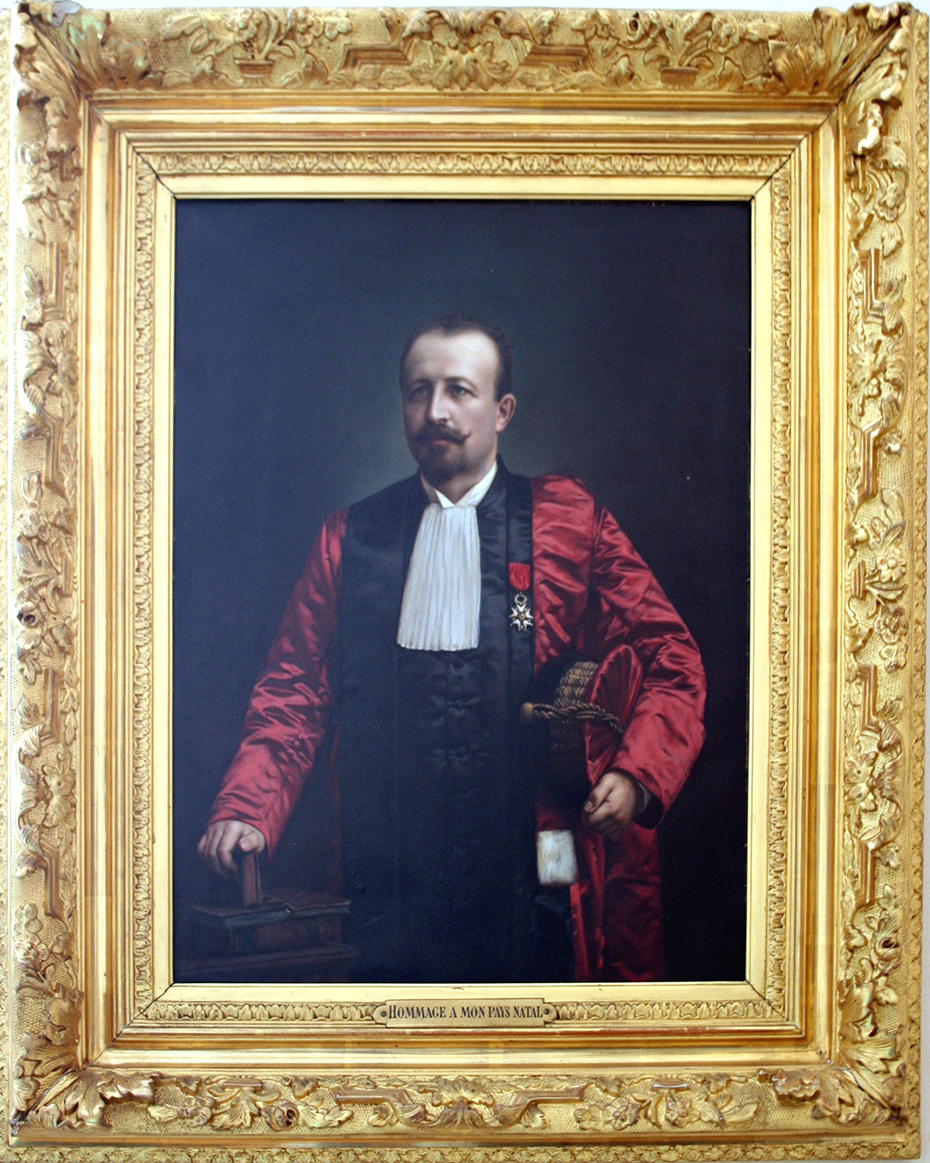
Professor Fulgence Raymond, Chair of Nervous System Diseases (Photo by the author. Painting kept in the Town Hall of Saint-Christophe sur le Nais).
Raymond set out to follow his illustrious predecessor’s path exactly, as he himself stated at his inaugural lesson: ‘Of course, faithful to tradition, I shall devote our Tuesday interviews to extemporaneous clinical examinations, to the study of the most interesting cases that arrive that day. The Friday lessons will be reserved for the dogmatic study of neuropathology’. 8
Having already suffered from aortic insufficiency, he experienced episodes of angina at the beginning of 1910 and had acute pulmonary oedema in early September while resting at his castle, Planche d’Andillé, in Poitou in west-central France. His relapse on 28 September 1910 was fatal. Only the base of his bust, which his second wife commissioned to honour his memory, remains in front of the town hall of his native village. Like the statue of Charcot in Paris, this bust in bronze was melted down during the requisitioning of metals by the French government in 1942 to support the German war effort 3 – another illustration of how history forgot Raymond.
Raymond the teacher
In reading the works left by Raymond, one is immediately impressed by the number of publications in all areas of neurology, both clinical and anatomopathological fields, and a few areas of psychiatry. 9 The 126 pages of his Titres et Travaux scientifiques from 1893, submitted for the competitive agrégation, listed all of his works to date, covering all branches of medicine, and already bore this out. 10 Examples of his publications include notices on embolism and thrombosis in the Dictionnaire encyclopédique des Sciences médicales, research on skin pigmentation during Addison’s disease, on the aetiology of tuberculosis, on liver abscesses, on syphilis and various cancers and so on.
His first objective was to teach. He did not neglect to publish his lessons, which he referred to as lectures, first those from 1890 to 1893 while he was working at Hôpital Lariboisière, then those given at the Clinic of Nervous System Diseases at La Salpêtrière from 1894 to 1910. Obviously, it is not possible to go into detail on both of these works, each comprising 600 to 800 pages. These lectures related one or two clinical cases, sufficiently demonstrative to be retained by his students. After the medical history and the clinical examination, he presented diagnostic hypotheses, followed by an aetiological and anatomical review. Often no therapeutic solution was proposed. He supported his teachings with a broad survey of French and foreign publications on the subject of interest. The format was that of a conversation, allowing digressions and a certain degree of chattiness. Here is how he explained his pedagogical ambitions: Start with a clinical fact in order to consider the problems of nervous pathology, as they present in reality, showing how often the pathological individualism of patients aligns poorly with the didactic descriptions of diseases, products of arbitrary or premature summarisation.
11
Raymond did not leave his name linked to descriptions of new diseases as did Charcot, Désiré Magloire Bourneville (1840–1909), Jules Dejerine (1849–1917), Pierre Marie (1853–1940) and Georges Gilles de la Tourette (1857–1904), among others. However, some of his works remain relevant and bear witness to his clinical perspicacity. We chose to examine his doctoral thesis and works on Raymond-Cestan syndrome, hereditary spastic paraplegia (HSP) and acute ascending paralysis. Each of these examples reveals the state of knowledge at the time, while remaining relevant today.
Hemichorea
Raymond defended his doctoral thesis on 23 May 1876: Étude anatomique, physiologique et clinique sur l’hémianesthésie, l’hémichorée, et les tremblements symptomatiques (Anatomical, physiological and clinical study of hemianaesthesia, hemichorea and symptomatic shaking). This subject was suggested to him by Charcot and Vulpian, at a time when the anatomoclinical elucidation of numerous pathologies was at its peak in the departments of La Salpêtrière. The unilateral nature of the problems studied allowed him to compare them, clinically and during autopsy, with the healthy side to determine the localisation of the damaged areas, then try to elucidate the pathophysiology. The cases of hemichorea that he chose were associated with hemiplegia of vascular or tumoural origin or due to sequelae of pathologies around birth. He used recent findings such as ‘the accurate descriptions of M. Duret’, more specifically the description ‘of the distribution of arterial blood’ territory by territory in the brain, in the carotid arteries, the brain stem and their branches, published by Henry Duret (1849–1921) 12 in 1874. 13 Based on 42 detailed observations, Raymond concluded that: ‘Symptomatic hemichorea is of great value in terms of localising the damaged area’. That is, it follows a lesion in a branch of the posterior cerebral artery, occupying ‘the tract that, in the foot of the corona radiata, is found in front, outside the sensitive fibres, composed of white masses in relation with the posterior part of the optical layer’. In 1874, the function of the basal ganglia was unknown, but Raymond does not seem far off by presciently considering ‘a disconnection’ of centres, still mysterious, in reality the interruption of a striato-thalamo-cortical pathway. Raymond carried out vivisection experiments on dogs in Vulpian’s laboratory in an attempt to provoke persistent involuntary movements similar to symptomatic hemichorea, without succeeding. It should be noted that at the time, the distinction between Sydenham’s chorea and Huntington’s disease was not established. To this day, involuntary abnormal movements, including chorea, ballism, hemichorea and hemiballism, remain a continual source of clinical case reports, which still investigate lesion localisation and aetiology, the very same issues faced by Raymond.
Raymond-Cestan syndrome
Raymond-Cestan syndrome, sometimes called superior Foville’s syndrome or upper dorsal pontine syndrome (affecting the medial and lateral lemnisci, superior cerebellar peduncle and supranuclear oculomotor pathways), 14 belongs to the family of alternating syndromes. The pontine localisation explains ‘these crossed symptoms’; that is, on one side pure hemicerebellar syndrome and paralysis of conjugate gaze towards the side of the lesion, and on the other side moderate hemiplegia, choreoathetosic involuntary movements and loss of thermoalgesic sensitivity, calling to mind syringomyelia. 15,16 In 1901, Raymond and Raymond Cestan (1872–1933) (Figure 4) published ‘three observations of conjugate gaze palsy’ in La Revue Neurologique. 17 After referring to the 1883 article of Henri Parinaud (1844–1905) on ‘conjugate gaze palsy’, 18 they introduced two cases of ‘horizontal conjugate gaze palsy’ associated with sensorimotor hemiplegia, the aetiology of which was the presence of a pontine tuberculoma. Following this article, presented to the Paris Society of Neurology in 1901, Raymond and Cestan published another case of alternating hemiplegia in La Gazette des Hôpitaux on 18 July 1903. Here again, a tuberculoma in the pontine tegmentum, ‘in the relatively vast space between the nuclei of the third and sixth pair’, provoked ‘a symptomatic complex that was neither Weber’s syndrome nor Millard-Gubler syndrome’. 4 In the absence of facial paralysis, ‘we clearly see horizontal conjugate gaze palsy; this paralysis affects both movements to the right and left but frequently predominates to one side’. Convergence remained normal as did raising and lowering of the eyes, but with nystagmic jerking. The pupils reacted normally to light. 19
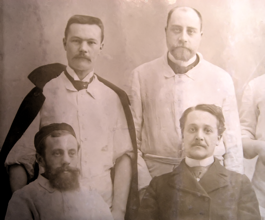
La Salpêtrière 1898: Henri Herbet (1873–1909) standing on the left, Maurice Lorrain on the right; Raymond Cestan seated on the left, Paul Froussard (1870–1927) on the right (Private collection of the author).
‘Associated with these ocular disturbances is sensorimotor hemiplegia on the side opposite the eye most damaged in its conjugate movement of abduction’. Raymond and Cestan added to their description the observation of ‘high-frequency static shaking of the hand and foot with athetosic movement of the fingers; this shaking is exaggerated in voluntary movements; finally, kinetic ataxia is increased by occlusion of the eyes’. Also presenting disorders in the lower limb, the patient showed ‘the characteristic clinical picture, described by Babiński, under the name of cerebellar asynergy’. 20 Disturbances in sensitivity were also present: ‘Sensitive alternating paralysis affecting the trigeminal nerve on one side and the arm and leg on the opposite side’. All modes of sensitivity were affected to variable degrees. The patients all suffered from a tuberculous tumour that gradually increased in volume, causing clinical progression then death. After the report from the histological examination, Raymond went on to discuss functional anatomy at length, referring to Babiński’s contributions to cerebellar and brain stem semiology, 21 then reviewed the literature on the various alternating syndromes published.
Based on their observations, Raymond and Cestan hypothesised that ‘linking fibres’ were likely to exist ‘that must follow the pathways of the pontine tegmentum, but it is not yet possible, despite the very interesting work of Pawlov [sic], to establish the exact role of each tract’, the fibres of which coordinate the movements of the head and eyes in association with the mobility and balance of the entire body. They concluded: ‘A syndrome of the tegmentum of the upper part of the pons can thus be described, or an upper pontine syndrome, which belongs alongside Weber’s syndrome or peduncular syndrome, and Millard-Gubler syndrome or lower pontine syndrome’. While the oculomotor nuclei were known at the time, the pathways between them were only suspected, notably the medial longitudinal fasciculus. The systems linking the cerebellum, reticular formations, labyrinths and vestibular pathways to the oculomotor nuclei were unknown, as well as the frontal oculomotor cortex and parieto-occipital oculomotor cortex. 22 Raymond-Cestan syndrome is rare and today is almost always vascular in origin. Recent tractography technology has led to a loss of interest in clinical aspects of stroke for shedding light on the pathophysiology and physiopathology of the brain, which Raymond and other 19th-century neurologists used to deduce the existence of deficits in intracerebral connections. Nevertheless, the use of this eponym keeps one of the few references to Raymond alive.
Hereditary spastic paraplegia
HSP is a group of rare diseases that have nonetheless been considerably researched because their genetic origin, still being analysed, offers a way to understand the links between the genome and cellular pathophysiology of the neuron. The implicated gene mutations could be responsible for cellular dysfunctions essential for maintaining axonal homoeostasis: permeability of the neuronal membrane, formation of the endoplasmic reticulum, physiology of lysosomes, myelination and so on.
Raymond focused on these diseases throughout his carrier, presciently proposing, in 1895, some of the theories being discussed today. Ten years earlier, in 1885, he authored the ‘spasmodic tabes’ entry of the Dictionnaire encyclopédique des Sciences médicales,
23
admitting that ‘this name induces regrettable confusion’ with the then-current use of the word tabes for syphilitic spinal damage. Raymond used this debatable term because it was used by Charcot in his 1875 lesson. Shortly before Charcot, Heinrich Erb (1840–1921) in Heidelberg had used a better expression: ‘spastic spinal paralysis’. Based on 16 clinical observations, Erb individualised a symptomatic association involving growing weakness in the lower limbs, later invading the upper limbs […]. There are multiple spasmodic phenomena which consist in more or less pronounced rigidity of the limbs with spontaneous jerking, tonic contractions, initially temporary, and clonic shaking in the lower limbs […]. His gait is hesitating and slightly vacillating. The soles of his feet stick to the ground and he drags his leg as he walks, which he does with small steps, keeping the legs held closely to one another. The front tip of his foot collides with the slightest obstacle […]. The tendon reflexes are almost always exaggerated.
24
In 1880, Adolf von Strümpell (1853–1925) published the first observation of hereditary spasmodic paraplegia in two brothers of the Gaum family in Estonia. In the older brother, onset was at around age 56 with slow progression. 26 The younger brother developed a pure form of the disease at around age 37 and died of tuberculosis at age 61. Strümpell published his autopsy in 1886: the spinal cord was normal to the naked eye, but under the microscope, in the dorsal and lumbar regions, there was ‘primitive combined sclerosis of the pyramidal tracts, spinocerebellar tract, and Goll tract [gracile tract]’ in the absence of any cerebral anomaly. 27 Strümpell compared his observation with that published by Raymond in 1882, 28 of a 78-year-old woman suffering from contraction in all limbs with exaggerated tendon reflexes and neuralgic pain in the lower limbs with onset before the paralysis. The anatomopathology indicated ‘sclerosis in the posterior tracts and lateral tracts’ whereas clinically sensitivity was intact. Raymond did not propose an aetiology.
Raymond’s 18 January 1895 lesson covered ‘spasmodic tabes’, faithfully keeping the name given by his teacher, and presented his audience with ‘two hereditary cases of childhood spasmodic paraplegia’.
29
He drew much material from the recent publication of his senior resident, Achille Souques (1860–1944), in La Revue Neurologique, for the clinical description of the two cases.
30
Focused on his responsibility as a teacher, a role he enjoyed, his main goal was to show how to distinguish this new nosographical entity, still uncertain for him, from Little’s disease. This lesson also served as an introduction to his subsequent lessons on ‘heredity in nervous pathology’. In 1895, Raymond and Souques observed a family in which two sisters suffered from spastic paraplegia. They compared their observation with those already published, which they accorded little credit, believing them to be ‘based on diagnostic errors’! Their conjectures on this pathology offer a sort of premonition: Spasmodic paraplegia could be considered a disease of the centrifugal protoneuron […] It is possible that the degeneration starts in the lumbar region, later reaching the dorsal and cervical regions. It thus appears to be ascending sclerosis, less evident in the cervical region than in the subjacent regions, ascending more or less according to the resistance of the pyramidal fibres and the duration of the disease.
31
In 1897, Raymond proposed ‘study of hereditary spasmodic paraplegia’ for his resident Maurice Lorrain’s thesis (1867–1956) (Figure 4). He presided over the thesis defence on 3 March 1898 32 (Figure 5). This thesis is the first summary of this subject, bringing together clinical aspects, as Charcot and Erb had established them, and a demonstration of heredity aspects through the addition of 29 observations. ‘The laws of hereditary are still too mysterious for us to attempt a study’. As a result, Lorrain did not use the term ‘hereditary disease’ but rather ‘familial disease’, the characteristics of which, according to Léon-Charles Pauly (1870–1936) and Charles Bonne (1872–?) in 1897, were:

Cover of Maurice Lorrain’s 1889 thesis (Private collection of the author).
Without changing form, it must affect several children of the same generation, start at around the same age in all children of this generation, and be clinically independent from any outside influence, from any acquired condition or intrauterine accident; these various characteristics must be the rule and not the exception. 33
After a summary of previous publications from which he excerpted 23 observations of spasmodic paraplegia, Lorrain added 6 personal observations, including 1 of 2 sisters, recorded at Hôpital Saint-Antoine by Georges Gilles de la Tourette (1857–1904). He proposed only one aetiology: heredity. Girls as well as boys could be affected, with the onset most often between age 8 and 15. Trauma or infectious disease seemed to be the notable aggravating factors, but motor difficulties preceded them.
Lorrain detailed the clinical aspects, highlighting clubfoot, the absence of sensitivity deficit, incoordination, speech difficulties, sphincteric problems, trophic problems and intellectual deterioration. Progression was very slow with periods of remission. He distinguished two forms: ‘One corresponding to spasmodic tabes, the other to multiple sclerosis’, that is, typical or pure forms or complicated (complex) forms. He reviewed in detail the result of the autopsy of Strümpell’s patient before presenting the one he carried out with the help of Claudien Philippe (1866–1903): There are lesions along the full length of the spinal cord, from the medullary cone up to the medulla. These lesions are clearly predominant in the white matter (anterolateral tracts and posterior tracts); they consist in more or less sclerotic areas.
Acute ascending paralysis
The book containing his 1895–1896 lessons opens with several lessons on ‘acute ascending paralysis’. 11 For anyone interested in the history of the individualisation of this syndrome, including the work of Octave Landry (1826–1865) and Guillaume-Benjamin Duchenne de Boulogne (1806–1875) 34 but also their predecessors, reading Raymond provides all the relevant information. Raymond gave an accurate clinical description of the symptoms: ‘Pins and needles and a numbing of the nerves are followed by ascending motor paralysis, generalising to all four limbs and part of the trunk over three days’. He did not neglect to mention abolition of reflexes and autonomic nervous system disorders, culminating in the risk of fatal apnoea. All that was missing was the analysis of cerebrospinal fluid, which was not yet practiced. The diagnostic discussion covers the clinical distinction between this syndrome and acute poliomyelitis, the infectious and contagious nature of which is detailed. Raymond goes on to describe the visual changes in the cellular body of the neurons in the anterior horn. He recognises the pre-existence of an acute intestinal disease and suspects the cause to be the release of bacterial toxins. Progression was generally favourable, and he tried to hasten it by ‘intestinal antisepsis’, ether, caffeine, strychnine and ‘dry suction cups’. Twenty years later, Pierre Marie (1853–1940) and Jean-Charles Chatelin (1884–1948), 35 as well as Georges Guillain (1876–1961), Alexandre Barré (1880–1967) and André Strohl (1887–1977), 36 would add the albuminocytological dissociation of cerebrospinal fluid as a diagnostic key. It should be noted that in 1900, Guillain served as a resident under Raymond, his first neurology teacher. Nevertheless, when Guillain and Barré gave their description of the ‘syndrome of radicular neuritis with hyperalbuminosis of the cerebrospinal fluid without cellular reaction’ in 1916, neither Landry nor Raymond were cited. This article, transcribed without commentary, initially published in the Bulletin de la Société médicale des Hôpitaux de Paris, 36 was reprinted in 1920, at the end of the ‘Varia’ chapter of their book Travaux Neurologiques de Guerre. It wasn’t until 1936 that Guillain would refer once again to the syndrome to which he owes much of his posthumous fame. 37 Major controversy then occurred, when the entity’s novelty was challenged in relation to Landry’s description. Guillain refused to accept cases described earlier, including those by Raymond.
Conclusion
The reputation of Fulgence Raymond, recognised by his contemporaries as an entirely worthy successor to his teacher, proved less resistant to time in the shadow of Charcot’s universal fame, which persists to this day. Nevertheless, Raymond was instrumental in enlarging and modernising the Charcot’s laboratories, particularly the one directed by Paul Richer (1849–1933), who created drawings and statues (La Parkinsonienne) useful during Raymond’s teaching 38 and now considered important artworks. Pierre Janet (1859–1947), who defended his doctoral thesis before a jury including Charcot 2 weeks before the master’s death, was able to practice as a clinical psychotherapist in La Salpêtrière thanks to Raymond and developed the concept of psychic trauma as the cause of hysteria at a time when ‘degeneration’ and ‘hereditarianism’ were accepted by almost all neurologists and alienists. With the support of Raymond, Janet was appointed in 1897 to the Experimental Psychology Chair at La Sorbonne University, the beginning of a fruitful career during which he worked in all domains of normal and pathological psychology. Unfortunately, his contributions were overshadowed by the success of psychoanalysis in France. 39 Raymond and Janet wrote two books together: Névroses et idées fixes in 1898 and Les obsessions et la psychasthénie in 1903. Other important topics on which Raymond wrote include syringomyelia, myasthenia gravis, progressive muscular atrophy, toxic-infectious diseases, brain tumours and spastic tabes. Raymond contended that tabes fell into the category of a syndrome and was always of syphilitic origin, even though these views contradicted those of his beloved master Charcot.
Three of his students went on to particularly distinguished careers: Jean Athanase Sicard (1872–1929) who was the first to perform spinal anaesthesia and initiated the use of Lipiodol as a contrast agent, Henri Claude who held the Chair of Mental Diseases and the Brain at Hôpital Sainte-Anne from 1922 to 1939 and Georges Guillain.
Taking to heart his duties as a teacher, Raymond passionately devoted himself to this most honourable of roles, but one accorded little posthumous fame. His lectures on neuropathology and clinical neurology served as a stimulus to many French and foreign students. In this way, he further enhanced the international reputation of La Salpêtrière as the great centre of neurology. With his strong interest in anatomopathology and his excellence as a clinician, he demonstrated many times, as exemplified above, his prescience in considering aetiologies for the diseases he studied, a gift other more well-known figures lacked. The handful of examples of his work presented here show that he has been unjustly overlooked.
Footnotes
Acknowledgements
The author thanks Jacques Poirier, Hubert Déchy and the reviewers for their critical reading and their thoughtful, pertinent comments.
Declaration of conflicting interests
The author declared no potential conflicts of interest with respect to the research, authorship, and/or publication of this article.
Funding
The author received no financial support for the research, authorship, and/or publication of this article.