
Abstract
Background:
Extensor retinaculum syndrome (ERS) is a relatively rarely diagnosed compartment syndrome-like entity caused by elevated pressures in the tissues deep to the superior extensor retinaculum (SER). ERS is identified as out-of-proportion anterior ankle pain, pain with passive toe plantarflexion, elevated SER pressures (>40 mm Hg), and ultimately toe extension weakness and first web space numbness. Although previously described in a pediatric population, this case series is the first to our knowledge in an adult population.
Methods:
Seven nonconsecutive cases over 18 years from 2 surgeons are reported who underwent complete SER release for ERS either through the direct lateral approach to the fibula or the anterolateral approach to the distal tibia. All were associated with traumatic injuries including 3 bimalleolar ankle fractures, 3 tibial pilon fractures, and 1 distal tibial/fibular shaft fracture. All patients developed writhing anterior ankle pain worsened with passive toe plantarflexion. SER compartment pressures ranged from 50 to >135 mm Hg. Five cases displayed decreased first web space sensation.
Results:
The diminished or absent first web space sensation uniformly improved post-release. Complications included 1 patient with complex regional pain syndrome type 1, 1 patient required hardware removal, and 2 had persistent but improved first web space sensation changes.
Conclusion:
Clinical suspicion for possible ERS should arise after distal tibial/fibular fractures when the excruciating pain localizes to the ankle instead of the classic anterior leg muscle bellies. If pain is worsened with passive toe plantarflexion, this diagnosis should be considered. Recommended treatment involves complete release of the SER anywhere on the anterior surface between the tibia and fibula depending on the approach needed for fixation of the associated fracture.
Level of Evidence:
Level IV, case series.
Keywords
Introduction
Following distal tibial/fibular fractures, some patients may develop intense, out-of-proportion anterior ankle pain that is unresponsive to opiate analgesia. Classically, surgeons would be suspicious of anterior leg acute compartment syndrome, but the patient’s pain localizes more distally over the anterior ankle without any discomfort over the more proximal muscle bellies of the anterior leg compartment. The pain is exacerbated with plantarflexion of the toes (particularly the great toe) and as the syndrome progresses, patients may experience numbness in the distribution of the deep peroneal nerve's first web space. However, on compartment pressure measurement, all 4 classic leg compartment (anterior, lateral, deep posterior, superficial posterior) pressures are normal, but pressures under the superior extensor retinaculum (SER) are elevated (>40 mm Hg). Although some of these symptoms are shared with anterior leg compartment syndrome, the starkly different pain location and lack of elevated traditional compartment pressure elevation indicates a different pathologic etiology.
In 2002, Mubarak 14 described this “extensor retinaculum syndrome of the ankle” in a case series of 6 pediatric patients after distal tibial physeal injuries. This was suspected to be a form of acute entrapment syndrome similar to acute carpal tunnel syndrome and was treated with the release of the SER, preserving the inferior extensor retinaculum (IER) to prevent extensor tendon bowstringing. Release of the SER led to relief of pain and return of motor and sensory function in the majority of the case series.
In his article, Mubarak further noted a personal, anecdotal adult case in which he encountered a necrotic extensor hallucis longus (EHL) during an approach to the tibial plafond. 17 He proposed that this is indicative of an equivalent syndrome in the adult population, but no case series has been published until this publication. Seven adult patients with distal tibial/fibular, exhibiting symptoms resembling those outlined in Mubarak’s article, are presented in this case series. Similar to compartment syndrome, extensor retinaculum syndrome (ERS) is suspected to be a progressive syndrome, and the patients presented at different points in the progression.
Compartment syndrome occurs when the interstitial pressure within a fascial compartment increases to the point that perfusion to that compartment is compromised as a result of outflow obstruction. This increase in interstitial pressure is usually caused by inflammation and edema after a traumatic injury, vascular compromise, an extended period of muscle compression, or surgery. A prolonged increase in pressure can result in ischemic damage to the compartment’s tissues, of which nerves and muscle fibers are the most vulnerable. 8
The SER of the ankle forms a tunnel as it spans between the distal tibia and fibula just proximal to the malleoli. Contrasted to a traditional muscle compartment, the contents of this tunnel are mostly tendinous (extensor digitorum longus and tibialis anterior). However the SER also contains the peroneus tertius (PT) muscle, the distal portion of extensor hallucis longus (EHL) muscle belly, 8 the deep peroneal nerve (DPN), and the anterior tibial vessels. Tendons are much more tolerant of ischemia than muscle fibers and nerves because the associated fibroblasts have a lower metabolic rate than muscle fibers and nerve tissues. Therefore, function of the PT, EHL, and DPN are most at risk in ERS.
Materials and Methods
Seven nonconsecutive patients were compiled from 2 independent surgeons over the span of 18 years at 6 different hospitals with a minimum of 3 months of follow-up. Institutional review board approval was obtained for the collection of the following data. Chart review was performed by the 2 independent surgeons (DC and KS) during which demographic and clinical data were compiled. The compartmental pressures were measured either with a Stryker (Kalamazoo, MI) pressure device or with the arterial line technique per Rorabeck et al. 15 The needle for measurement was placed approximately 4 cm proximal to the tip of the lateral malleolus and just lateral to the EDL to protect the anterior neurovascular bundle. Normal pressures under the SER were presumed to be equivalent to those in a traditional leg compartment. If the SER pressures were elevated (>40 mm Hg) and the clinical findings coincided with ERS, the patients were emergently taken to the operating room for surgical release with or without definitive fixation of the fractures. No patients with similar symptoms were treated nonoperatively to preserve viability of muscular and nervous structures.
Surgical Treatment
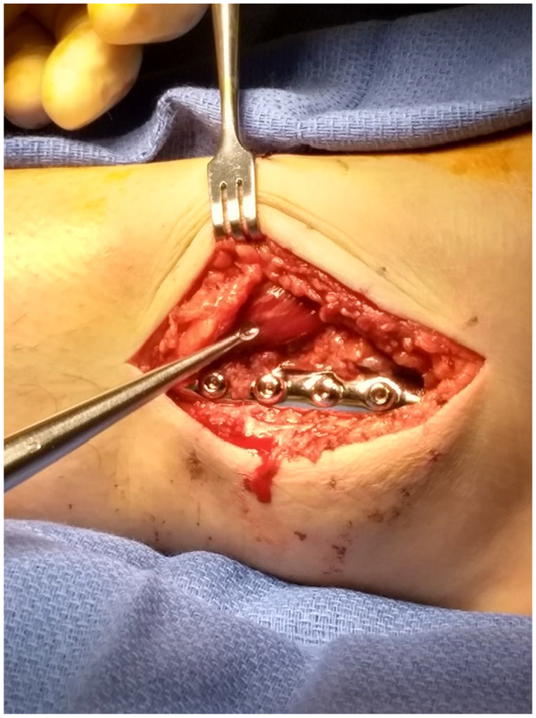
The goal of surgical treatment for ERS involved transection of the SER to release the elevated pressure and restore blood flow to the underlying muscular and nervous structures. The execution of this goal can be achieved with different approaches. The two main approaches to release the SER involve modifications of the direct lateral approach to the distal fibula and the anterolateral approach to the distal tibia that is dictated by the underlying fracture (Figures 1 and 2).

Anatomy of the superior extensor retinaculum (SER) and surrounding structures. Yellow band indicates location of SER release through the anterolateral approach to the distal tibia, and the green band indicates the release through the direct lateral approach to the distal fibula. EDL, extensor digitorum longus; EHL, extensor hallucis longus; IER, inferior extensor retinaculum; PT, peroneus tertius; TA, tibialis anterior.

Cross-sectional anatomy of the leg at the level of the superior extensor retinaculum. AT vessel, anterior tibial vessel; DPN, deep peroneal nerve; EDL, extensor digitorum longus; EHL, extensor hallucis longus; PT, peroneus tertius; SER, superior extensor retinaculum; SPN, superficial peroneal nerve; TA, tibialis anterior.
During the direct lateral approach to the distal fibula, a standard linear incision centered over the fibula is created. Dissection down to the fibula is performed with care to protect the superficial peroneal nerve (SPN). The dissection is then carried anteriorly to access the lateral attachment of the SER that is found approximately 3 cm proximal to the tip of the lateral malleolus and extends superiorly for approximately an additional 6 cm. 1 The SER is sharply released just anterior to the fibula along the entire length of the retinaculum. Complete release of the SER is ensured by placing either a finger or a similar surgical instrument parallel to the tendons under the SER to palpate for any tethering fibrous bands. In some cases, the peroneus tertius muscle belly will be visualized under the released SER, and it may be noted to bulge in a similar fashion to leg muscles after fasciotomy (Figure 3). Cadaveric studies have not identified any deep extensions of the retinaculum, which would theoretically subdivide the space under the SER into multiple subspaces. 1
Intraoperative clinical photograph of the swollen peroneus tertius muscle belly after SER release.
SER release can similarly be performed through the anterolateral approach to the distal tibia, which utilizes the interval between the anterior leg compartment tendons and the fibula. While taking care to protect the deep peroneal nerve and anterior tibial vessels, the SER is released in line with the skin incision between the fibula and anterior leg compartment tendons. Ensure complete release by palpating for any persistent restriction points.
Five of 7 patients in our series underwent SER release through the direct lateral approach to the distal fibula, and patients 3 and 7 were released through the anterolateral approach to the distal tibia. The approach for the release was determined based on the needed approach for fracture fixation.
Average patient age was 30 years (range 24-35) with 6 females and 1 male (Tables 1 and 2). Associated fractures included 3 bimalleolar ankle fractures, 3 tibial pilon fractures, and 1 distal one-fourth tibial/fibular shaft fracture. Six of the 7 patients had an associated Weber C fibula fracture and the remaining patient had a Weber B fibula fracture. Mechanisms of injury included motor vehicle accident, pedestrians struck by a motor vehicle, roller derby, and falls on ice. All 7 had elevated pressures (50 to >135 mm Hg) measured under the SER using the technique described above. Five of 7 patients had diminished or absent first web space sensation. Five of 7 patients’ release incisions were left open and returned later for closure of wound.
Patient Demographics (N = 7).

Clinical Findings.

Abbreviations: Ant, anterior compartment of the leg; Auto vs ped, automobile versus pedestrian; CRPS-1, complex regional pain syndrome 1; ED, emergency department; f/u, follow-up; Fx, fracture; Lat, lateral compartment of the leg; MOI, mechanism of injury; MVC, motor vehicle collision; OR, operating room; POD, postoperative day; ORIF, open reduction internal fixation; SER, superior extension retinaculum.
Patient 7 had the diagnosis of ERS in the recovery room after anterior tibial pilon fracture open reduction and internal fixation. We suspect that this was an iatrogenic ERS from the tight closure of the SER in a patient with swelling and robust fascia. She had an early return to the OR for release of the superior extensor retinaculum and had only the skin closed. Her writhing pain was completely resolved on waking from anesthesia.
Results
The diminished or absent first web space sensation uniformly improved postrelease.
Complications included 1 patient with complex regional pain syndrome type 1, 1 patient required hardware removal, and 2 had persistent but improved first web space sensation changes. During chart review, 4 patients were noted to have plantar foot sensation abnormalities that are unexplained by ERS. Two patients (patients 3 and 4) had elevated anterior leg compartment pressures, with one of these also having elevated lateral leg compartment pressures. Patient 3 had anterior compartment fasciotomies in conjunction with SER release. Anterior and lateral compartment pressure measurements were repeated again after SER release on patient 4, and both compartment measurements had normalized. Therefore, anterior or lateral leg compartment fasciotomy was not performed on patient 4.
Discussion
Extensor retinaculum syndrome of the ankle is a severely painful entity with the potential for long-term sequelae such as toe extension weakness or contractures and persistent first webspace sensory alterations. Our case series of 7 adult patients with the painful symptoms described by Mubarak suggests that ERS is not solely a pediatric condition. We suspect that ERS is often misdiagnosed as simply poor pain control after distal tibia/fibula fractures, and the resultant sensory changes and weakness are dismissed as unavoidable local soft tissue damage.
All 7 patients had an associated fibula fracture at or slightly above the ankle syndesmosis (1 Weber B and 6 Weber C fibula fractures). A Weber C fibula fracture’s association with ERS is logical as the SER is found between 3 and 9 cm proximal to the tip of the lateral malleolus. 1 Mubarak’s patients all had anterior translation of the proximal fragment, which decreased the potential space under the SER. 13 However elevated pressures in the SER were noted in this case series with fractures not specifically associated with anterior translation of a bony fragment. This suggests that the inflammatory response to the nearby trauma is more responsible for the development of ERS than direct bony compression of the compartment. A hemorrhagic etiology could also be a cause; however, no bleeding or hematoma was encountered under the SER in any of the 7 cases. Regardless of the actual cause, we propose that fractures near the SER, such as tibial fractures with an associated Weber C fibula fracture, may be risk factors for the development of ERS.
Similar to traditional ACS, we propose that ERS is a progressive condition with more symptoms developing with prolonged ischemia. Two patients were diagnosed early (within hours of injury) and therefore were not ischemic for enough time to develop first web space sensation changes. After surgical release of the SER, each patient had relief from the writhing pain and subjective improvement in first web space sensation changes.
Also like the treatment of ACS, the skin incisions in this series were not acutely closed in 5 of the 7 patients except for patients 5 and 7. However, only 1 patient (patient 6) had muscle belly bulging similar to what would be expected after ACS compartment release (Figure 3). Thus the skin was generally not a constricting force as it is in the more muscular compartments of the leg, thigh, and forearm. In this respect, ERS appears to be similar to acute carpal tunnel syndrome where the skin is not a pressure-inducing constriction, making skin closure potentially not a problem in many cases. Patient 5’s skin was closed after the release of the SER with the hope of accelerating the timeline to open reduction and internal fixation of the tibial pilon fracture. The pressure in the subretinacular space after skin closure was measured and found to have normalized. It was planned to release the sutures if all symptoms were not resolved in this patient. On awakening from anesthesia, the pain out-of-proportion resolved, and the first web space sensation had partially returned.
When it is noted that the skin is causing increased pressure or if there is increased pressures in the remaining compartments, we would recommend leaving the skin open for later closure when the swelling resolves. If the skin was closed after release of the SER, it might be indicated to remeasure the pressure in the compartment as we did in our patients where we closed the skin in the same surgical setting as the release.
Although the exact natural history of ERS is not known, the most likely sequelae include long-term hypoesthesia of the first web space and EHL weakness due to the distal extent of EHL muscle fibers under the SER. The ischemic damage to the EHL could result in a checkrein deformity, which has been described in case reports after distal tibial/fibular injuries where the great toe extends with ankle plantarflexion.7,16 The case reports noted decreased mass of the EHL on magnetic resonance imaging. Although not definitely a result of ERS, these findings would correlate closely with what would be expected in a missed ERS, resulting in tethering or ischemic contracture of the EHL. The loss of function of the muscles innervated by the DPN may be asymptomatic as patients are able to retain normal function due to EDL compensation2,11; however, the functional deficits from denervation of Extensor Digitorum Brevis (EDB) and Extensor Hallucis Brevis (EHB) are unclear.
It is unclear whether ERS is a form of early anterior leg compartment syndrome or if elevated pressures under the SER have impact on the surrounding compartments. Patient 3 had elevated anterior compartment pressures, and patient 4 had elevated pressures in both the anterior and lateral compartments. Although patient 3 was treated with anterior leg compartment fasciotomies, the anterior and lateral compartment pressures were re-measured in patient 4 after release of the SER, and the pressures had normalized. This relationship between release of the SER and the normalization of the adjacent compartment pressures is not readily apparent. One might speculate that because of the continuity of the SER with the anterior compartment severely elevated SER pressures could transfer to the anterior leg compartment, but this does not explain the normalization of lateral compartment pressures. Of course, SER release is not a reasonable treatment for anterior leg compartment syndrome, and the pressure changes may have been only coincidental.
Hypothetically, ERS could also be a precursor to anterior leg compartment syndrome especially when associated with distal fractures. This combined with the multiple case reports in the literature of anterior and lateral compartment syndromes after ankle fractures make one wonder if the superior extensor retinaculum syndrome might be the initial problem with later extension of the swelling proximally as the injuries are located more distally in these patients.3 -6,9,10,14,17 -19 It is possible, though, that the superior extensor retinaculum syndrome and anterior and/or lateral compartment syndromes occurred simultaneously and there were no associative effects. Further research is needed to evaluate the true incidence and natural history of ERS.
Similar to ERS is the chronic neuropathy called anterior tarsal tunnel syndrome (ATTS) which is usually caused by an external compressive force such as a shoe strap. Some might suggest that these ERS and ATTS are the same entity. However, ATTS is usually the result of chronic compression under the IER instead of the SER. 12 ATTS is treated with the release of the IER, whereas ERS is a traumatic condition whose symptoms improve with release of the SER.
During the retrospective chart review in preparation for this manuscript, 4 of 7 patients were noted to have documented plantar sensation changes which does not anatomically correlate with ERS as the tibial nerve does not pass under the SER. We suspect that an associated condition was present but its exact etiology is unclear. There is a possibility there was also an acute tarsal tunnel syndrome that was concurrent with the ERS. Of the 4 patients with plantar sensory changes, 2 ultimately had complete resolution of the plantar sensory deficit, 1 had continued resolution of the numbness to the level of the metatarsal heads at 6 months postoperatively, and patient 4 had residual plantar hypersensitivity and the appearance of type 1 complex regional pain syndrome in that foot. At the subsequent follow-up appointment, nerve conduction studies for patient 4 were ordered to evaluate for chronic tarsal tunnel syndrome but were not completed because of patient loss to follow-up. More research is needed to evaluate for the possibility of other local concomitant entrapment syndromes that could be associated with ERS.
ERS has been previously recognized in the pediatric age groups with distal tibial physeal injuries. 13 When patients present with high-energy distal tibia and/or fibula fractures, one should be alert to the possible development of ERS, particularly in patients with involvement of both the distal tibia and fibula. If a patient reports persistent excruciating anterior ankle pain that is unresponsive to opiate analgesia and is worsened with toe plantarflexion, it may be prudent to measure the pressure under the SER as described above, in addition to evaluation for other leg ACS. More than likely, ERS has not been recognized by surgeons in the past, and the resultant sequelae were attributed to blunt soft tissue damage. When ERS is clinically suspected and pressures under the SER are elevated (>40 mm Hg), emergent release of the SER, as described earlier, appears to be indicated. More data are needed to better understand this intriguing condition that was previously not recognized.
Supplemental Material
sj-pdf-1-fao-10.1177_24730114241265342 – Supplemental material for Extensor Retinaculum Syndrome of the Ankle: An Adult Case Series
Supplemental material, sj-pdf-1-fao-10.1177_24730114241265342 for Extensor Retinaculum Syndrome of the Ankle: An Adult Case Series by Bradley Carlson, DuWayne Carlson, Tadiwanashe Chirongoma and D. Kevin Scheid in Foot & Ankle Orthopaedics
Footnotes
Ethical Approval
Institutional review board approval was obtained.
Declaration of Conflicting Interests
The author(s) declared no potential conflicts of interest with respect to the research, authorship, and/or publication of this article. Disclosure forms for all authors are available online.
Funding
The author(s) received no financial support for the research, authorship, and/or publication of this article.
References
Supplementary Material
Please find the following supplemental material available below.
For Open Access articles published under a Creative Commons License, all supplemental material carries the same license as the article it is associated with.
For non-Open Access articles published, all supplemental material carries a non-exclusive license, and permission requests for re-use of supplemental material or any part of supplemental material shall be sent directly to the copyright owner as specified in the copyright notice associated with the article.