
Abstract
Keywords
Introduction
The accessory soleus muscle may cause swelling and compression of the tibial nerve and may present in the second decade of life because of increased muscle mass and activity. 7 Vascular disorders have also been reported to physically restrict the tibial nerve.3,6 Additionally, muscle swelling from injury can also lead to an accumulation of connective tissue fluids that may progress to compartment syndrome. 8 Furthermore, exercise-induced compartment syndrome is a rare clinical diagnosis that can have long-term ramifications if not diagnosed in a timely fashion. 4
We present a report of a patient with a gastrocnemius fascial tear, soleus muscle edema, and tibial neuritis treated with superficial and deep compartment fasciotomies, gastrocnemius fascial repair, ligation of a vascular leash, and tibial nerve neurolysis.
Case Report
An 18-year-old female lacrosse player with no personal or family history of hypercoagulability or leg injury presented with left calf pain and paresthesia in the plantar aspect of her foot that started after she fell from 10 feet landing on her leg.
On initial examination, her posterior tibial and dorsalis pedis pulses were intact. All sensation was grossly intact. She had bruising over the dorsum of the calf and her entire calf was tight to palpation. She had tenderness along the medial gastrocnemius musculature extending into the popliteal space but full range of motion and full strength. Ultrasonography showed no sign of deep vein thrombosis in the left lower extremity. She was initially treated for a calf strain with naproxen, muscle relaxers, and focused therapy with athletic training staff.
Despite 2 weeks of conservative treatment, she continued to have significant pain, especially in the medial gastrocnemius muscle. Pain was worse with prolonged ambulation and calf activation, but she denied swelling or bruising. There were palpable knots within the medial gastrocnemius muscle belly but no palpable defect.
Radiographs showed no fractures or dislocations. A tibia/fibula magnetic resonance imaging (MRI) was ordered to evaluate her soft tissues, and the left calf showed inflammation within the superficial soft tissues surrounding the saphenous vein. There was concern for superficial thrombophlebitis and she was prescribed Aspirin and Keflex. However, at her next visit 2 weeks later, she reported pain and numbness of the plantar aspect of her foot and in the fascia of the gastrocnemius-soleus muscle junction. She had a negative Thompson test but a positive Tinel over her deep compartment and tibial nerve. A diagnostic left ankle MRI was performed 3 weeks after the previous MRI, which showed interstitial tearing of the gastrocnemius fascia all the way to the distal Achilles, inflammation along the plantaris, and a grade 1 strain of the Achilles and soleus muscles with edema (Figure 1). There was no evidence of tibial neuritis or compression. This was managed nonoperatively with a nonweightbearing cast and steroid dose pack.

Left lateral ankle magnetic resonance imaging scan with the yellow line demonstrating Achilles edema on its insertion site on the calcaneus that resulted from a tear. Proximal to the yellow line, an interstitial gastrocnemius fascial tear is visualized.
Upon return, her symptoms improved but did not resolve. An electromyographic nerve conduction study was ordered but came back normal. She underwent an ultrasonography-guided corticosteroid injection in her left posterior calf along the soleus and gastrocnemius musculotendinous and around the tibial nerve. The injection significantly decreased her numbness and tingling of the foot, but she still experienced calf pain. At this time, a third MRI was obtained of the left ankle 3 months after her previous MRI to assess whether any new pathology could be visualized and to help guide treatment with further conservative management such as platelet-rich plasma injections vs surgery, which showed significant edema within the soleus muscle.
Given her symptoms along with edema within the soleus, it was felt she was having a localized compartment syndrome compressing her tibial nerve. Surgery was discussed and the plan was a superficial and deep compartment fasciotomy. On the day of surgery, she stated that her tibial nerve paresthesia returned about 50% over the last few weeks and was getting worse.
Surgery
The patient was positioned supine with a popliteal femoral block and a thigh tourniquet. A vertical 4-cm incision was made over the medial border of the calf at the point of maximal tenderness (Figure 2). The crural fascia was opened and superficial fascia released, and immediately the soleus muscle belly extruded from the wound, indicating significant swelling of the muscle. We found substantial tenosynovitis of the gastrocnemius fascia, and a tenosynovectomy was performed. We then identified a complete tear of the gastrocnemius with degenerative and unhealthy tissue within the tendon, which was repaired and debrided (Figure 3). The deep compartment was then released, and the tibial nerve was identified immediately in the area of maximal tenderness. There was also a vascular leash from the soleus muscle over the tibial nerve compressing it. The vascular leash was cauterized and released, and an external neurolysis of the tibial nerve was performed.

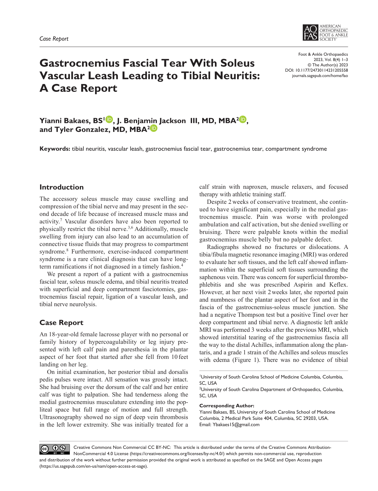
Left leg marked for surgery, with the vertical line showing the area of maximal tenderness where the incision was made.
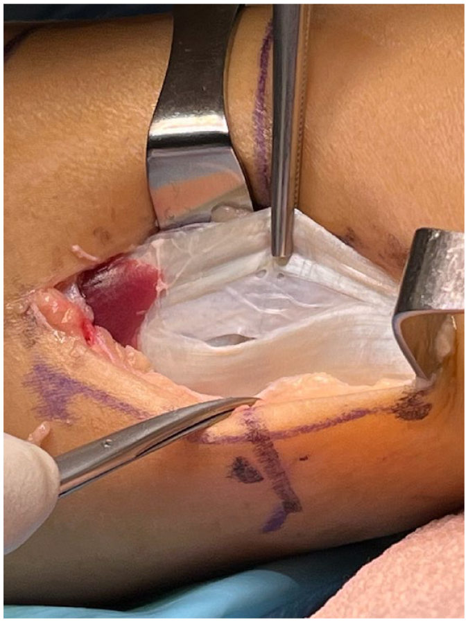
Left medial gastrocnemius tear.
Postoperatively, the patient was made nonweightbearing in a splint for 2 weeks, then progressed to weightbearing as tolerated in a boot for 2 weeks and began physical therapy at 4 weeks in a regular shoe. At her 4-month postoperative follow-up appointment, she was doing well with improved numbness and pain. She was improving in physical therapy and her activities were advanced to running, agility, and return to sports.
Discussion
Neuritis can occur from a variety of causes such as injury, vascular deformities, and an accessory soleus muscle.1,5 The mechanism of this patient’s pain and tibial neuritis was a combination of the gastrocnemius fascial tear, muscle swelling, and vascular leash compressing the tibial nerve.
Ultimately, the MRI report and clinical severity of the patient’s condition led to the decision for surgery. However, the extensive degree of her gastrocnemius fascial tear and associated degeneration was surprisingly not appreciated on MRI. This clinical variable as well as the vascular leash were found during surgery and corrected. The patient’s tibial neuritis symptoms may be important for surgeons to consider when evaluating lower extremity pain that persists despite conservative measures. Compression of the tibial nerve usually leads to pain with prolonged standing or walking. It is characterized by foot pain at rest and burning, numbness, or tingling along the plantar medial aspect of the foot.1,5 Patients may also have swelling and tenderness at the posteromedial ankle or foot, Tinel’s sign over the tibial nerve, and hypoesthesia of the toes.1,5
Nonoperative treatment such as activity modification and anti-inflammatory medications have been proven to provide relief in some cases and should be tried first. 2 However, for patients who do not experience improvement with nonoperative modalities, surgery is indicated.1,5
In this report, the gastrocnemius fascial tear, soleus swelling, and vascular leash from the soleus were clinically evaluated in the same manner as would an accessory soleus muscle because the mechanism is similar with the issue of physical compression on the tibial nerve with presentation resembling a localized compartment syndrome. We hope this report helps surgeons isolate tibial neuritis symptoms and consider the variety of factors that may cause this when conservative treatments fail. It may be important to consider a broad differential including factors that may be missed on MRI such as a gastrocnemius facial tear along with patient-unique anomalies such as a soleus-derived vascular leash to efficiently reach the correct diagnosis and ultimately reduce patient morbidity.
Supplemental Material
sj-pdf-1-fao-10.1177_24730114231205558 – Supplemental material for Gastrocnemius Fascial Tear With Soleus Vascular Leash Leading to Tibial Neuritis: A Case Report
Supplemental material, sj-pdf-1-fao-10.1177_24730114231205558 for Gastrocnemius Fascial Tear With Soleus Vascular Leash Leading to Tibial Neuritis: A Case Report by Yianni Bakaes, J. Benjamin Jackson and Tyler Gonzalez in Foot & Ankle Orthopaedics
Footnotes
Ethical Approval
Ethical approval for this study was obtained from the Prisma Health Institutional Review Board [2062248-1]
Declaration of Conflicting Interests
The author(s) declared no potential conflicts of interest with respect to the research, authorship, and/or publication of this article. ICMJE forms for all authors are available online.
Funding
The author(s) received no financial support for the research, authorship, and/or publication of this article.
References
Supplementary Material
Please find the following supplemental material available below.
For Open Access articles published under a Creative Commons License, all supplemental material carries the same license as the article it is associated with.
For non-Open Access articles published, all supplemental material carries a non-exclusive license, and permission requests for re-use of supplemental material or any part of supplemental material shall be sent directly to the copyright owner as specified in the copyright notice associated with the article.