
Abstract
Introduction
Blastomycosis is a rare disease caused by the environmental fungi species Blastomyces dermatitidis, and Blastomyces gilchristii. In the United States, it is mainly found in the midwestern, south-central, and southeastern states, particularly in areas surrounding the Ohio and Mississippi River valleys, the Great Lakes, and the Saint Lawrence River. 8 In 2021, the CDC analyzed surveillance data from 4441 cases from 1987 to 2018 and found only 5 US states in which blastomycosis was reportable: Arkansas, Louisiana, Michigan, Minnesota, and Wisconsin, with Northern Wisconsin having the highest incidence of cases. 2 Few cases have also been reported in Canada, Africa, and India. 8
Infection begins with inhalation of spores leading to pulmonary disease that may disseminate to other organ systems. In rare cases, infection has resulted from direct cutaneous inoculation via a puncture wound. Once the spores have inoculated the patient, the incubation period varies from 2 to 15 weeks. 2 Rare but serious manifestations of blastomycosis infection result from hematogenous dissemination to other organ systems. 1 Individuals most at risk for disseminated infection by fungi are the immunocompromised, those with active malignancies, and patients who have received a solid organ transplant. In this report, we describe a case of disseminated blastomycosis affecting the lung, bone, skin, and ovary after almost 2 years of the first symptom onset.
Case Presentation
We present a 49-year-old woman, tobacco and cannabis smoker, who recently relocated to Chicago, IL, from Racine, WI, where she had been residing for many years in a wooded area next to the Root River. She presented to the emergency department (ED) complaining of right heel pain and chronic wounds to the right anterior leg and right posterior heel. The patient had acquired the wounds over the past 6 months following a series of syncopal episodes resulting in falls and reported spontaneous rupture of a blister overlying the right heel with purulent drainage after a fall the day prior to presenting to the ED. She also noted unintentional 23-kg weight loss over the past 3-4 months and night sweats, with no changes in her diet or exercise regimen.
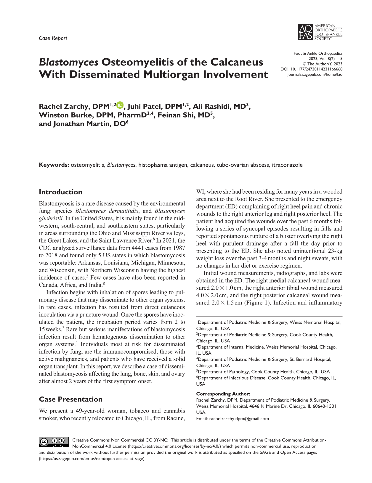
Initial wound measurements, radiographs, and labs were obtained in the ED. The right medial calcaneal wound measured 2.0 × 1.0 cm, the right anterior tibial wound measured 4.0 × 2.0 cm, and the right posterior calcaneal wound measured 2.0 × 1.5 cm (Figure 1). Infection and inflammatory markers were within normal limits. Plain radiographs of the foot and ankle revealed a well-demarcated cystic lesion in the posterior plantar aspect of the calcaneus that did not cross any joint lines (Figure 2). There were no signs of erosions, active destruction, or osteopenic changes near or around the cystic lesion. The patient was discharged home from the ED on oral clindamycin and advised to follow up in the podiatry outpatient clinic.

Initial photographs of the right anterior leg and heel ulcerations. (A) Anterior leg ulceration after sharp debridement of necrotic and fibrotic tissue. (B) Posterior heel and medial ankle ulcerations, weeks after rupture of posterior heel abscess.

Nonweightbearing plain radiograph of right ankle. Lateral view showing bulky and sclerotic appearance of the calcaneus with well-demarcated cystic lesion.
In the podiatry clinic, based on clinical and radiologic findings, and to rule out benign and malignant diagnoses, a magnetic resonance imaging scan was ordered. Magnetic resonance imaging of the right ankle revealed a focal 0.7 × 0.5 cm area in the posterior calcaneus of increased signal intensity on T2-weighted image and decreased signal intensity on T1-weighted image (Figure 3).

(A) T2-weighted sagittal image. (B) T1-weighted sagittal image. (C) T2-weighted axial image. (D) T1-weighted axial image. Magnetic resonance imaging scan of the right ankle revealed increased T2 signal in the calcaneus with decreased signal on T1-weighted images, most of which enhances on postcontrast imaging (Figure 3). A focal area measuring approximately 0.7 × 0.5 cm; however, showed no enhancement on postcontrast imaging.
Because of concern for concomitant underlying systemic illness, including tuberculosis, malignancy, and fungal infection, the patient was referred to an infectious disease physician, who expressed concerns about autoimmune diseases, lung cancer, syphilis, and fungal infection. Accordingly, laboratory testing and imaging studies were ordered. The lab tests demonstrated a weakly positive urine histoplasma antigen and microcytic anemia.
Chest radiograph (CXR) displayed blunting of left costophrenic angle. Computed tomography (CT) of the chest, abdomen, and pelvis with IV contrast revealed slight air bronchograms and moderate left-sided pleural effusion (Figure 4). Given her weakly positive urine histoplasma antigen test, the patient was given the tentative diagnosis of pulmonary fungal infection. Accordingly, infectious disease and podiatry teams agreed on a biopsy with periodic acid–Schiff (PAS) and Grocott methenamine silver (GMS) stains with fungal culture.

(A) Anterior-posterior portable plain radiograph of the chest displaying blunting of the left costophrenic angle. (B) Computed tomography of the chest displaying linear consolidation with air bronchograms and moderate-sized left-sided pleural effusion with borderline bilateral axillary lymphadenopathy.
The patient was taken to the operating room for radical debridement of the right tibial crest and calcaneus, and intraoperative biopsies of the right calcaneus and heel soft tissue. Attention was first directed to the anterior leg ulceration. With a 2- to 3-mm margin, the wound was excised to the level of subcutaneous tissue with a No. 10 blade. Two punch biopsies were obtained from the center of the specimen for bacterial and mycotic culture and sensitivity testing. The remaining tissue was sent in formalin for histocytologic examination. Next, attention was directed to the medial heel. An oblique incision was placed along Langer’s lines after using the C-arm to identify the exact area of the bone lesion. Caution was taken to avoid the cutaneous ulceration while making this skin incision. Upon incision, yellow purulence was noted. The medial cortex of the bone cyst was necrotic. Intraoperatively, the cyst measured to a depth of 3.2-cm. A bone biopsy needle was used to harvest a specimen from the medial calcaneus for histocytologic examination. The 0.5-cm trephine tip of the outer cannula was placed on the desired area of biopsy. With little resistance, the biopsy needle was advanced through the medial cortex of the calcaneus to the lateral cortex.
Biopsy taken from the medial-posterior heel revealed soft tissue fragments with necrosis and granulomas with budding yeasts and skin and soft tissue biopsies showed superficial granulomatous inflammation with Langerhans' giant cells containing thick-walled broad-based budding yeasts, suggestive of B dermatitidis (Figure 5). Based on the clinical, imaging, and pathology findings, the diagnosis of blastomycosis involving lung, bone, skin, and ovary was confirmed and treatment was initiated by itraconazole for a total of 2 years to ensure complete eradication and prevent recurrence. Cutaneous ulcerations closed and osseous healing was observed on radiographs approximately 6 months after surgery and initiation of itraconazole (Figure 6).

(A) GMS stain of skin and subcutaneous tissue from right upper leg with focal pseudoepitheliomatous hyperplasia, chronic inflammation, and superficial granulomatous inflammation with Langerhans’ giant cells containing thick-walled broad based budding yeasts, morphologically suggestive of blastomycosis. (B) PAS stain of skin and subcutaneous tissue from right upper leg with thick-walled budding yeasts located in multinucleated giant cells. (C) GMS stain of bone fragments from right calcaneus with necrosis, granulation tissue, acute and chronic inflammation, and granulomas with budding yeasts located in osteocytes. GMS, Grocott methenamine silver; PAS, periodic acid–Schiff.

Photographs of the anterior leg and heel obtained 4 months postoperation. (A) Anterior leg ulceration epithelialized. (B) Posterior heel ulceration, medial ankle ulceration, and surgical incision all epithelialized.
Discussion
This report presents a case of disseminated blastomycosis affecting the lung, bone, skin, and ovary. The clinical similarities between blastomycosis and other pulmonary infections often result in diagnostic delays and inappropriate empiric antimicrobial drug treatment. 2 CXR can reveal nonspecific findings that are often indistinguishable from pneumonia and may mimic other diagnoses, such as lung neoplasm or tuberculosis. 7 Thus, blastomycosis is often not included in the differential diagnosis unless the patient presents with other clinical findings, or if the patient has failed to respond to antibiotic therapy.
Fungi are detected in clinical specimens either by direct visualization of the organisms, or by detection of substances produced by the organism. The most common methods for visualizing fungi directly are by Gram stain, including periodic acid–Schiff stain (PAS) and Grocott methenamine silver stain (GMS), potassium hydroxide (KOH) wet mount, or a calcofluor white stain. 7 Specimens for these tests are obtained from nail clippings, skin scrapings, hair, vaginal swab, and sputum.
Antigen testing allows for earlier detection of most dimorphic molds, like blastomycosis. The sensitivity of antigen testing has been reported to be >90% and specificity has been reported to be 100% in healthy subjects. 4 Urine antigen testing is one of the fastest and cheapest diagnostic modalities for detecting blastomycosis. In patients with blastomycosis, the urine may contain cross-reactive or shared antigens with Histoplasma capsulatum. This is in accordance with our finding in this case, who had a positive urine histoplasma antigen. In these situations, cytologic preparations would be distinguishing.
Untreated pulmonary blastomycosis may progress to acute respiratory distress syndrome, vertebral osteomyelitis, paraspinal abscess, and ultimately spinal cord compression. 5 The most common extrapulmonary sites of blastomycosis dissemination are skin, bone, and the genitourinary (GU) system. 7 Thus, maintaining a high level of clinical suspicion is critical.
Treatment of blastomycosis infection is achieved with Azole antifungals, or Amphotericin B (AmB) depending on the site and severity of the infection, host immune status, and pregnancy.6,7 It is important to note that with treatment with itraconazole, it is recommended to obtain therapeutic levels and adjust the dosing as needed as there is a narrow therapeutic window with these azoles. Patients with osteomyelitis secondary to blastomycosis will be more prone to relapse of infection as fungus is slower growing than bacteria, and should receive antifungal treatment for a minimum of 12 months. 3
Conclusion
With increasing numbers of immunocompromised patients, the incidence of fungal infections has dramatically increased over recent years. A high level of clinical suspicion for fungal infection in patients with unresolving disease courses or patients with known risk factors who fit the geographic profile for fungal infection should be maintained, specifically in patients with musculoskeletal signs and symptoms.
Footnotes
Ethical Approval
Ethical approval was not sought for the present study because our institution does not require ethical approval for reporting individual cases or case series. This study has been performed in accordance with ethical standards.
Declaration of Conflicting Interests
The author(s) declared no potential conflicts of interest with respect to the research, authorship, and/or publication of this article. ICMJE forms for all authors are available online.
Funding
The author(s) received no financial support for the research, authorship, and/or publication of this article.