
Abstract
Hydrogels are homogenous materials that are limited in their ability to form oriented multilayered architecture in three-dimensional (3D) tissue constructs. Current techniques have led to advancements in this area. Such techniques often require extra devices and/or involve complex processes that are inaccessible to many laboratories. Here is described a one-step methodology that permits reliable alignment of cells into multiple layers using a self-assembling multidomain peptide (MDP) hydrogels. We characterized the structural features, viability, and molecular properties of dental pulp cells fabricated with MDP and demonstrated that manipulation of the layering of cells in the scaffolds was achieved by decreasing the weight by volume percentage (w/v%) of MDP contained within the scaffold. This approach allows cells to remodel their environment and enhanced various gene expression profiles, such as cell proliferation, angiogenesis, and extracellular matrix (ECM) remodeling-related genes. We further validated our approach for constructing various architectural configurations of tissues by fabricating cells into stratified multilayered and tubular structures. Our methodology provides a simple, rapid way to generate 3D tissue constructs with multilayered architectures. This method shows great potential to mimic in vivo microenvironments for cells and may be of benefit in modeling more complex tissues in the field of regenerative medicine.
Keywords
Introduction
Despite a growing body of evidence demonstrating that hydrogels are well suited for three-dimensional (3D) tissue constructs and cell delivery, challenges still remain to enable the efficient manipulation of fabricated cells within hydrogels for such applications in terms of their alignment and the development of cellular networks.1–3 When fabricating cells within hydrogels, it is difficult to manipulate the ratio of contact between cells and hydrogel structure. This results in inadequate control of cell–cell distance and the loss of multiple-layered cell integrity.4,5 The oriented microstructure of multiple cell layers is crucial for communication among neighboring cells and cross-talk between cells and their surrounding extracellular matrices (ECMs).6–9 Hence, understanding the way in which hydrogel nanofibrillar elements interact with fabricated cells is critical to understanding their potential to mimic native ECM.
Currently, surfactant chemistry and mechanical engineering techniques have facilitated the control of cell–cell distance and alignment of cells into microscaled multiple layers in 3D tissue constructs.10–14 However, these techniques generally rely on additional devices and complex procedures in order to achieve desired effects.10–14 In addition, either chemical or physical cross-linking is required for these fabrication techniques. Such approaches can cause damage to fabricated cells or influence the properties of the materials themselves due to the use of potentially cytotoxic agents or relatively dramatic changes in temperature or pH during the process of cross-linking.12,15,16 For example, covalent cross-linking of a hydrogel system sometimes requires in situ cross-linking, which has pronounced effects on mechanical properties, such as shear thinning, Young’s modulus, viscoelasticity, and recovery. The alteration of these mechanical properties could lead to a lack of ability to fill defects with irregular shapes or apply to scaffolds with complex structures.
Self-assembling hydrogels have been utilized in a number of tissue engineering applications and have been proposed as biocompatible and biomimetic scaffolds to drive tissue regeneration in the oral cavity.17–21 In contrast to covalently cross-linked hydrogels, multidomain peptides (MDPs) are self-assembling nanofibrillar scaffolds that show shear thinning and stress recovery due to their noncovalent nature.17–21 These well-characterized MDPs possess structural properties generally comparable to soft tissues in terms of their stiffness. They can also easily fill irregular defects due to their unique physiochemical properties.22–24 Such properties allow them to be extensively used as tissue engineering scaffolds within various types of tissues, mimicking the native ECM.25–27 The integrin-binding sequence RGD has been added into the base sequence of our MDPs. The RGD sequence has long been known to facilitate integrin-dependent functions, such as promoting cell adhesion and proliferation.17,18,28–30 In this article, we report that this particular sequence probably allows cells to be coated with a controlled thickness of MDP at the single-cell level through RGD peptide–integrin interactions in a density-dependent manner.
Here, we present a simple and reliable single-step self-assembly approach to create aligned and multilayered 3D tissue constructs by decreasing the ratio of nanofibrillar elements to cells in MDP scaffolds. The results of this study elucidate the ways in which MDP scaffold density dramatically impacts the behavior of fabricated mouse dental pulp cells and 3T3 fibroblasts. Further, we have demonstrated that 3D tissue constructs with different architectural configurations may also be simply generated by using this approach. This relatively simple method for creating multilayered 3D cellular constructs provides the basis for understanding how cells interact within these biocompatible nanofibrillar scaffolds and may have significant applicability for their use in tissue regeneration.
Materials and Methods
MDP Preparation
The MDP hydrogel was prepared as previously described.17,18,28 Briefly, MDP peptides ( Suppl. Table S1 ) were synthesized with Focus XC Solid Phase Peptide Synthesizer (AAPPTec, Louisville, KY). After synthesis, the correct molecular weight of peptide was verified by MALDI-TOF mass spectrometry. Lyophilized MDP was suspended in 29 8mM sterile filtered sucrose at 2% w/v MDP. To form a 1% w/v hydrogel, 2% w/v MDP was mixed with equal volumes of HBSS (1×; Mediatech, Inc., Manassas, VA) containing 298 mM sucrose.
Cell Culture
The mouse dental pulp cells (MDPC-23) are mouse dental pulp mesenchymal cells with the potential to differentiate into odontoblast-like cells (a gift from Dr. Yongbo [Bob] Lu, Texas A&M University),31,32 and NIH3T3 cells (ATCC CRL-1658) were cultured in DMEM containing 10% fetal bovine serum (FBS). When the monolayer of cells reached 90% confluence, 0.25% trypsin was used to harvest cells from petri dishes. The cell solution was transferred to a fresh 50 mL conical tube, leaving the larger tissue pieces behind, and centrifuged for 5 min at 1000g. The cell pellet was resuspended in 10 mL of DMEM in the appropriate volume before use.
MDP Fabrication Procedures for 3D Tissue Engineering
Mouse dental pulp cells were coated with MDPs at various densities: 0.04%, 0.4%, and 1% w/v. Briefly, cells were trypsinized and suspended in HBSS at 1.25 × 10⁷ cells/mL. A total of 2% w/v peptide solutions were prepared by dissolving 20 mg of peptide in 1 mL of a sucrose solution (298 mM). To prepare 1% w/v MDP fabricated cells, a cell suspension containing 1.25 × 10⁷ cells/mL was mixed in equal volume with the 2% w/v peptide solution to fabricate the cells with MDPs. For generating the 0.4% and 0.04% w/v MDP fabricated cells, 50 and 15 µL of 1% w/v peptide stock solution were added to 1.25 × 10⁷ cells/mL for final volumes of 125 and 375 µL, respectively. To obtain the final 3D tissue constructs, the mixture of cells and MDP solution was added to 24-well Transwell inserts with or without additional molding (e.g., capillary tubes for tubular-like structure) and incubated for 3 days at 37 °C, 5% CO2. To obtain tissue constructs with stratified multilayered architecture, 1.25 × 10⁷ cells/mL mouse dental pulp cells and NIH3T3 cells were fabricated in 0.04% w/v for final volumes of 90 and 275 µL of solutions, respectively.
Fluorescent-Activated Cell Sorting
Mouse dental pulp cells fabricated in different densities of MDPs were collected. Harvested cells were centrifuged at 150g and washed twice with 0.5% bovine serum albumin (BSA) in phosphate-buffered saline (PBS). Subsequently, 1 × 10⁶ cells were stained with 5 µM carboxyfluorescein succinimidyl ester (CFSE) dye. Labeled cells were then washed with 0.5% BSA in PBS three times and then resuspended in 0.5 mL of 0.5% BSA in PBS and kept on ice until analysis. Cells were then stained with 1 µg/mL of 4′,6-diamidino-2-phenylindole (DAPI) to positively identify dead cells after labeling with CFSE. Finally, stained cells were counted on a fluorescent-activated cell sorting (FACS) Canto II (BD Biosciences, San Jose, CA). Results were analyzed using the FlowJo software (Tree Star, Ashland, OR).
Scanning Electron Microscopy
Mouse dental pulp cells fabricated in different concentrations of MDPs were fixed with 4% paraformaldehyde at room temperature (RT) for 20 min. After fixation, cells were rinsed with deionized (DI) water and dehydrated with serial alcohol solutions (30%, 50%, 70%, and 100%) at RT for 30 min each step. Dehydrated samples were then mounted on carbon tape and coated with a thin layer of gold to reduce charging. After fabrication, samples were examined with a FEI Quanta 600 scanning electron microscope in low-vacuum mode (15 kV) at the University of Utah Surface and Nanoimaging Facility using a secondary electron detector.
Transmission Electron Microscopy
Mouse dental pulp cells fabricated in different concentration of MDPs were fixed overnight in 2% glutaraldehyde and 1% paraformaldehyde in 0.1 M cacodylate buffer (pH 7.4). Fixed samples were treated with 2% OsO4 in cacodylate buffer and then rinsed in DI H2O. The samples were stained with 4% uranyl acetate, dehydrated with serial ethanol solutions, washed with acetone, and then embedded in epoxy Embed-812 (Electron Microscopy Sciences, Inc., Hatfield, PA) following the manufacturer’s instructions. Polymerized blocks were cut into approximately 70 nm sections with a diamond Ultra 45° knife (Diatome, Inc., Hatfield, PA). Sections were mounted on copper grids and stained with saturated uranyl acetate, rinsed with water, and stained with Reynold’s lead citrate. The sections were imaged by transmission electron microscopy (TEM) in a Tecnai 12 (FEI Technologies, Inc., Hillsboro. OR) microscope.
Quantitative Polymerase Chain Reaction
Mouse dental pulp cells fabricated in the varying densities of MDPs were immersed in RNA stabilization solution (Invitrogen, Waltham, MA) at 4 °C overnight. The following day, the RNA stabilization solution was removed and total RNA was extracted using the RNAeasy Kit (QIAGEN, Germantown, MD) according to manufacturer’s instructions. Total RNAs were reverse transcribed into cDNA according to the manufacturer’s instructions (Invitrogen). Finally, total cDNA was diluted at 1:50 ratios and used as templates for qPCR. Briefly, the reactions were carried out by adding the following reagents: 2.5 µL of each primer ( Suppl. Table S2 ), 5 µL of 1:50 cDNA dilutions, and 10 µL of 2× SYBR Green master mixes (Bio-Rad, Hercules, CA). PCR conditions were performed on 96-well plates at the following temperature cycles: step 1, 95 °C for 5 min; step 2, 95 °C for 30 s, 60 °C for 30 s, and 72 °C for 35 s for 35 more cycles; step 3, 72 °C for 5 min; and add melting curve detection at the final step to verify the specificity of amplicons. Relative fold changes of gene expression were normalized using 18s rRNA, and results were plotted and analyzed using the Prism software (GraphPad Software, Inc., LA Jolla, CA).
Immunofluorescence
MDP-fabricated mouse dental pulp cells were fixed in 4% paraformaldehyde at RT for 30 min. After fixation, samples were rinsed with PBS three times and divided into two groups for further analysis. The first batch of samples was used for actin staining, and the second batch of samples was cryosectioned to 10 microns and stained for caspase-3, actin, and TUNEL (terminal deoxynucleotidyl transferase dUTP nick end labeling). For actin staining, samples were incubated in phalloidin solution at a 1:25 dilution (Invitrogen) in 1% BSA at RT for 30 min. Samples were then counterstained with DAPI (Invitrogen) at RT for 15 min at 1:1000 dilutions, and then washed three times with PBS for 5 min. Stained samples were then mounted with gold prolong antifade mounting medium (Invitrogen), and fluorescent signals were captured by fluorescence microscopy (Olympus, Center Valley, PA).
For caspase-3 staining, samples were permeabilized with 0.1% Triton X-100/PBS at RT for 45 min, and then blocked in 1% goat serum diluted in PBS for 30 min at RT. Samples were then incubated with anti-caspase-3 primary antibody (Cell Signaling, Danvers, MA) at a 1:200 dilution in blocking solution at 4 °C overnight. Samples were washed three times for 5 min with PBS. The next day, they were incubated for 1 h with anti-rabbit Alexa Fluor 594 secondary antibody 1:400 dilutions (Invitrogen) at RT. Samples were then washed three times with PBS for 5 min. Subsequently, samples were counterstained with DAPI (Invitrogen) at RT for 15 min at 1:1000 dilutions, and then washed three times with PBS for 5 min each. Stained samples were then mounted with gold prolong antifade mounting medium (Invitrogen), and fluorescent signals were captured by fluorescence microscopy (Olympus).
TUNEL Analysis
Ten micrometers of cryosections from each group was permeabilized with 0.25% Triton X-100/PBS at RT for 25 min. TUNEL reaction was carried out according to the manufacturer’s instructions (Invitrogen) in the dark at all times. Slides were then mounted, and fluorescent signals were captured by fluorescence microscopy (Olympus). Fluorescent intensities were analyzed with ImageJ software (NIH, Bethesda, MD), and statistical results were analyzed using Prism software (GraphPad Software, Inc.).
Gel Formation and Rheological Properties
The lyophilized peptides were dissolved in DI water containing 298 mM sucrose at 20 mg/mL, and the pH was adjusted to 7.4. Gelation was induced by addition of phosphate buffer to a final peptide concentration of 10 mg/mL (1% by weight). To evaluate viscoelasticity and gelation behaviors of MDP hydrogels, oscillatory stress sweep analysis was performed 24 h after induction of gelation (AR-G2, TA Instruments, 12 mm parallel steel plates). Aliquots (50 µL) of gel were pipetted onto the center of the plate and a gap of 250 µm was established. Storage modulus (G′) and loss modulus (G′′) were measured as a function of oscillatory stress ranging from 0.01 to 1000 Pa at an angular frequency of 0.5 rad/s.
Statistical Analyses
Data are mean ± SD of the results from three or more experiments. p values less than 0.05, calculated from a two-way ANOVA with Prism (GraphPad Software, Inc.), were taken to represent significant differences.
Results and Discussion
Coating Thickness of Individual Cells Is Dependent on the Density of MDP
MDPs were designed as a biocompatible injectable material for tissue engineering, self-assembling into 6 nm in diameter nanofibers.17,18,28 It is postulated that the fibrillar nature of these hydrogels enables MDPs to better mimic native ECM, although much about the specific nature of their interactions with individual cells remains to be elucidated ( Fig. 1A ). Therefore, in this present study, we set out to characterize the ways in which fabricated cells interact with MDP hydrogel scaffolds and to evaluate their efficacy in easily and efficiently constructing aligned cell layers.
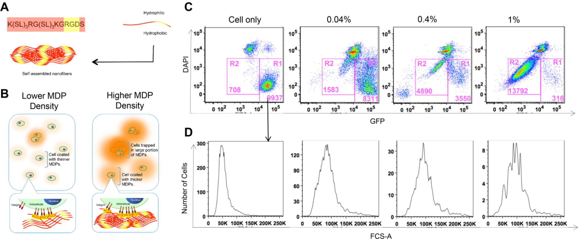
A one-step approach for coating thin layers of nanofiber matrices on cell surfaces by MDPs. (
Here, we demonstrate a one-step approach to align cells into multiple cell layers by varying the density of fabricating MDP hydrogel using a controlled ratio of MDP hydrogel to cells ( Fig. 1B ). Mouse dental pulp cells fabricated in various densities of MDP hydrogels were stained with CFSE and analyzed by using FACS. The R1 regions of Figure 1C reveal that at increased densities of MDPs, larger sizes of mouse dental pulp cells are present according to their forward scatter profiles when compared with MDP hydrogels with lower density ( Fig. 1C , D ). Increased coating layer thickness results in larger clusters of mouse dental pulp cells and a decreased fluorescent intensity emitted by each fabricated cell ( Fig. 1C , R2). The quantities of 1% and 0.4% w/v MDP-coated cells found at around 105 fluorescent intensity were greatly decreased by 26- and 2-fold, respectively, compared with 0.04% w/v MDP hydrogel-coated cells ( Fig. 1C , R1). The lowest density of fabricating MDP hydrogel, 0.04% w/v, also yields fewer hydrogel–cell aggregates and a lower cell-to-coating MDP ratio. In addition, the quantities of coated cells found in R1 were similar when cells were encapsulated with different densities of control peptide (CP) hydrogel without the integrin-binding sequence RGD ( Suppl. Fig. S1 and Suppl. Table S1 ). Increasing densities of fabricating CP hydrogel did not dramatically alter hydrogel–cell aggregates (R2) or the number of cells at 105 fluorescent intensity compared with 1% and 0.4% w/v MDP-coated cells, in contrast to the MDP containing the RGD sequence. Taken together, these data suggest that higher densities of the MDP scaffold lead to more nanofibers coating each cell surface, and that this is likely due to the presence of the RGD cell adhesion sequence contained within the MDP ( Fig. 1B ). Conversely, thinner coats of nanofibers on cell surfaces seemed to result from decreasing densities of fabricating MDP hydrogel to 0.04% w/v.
We next investigated the influences of MDP hydrogel density on the morphology and nanofiber–cell interaction by TEM and scanning electron microscopy (SEM). The high-magnification TEM and SEM images further confirm that MDP scaffolds coat cells with nanofibers, forming a microlayer around fabricated cells ( Fig. 2 ). Cell surfaces appear to be more heavily covered by this microlayer at higher densities of fabricating MDPs, 0.4% and 1% w/v ( Fig. 2C,DG,H,K,L ), compared with 0.04% w/v MDP ( Fig. 2B,F,J ), with cell surface structures still visible on the 0.04% w/v MDP-fabricated cells. Nanofibers of the 0.4% and 1% w/v samples form thicker microlayers around the cell surfaces, resulting in increased diameters of visualized cells ( Fig. 2K,L ). It also appears that cells fabricated in higher densities (0.4% and 1% w/v) of MDPs have greater disruption of their cellular organelles, suggesting greater levels of cellular stress ( Fig. 2C,D,G,H ). In contrast, the 0.04% w/v MDP nanofibers more sparsely attached to the cell surfaces ( Fig. 2B,F,J ), and their size and the morphology of the cells corresponded to unfabricated controls ( Fig. 2A,E,I ). These microscopy data are consistent with the FACS data ( Fig. 1C,D ), indicating that 0.04% w/v MDP produces a thinner coating on cell surfaces, which may be more favorable to normal cellular function. Thus, decreasing the density of fabricating MDP scaffolds could avoid an excessive thickness of cell coating, potentially improving cell viability and cell–ECM interactions.
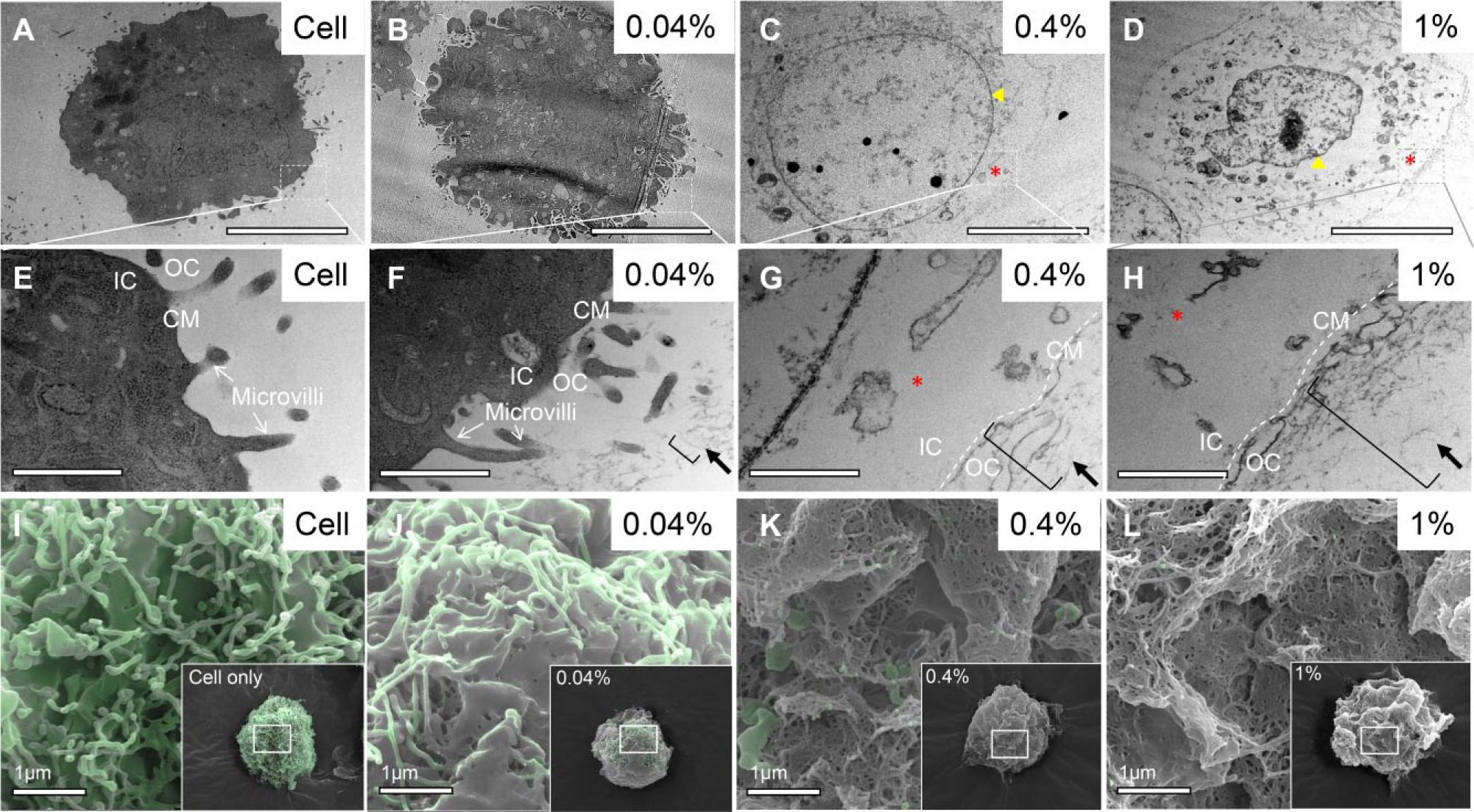
Higher-magnification surface SEM and TEM views of MDPs coating on mouse dental pulp cell surfaces. (
Cell Viability Is Inversely Proportional to the Density of MDP Scaffolds
TEM images indicate that a sizable proportion of the cells coated with higher densities of MDP (0.4% and 1% w/v) appeared ultrastructurally different from the unfabricated cells and cells fabricated with 0.04% w/v MDP. Alteration of morphology is seen in cells fabricated with the higher densities of MDPs (0.4% and 1% w/v), including a loss of intact cell membrane, abnormally shaped nuclei, and intracellular contents of ruptured organelles ( Fig. 2C,D,G,H ). In addition, we noted that cells aggregated and formed a number of large aggregates within the higher densities of MDP (0.4% and 1% w/v) tissue constructs ( Fig. 4B,C,E,F ).
These morphological alterations and structural changes observed in cells fabricated in higher-density MDP scaffolds suggest that excessive coating by MDP nanofibers might reduce cell viability. To assess the survival of cells in 3D tissue constructs with different densities of MDPs, apoptosis in fabricated cells was quantified with TUNEL assay. Apoptosis was additionally characterized using the cleaved caspase-3 (Asp175) antibody to detect the activated caspase-3 in dying cells. Taken together, the TUNEL and activated caspase-3 staining both suggest that higher densities of MDP scaffolds were associated with decreased cell viability in fabricated mouse dental pulp cells ( Fig. 3B–D,F–H ) when compared with lower density (0.04% w/v) MDPs ( Fig. 3A,D,E,H ).
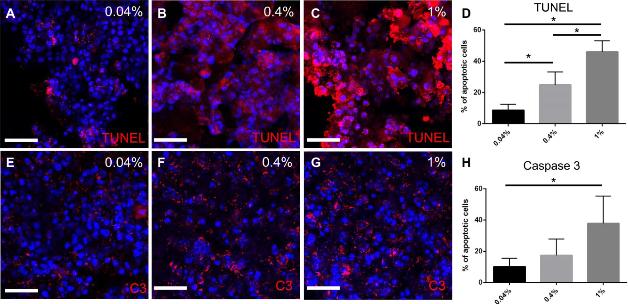
Cell viability of mouse dental pulp cells in 3D tissue constructs with different densities of MDPs. (
Thick layering of nanofibers at the cell surface could restrict nutrient delivery and waste removal at the cellular level, resulting in increased cell death. Diffusion of nutrients and gases at the center of 3D tissue constructs is typically decreased relative to the periphery due to the diffusional limitation in thicker tissue constructs.33–35 Therefore, 3D tissue construct systems that enable nutrients and gas exchange homogenously within the 3D tissue constructs are highly desired. In the cross section images of 0.04% w/v MDP tissue constructs ( Fig. 3A,E ), most of the cells were vital either from the periphery or toward the center areas of the constructs. These data suggest that the lower density of MDP in the 3D tissue constructs caused the system to deliver the nutrient and remove the waste in a more efficient way compared with the higher densities of MDP.
Lower-Density (0.04% w/v) MDP Facilitates the Rapid Formation of Aligned and Multilayered 3D Cell Constructs
Data collected by FACS and electron microscopy shown in
Figures 1
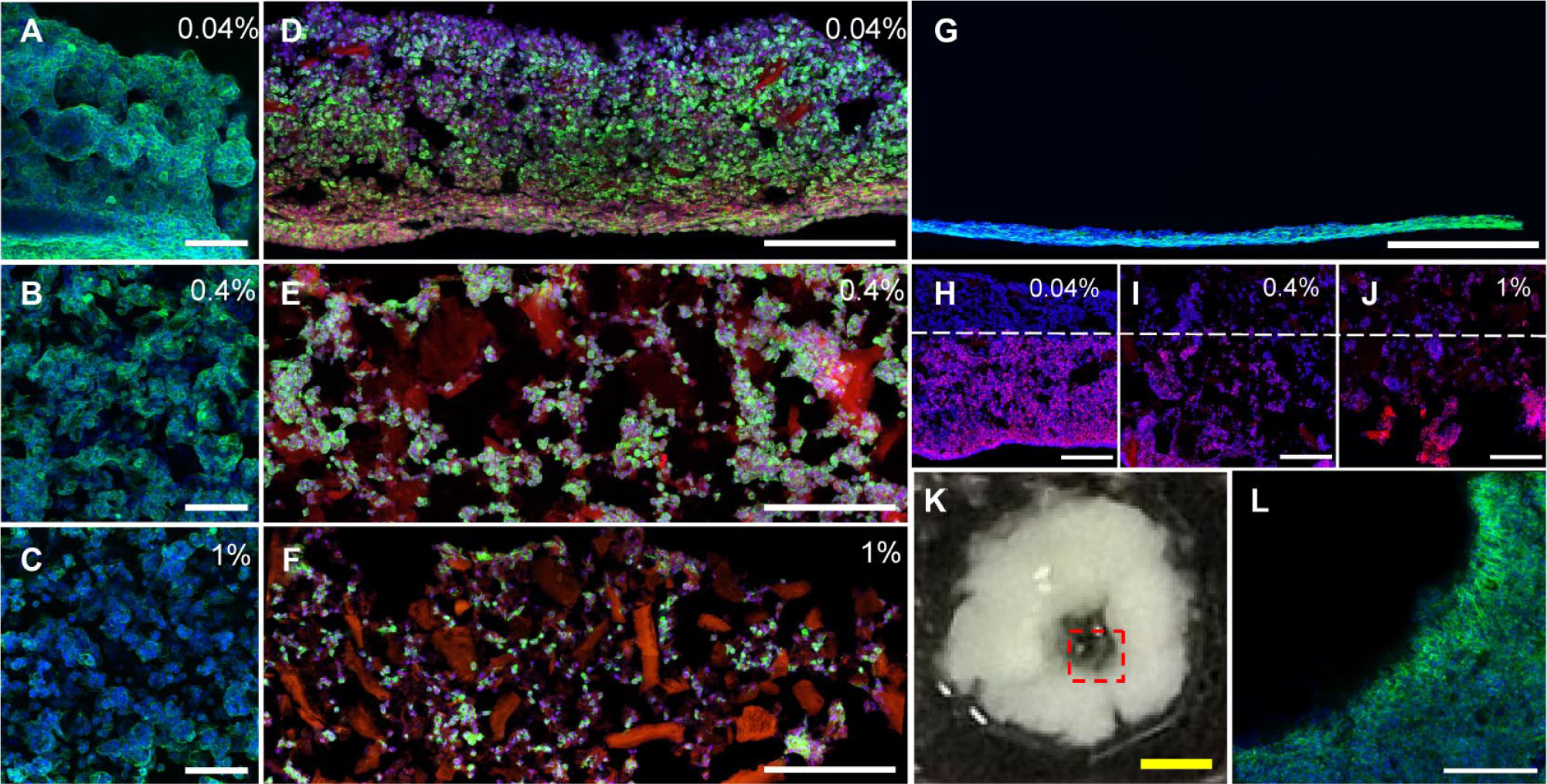
Cellular morphology and alignment of murine cell lines in 3D tissue constructs with different densities of MDPs. (
At low-density (0.04% w/v) MDP, organized arrangements of cells are formed in a rapid, controllable, and reproducible fashion. We also attempted a serial dilution of fabricating MDPs and found that aligned and multiple layered 3D tissue constructs can be formed efficiently in the range from 0.08% to 0.03% w/v MDP with 1.25 × 10⁷ cells/mL (data not shown). Similar numbers of cells fabricated with densities of MDPs more than 0.1% w/v resulted in the formation of more uneven cell distribution and cell aggregation within the layered scaffolds (data not shown). Control experiments using CP nanofibers at 0.04% w/v exhibited similar patterns of cell layering as the higher densities of MDPs (0.1% and 1% w/v), indicating the significance of the cell adhesion sequence ( Suppl. Fig. S2A,C ). These results suggested that low-density MDP offers advantages in fabricating 3D tissue constructs for potential applications in tissue regeneration. Excessive coating by MDP, in contrast, generating a higher level of nanofiber-to-cell interaction, may result in the disruption of cellular interactions among the neighboring cells and surrounding microenvironment.4,5
Here we have demonstrated that 3D multilaminar cell constructs can be generated by coating cells in low-density MDP. This raises the intriguing possibility that more complex patterns, such as microscale vascular structure, could also be constructed in the more favorable extracellular environment provided by lower-density MDP scaffolds. Thus, mouse dental pulp cells were fabricated with 0.04% w/v MDP and molded around a glass capillary tube. Confocal imaging reveals that cells associated with lower-density MDP align and can be molded into a tubular structure with an inner diameter of 1 mm ( Fig. 4K ). Furthermore, these cells formed multiple layers with a specific organization of the actin filament networks ( Fig. 4L ).
Tissues found in vivo consist of alternate layers of cells, which often have varying cell density and ECM physiochemical properties. Thus, we set up a simple co-culture experiment to observe the 3D interactions of mouse dental pulp cells with NIH3T3 fibroblasts. MDP-fabricated mouse dental pulp cells prestained red with Qtracker were layered on top of MDP-fabricated NIH3T3 fibroblasts. Immunofluorescence staining reveals a clear boundary line between the two tissue layers in the lower-density (0.04% w/v) MDP construct ( Fig. 4H ). Additionally, cellular alignment and cell distribution on both sides of the boundary line appear to be more profound and homogeneous when using 0.04% w/v MDP ( Fig. 4H ) compared with 0.4 or 1% w/v MDP, which had a poorly defined boundary line between the two scaffold layers and a more uneven cell distribution within the layered scaffolds ( Fig. 4I,J ).
Here, we demonstrate that such a complex tissue architecture can be achieved by alteration of fabricating MDP scaffold density, which presents particularly intriguing possibilities in establishing co-culture systems with multiple cell types and/or bioactive factors. Growing single or different cell types in 3D with low-density (0.04% w/v) MDP scaffolds provides several advancements to re-create such tissue structures; for example, it can provide a platform to study different cell–cell interactions, which more precisely mimic the complex in vivo microenvironment.
Fluid-Like Low Density of MDP Fabricated on Cell Surfaces Provides Structural Support and Spaces for Cell Attachment and Organization
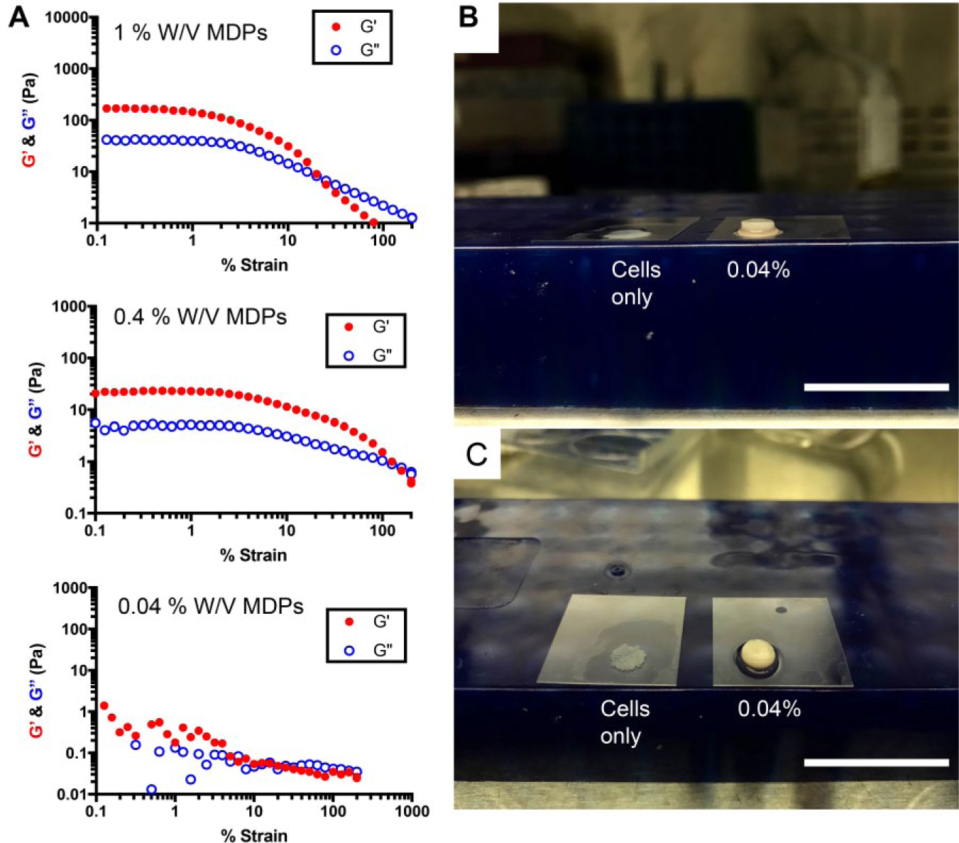
The viscoelastic property of MDPs at various densities, 0.04%, 0.4%, and 1% w/v, was explored by rheological studies. Strain sweep oscillation tests were performed in order to determine the linear viscoelastic region (LVR). The average storage modulus (G′) for 0.4% and 1% w/v MDPs is around 20 and 200 Pa, respectively (
Fig. 5A
). These results indicate that increased densities of MDPs are more “solid,” mechanically speaking (greater G′, greater strength). Since it has been shown that cellular responses to matrix stiffness are different among different cell types, it is very likely that MDP could be applied as a substrate for different cell types due to its ability to create a wide range of stiffness by simply adjusting the density of the encapsulating MDP scaffold. The flowable behavior of MDPs was observed at 0.7 and 25 Pa for 0.4% and 1% w/v MDPs, respectively (
Fig. 5A
). These results suggest that both 0.4% and 1% w/v MDPs can tolerate a wide range of shear stress and still behave as a hydrogel. It is interesting to note that the irreversible network deformation of MDPs is highest for the softer hydrogels, 0.4% w/v. It is not surprising that 0.04% w/v exhibits fluid-like behavior, having G′ and G′ values that are close to zero (
Fig. 5A
). However, decreasing the ratio of nanofibrillar elements to 0.04% w/v is still sufficient to hold and support cells growing into 3D structures, based on the observations from cell culture experiments. Low-density MDP scaffolds fabricated on cell surfaces appear to provide structural support and spaces for cell attachment and organization. Unlike unfabricated controls where cells sediment due to gravity, cells fabricated with 0.04% w/v MDP nanofibers can stack and form multilayered tissue-like structures (
Figs. 4A,D
(
Lower-Density (0.04% w/v) MDP Scaffolds Promote the Upregulation of Genes Related to Cell Proliferation, Angiogenesis, and ECM Remodeling
In agreement with the view that structure ultimately relates to function, so too is cell distribution and alignment crucial to the functionality of engineered tissues.15,36 As MDP scaffolds are formed from amino acid subunits mimicking native ECM, cells can locally degrade and remodel these scaffolds, and it is expected that such cellular interactions will influence gene expression profiles. Therefore, cells fabricated with different densities of MDP were analyzed in terms of their expression profiles of genes involved in proliferation, angiogenesis, and ECM remodeling by qPCR. Mouse dental pulp cells fabricated with lower-density (0.04% w/v) MDP scaffolds promote numerous gene expression profiles related to proliferation, angiogenesis, and ECM remodeling, including Fgfr2, Vegf a, Col1 a1, Mmp2, Opn, and actin, compared with higher densities of MDP (0.4% and 1% w/v) ( Fig. 6 ).
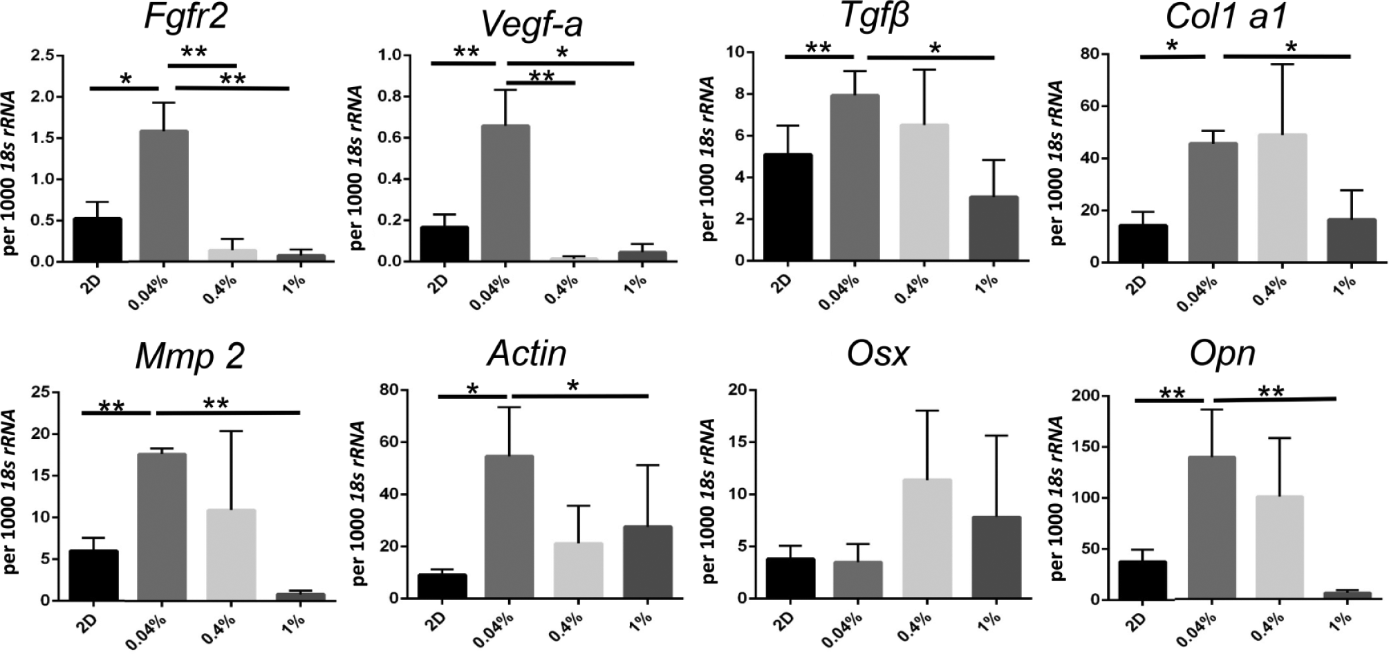
Surface thickness and structural organization influence gene expression profiles of mouse dental pulp cells. Quantification of gene expression by qPCR. Mouse dental pulp cells were cultured for 3 days as described in the text and harvested for RNA extraction. Real-time polymerase chain reaction was performed and total cDNA from each sample was normalized to 18s rRNA. N = 5 was used for each experimental group. Data are given as mean ± SD. *p < 0.05; **p < 0.01.
As the MDP scaffolds include an incorporated matrix metalloprotease-2 (Mmp2) vulnerable cleavage site (LGR), 36 it should facilitate the ultimate degradation of hydrogel scaffolds in these 3D cell constructs, as fabricated cells would be expected to remodel their extracellular environment. Interestingly, expression of Mmp2 and Col1 a1 is upregulated in the 0.04% w/v hydrogel scaffolds ( Fig. 6 ), suggesting that cells can locally degrade and remodel the MDP-based matrix more efficiently when fabricated with lower-density hydrogel scaffolds.
We also found that cells fabricated in lower-density (0.04% w/v) MDP show a significant increase in gene expression of Opn ( Fig. 6 ), which is a phosphoprotein component of bone and dentin natively synthesized by osteoblasts and odontoblasts, both of which may derive from the mesenchymal lineage of the mouse dental pulp cells. Opn has various diverse cellular functions, including modulating cell adhesion, migration, and survival. It also has been shown to play a positive role in regulating wound healing as well as ECM remodeling.37–39 Specifically with regard to its role in ECM remodeling, its enhanced expression highlights the importance of low-density MDP-based tissue constructs that are capable of providing structural support and mediating biological cues for ECM synthesis.
Conclusion
Here we provide a rapid and simple method to generate 3D cellular constructs by lowering the density of fabricated MDP scaffolds. Additionally, we offer insights into the nature of the interactions between cells and these biomimetic scaffolds. This approach does not require multiple steps and additional cross-linking processes to create networks of layered cells in 3D. Hence, the model provides an interesting platform for tissue regeneration research but also is potentially useful for future clinical translation, such as the creation of complex tissue structures. At a lower density (0.04% w/v), the MDP scaffold appeared to provide an optimal coating ratio of cell to nanofibers, which not only promotes cell proliferation and viability but also allows the formation of 3D multilaminar cell constructs. Additionally, lower-density MDP scaffolds upregulate the expression of genes involved with tissue growth and remodeling compared with denser MDP scaffolds. These effects may well be due to the RGD domain present on MDP nanofibers driving efficient coating of scaffold-fabricated cells, with biological effects influenced by the ratio of coating nanofibers to cells. These findings are particularly important when considering the control over the composition of single or multiple cell types within a proper ECM microenvironment in order to recapitulate native tissue architecture and functions when broadly considering tissue engineering applications both for the oral cavity and, more broadly, in the body.
Supplementary Material
Supplementary Material, Supplemental_Info – A Single-Step Self-Assembly Approach for the Fabrication of Aligned and Multilayered Three-Dimensional Tissue Constructs Using Multidomain Peptide Hydrogel
Supplementary Material, Supplemental_Info for A Single-Step Self-Assembly Approach for the Fabrication of Aligned and Multilayered Three-Dimensional Tissue Constructs Using Multidomain Peptide Hydrogel by Yinshen Wee, Amanda N. Moore, Shihai Jia, Jing Zhou, John S. Colombo, and Rena N. D’Souza in SLAS Technology
Footnotes
Acknowledgements
We thank Linda Nikolova for helping with TEM and Dr. Paulo Perez for helping with SEM.
Supplemental material is available online with this article.
Declaration of Conflicting Interests
The authors declared no potential conflicts of interest with respect to the research, authorship, and/or publication of this article.
Funding
The authors disclosed receipt of the following financial support for the research, authorship, and/or publication of this article: This work is greatly supported by NIH R01DE021798 and NIH F32DE027866. The PIs for NIH R01DE021798 were Jeffrey Hartgerink and Dr. Rena N. D’Souza, while F32DE027866 supports Yinshen Wei, co-mentored by Rena D’Souza and John Colombo.
References
Supplementary Material
Please find the following supplemental material available below.
For Open Access articles published under a Creative Commons License, all supplemental material carries the same license as the article it is associated with.
For non-Open Access articles published, all supplemental material carries a non-exclusive license, and permission requests for re-use of supplemental material or any part of supplemental material shall be sent directly to the copyright owner as specified in the copyright notice associated with the article.