
Abstract
Introduction
Workers and students are often stressed and distressed by a variety of factors, including work and study. However, support for individual psychological stress is not yet well established.
Objective
This study aimed to evaluate the effect of mindfulness breathing on the mental load of college students using an electroencephalogram (EEG).
Methods
Twenty participants were randomly allocated into treatment or control groups, with 10 participants in each group. Mindfulness breathing was applied to participants in the treatment group, while those in the control group received no treatment. The regression equation was evaluated from the EEG, and time series analysis was performed based on autocorrelation and partial autocorrelation.
Results
In the After condition after mindfulness breathing exercises in the Mi group, the alpha wave content of the regression equation at eye closure after task performance showed an upward trend, and the autocorrelation coefficient showed repeated upward and downward fluctuations.
Conclusion
It was suggested that alpha wave content may increase over time with mindfulness breathing exercises. The EEG after mindfulness breathing exercises was shown not to be constant and to have non-linear characteristics. This suggested that the effects of mindfulness breathing exercises could be evaluated using time series data.
Introduction
Many findings have been reported on the stresses and worries of workers and students and their factors (Kou, 2020; Otaki, 2019). However, it is difficult to say that support for individual psychological stress has been established. Recently, psychotherapy based on mindfulness has been attracting attention as an intervention to relieve stress, improve brain function, and reduce recurrence of depression and other disorders (Kabat, 1990; Probst et al., 2020; Segal et al., 2002).
Review of Literature
Theoretical Framework
Physiological studies on the effects of mindfulness have revealed a relationship with the amygdala and prefrontal cortex (Murakami, 2012). Research on brain networks suggests that mindfulness training may be associated with changes in brain white matter (Tang et al., 2012), and there are reports of increased grey matter density with mindfulness training (Hölzel et al., 2011). Empirical studies of mindfulness with university students have reported an interaction between mindfulness and academic assessment using interviews (Bóo et al., 2020). Previous studies have mainly analyzed brain structural changes, such as changes in brain networks, due to mindfulness interventions, or evaluated mindfulness using subjective measures of subjects. An unresolved point in these studies is the evaluation of the effect of mindfulness breathing exercises during mental workload and its influence over time.
As an analysis of time series data, it has been reported that there is a temporal effect on EEG after emotional stress loading using regression analysis (Mizuno-Matsumoto et al., 2012). A study investigating the effective duration of EEG alpha wave responses to sounds from neighbors, using an autocorrelation function, found significant differences between the sounds heard (Shigeyasu, 1994).
The novelty of the present study is to evaluate the effects of mindfulness breathing exercises on EEG changes over time during mental workload in nursing students and to estimate the relationship between the effects of mindfulness breathing exercises and time.
Objective
Therefore, in order to investigate the correlation between the effects of mindfulness breathing exercises and time, this study aims to capture changes in EEG over time using regression equations and autocorrelation and partial autocorrelation coefficients calculated based on the EEG of the subjects who applied mindfulness breathing exercises and those who did not apply mindfulness breathing exercises.
Method
Design
The design of the study used a randomized controlled trial.
Research Question
As a hypothesis, we hypothesized that subjects who practiced mindfulness breathing exercises would show characteristics of EEG when compared to subjects who did not practice mindfulness breathing exercises.
Sample
Subjects were 20 healthy university students (mean age: 21.5 ± 0.9 years). Subjects were divided into two groups: a mindfulness implementation group (hereinafter referred to as the “Mi group”) and a mindfulness non-implementation group (hereinafter referred to as the “nMi group”). The number of subjects in each group was randomly assigned, with an equal number of 10 subjects in each group.
Inclusion/Exclusion Criteria
Two criteria for inclusion of subjects were set: the criteria of the subject and the criteria of the experimental environment. The exclusion criteria for subjects were students with previous meditation and mindfulness experience. The experimental environment was measured in the same laboratory at the university to ensure that the experimental conditions for both groups were as identical as possible. The environmental settings were set at the time of implementation from 16:00 to 19:00 and a room temperature of 24 °C in order to reduce the influence of circadian cycles and temperature differences on autonomic function.
Institutional Review Board Approval
Before the study began all subjects were fully informed of the purpose and objectives of the study, protection of their privacy, anticipated risks, the right to withdraw during the course of the study and no disadvantages associated with this, publication of the results, guarantee of anonymity and voluntary participation, and that the results would not be used for any purpose other than research, and consent was obtained in writing. The EEG measurement experiment was conducted in a private room, one by one, to protect privacy.
This study was approved by the Ethics Review Committee of Hyogo University, to which the author belongs, and was conducted from May 2019 to February 2020 (approval number 19001).
Mindfulness
The term mindfulness is derived from Buddhist scriptures and is understood to be central to the practice of meditation (Dreyfus, 2013). In recent years, mindfulness has become a psychotherapy that is expected to be effective for stress reduction, relaxation, attention, and awareness. The definition of mindfulness in the Mindfulness-Based Stress Reduction (MBSR) method used in this study is “to focus one's awareness on the present moment, the ‘now’” (Kabat, 1990). In this study, the author practiced mindfulness breathing exercises using an audio guide created by the author using Windows Movie Maker®, based on the MBSR and Mindfulness Meditation Guide CD developed by Kabat (1990). The procedure of the mindfulness breathing technique was as follows: (1) Sit in a chair and assume a meditative posture. (2) Take abdominal breaths and focus attention on the movement of the breath. (3) When the mind is able to focus on the breath, expand the awareness to each part of the body. (4) When the mind leaves the breath, refocus attention on the sensation of the breath. The duration of the mindfulness breathing exercise in the laboratory was 5 min.
Previous studies have reported that reduced time in MBSR can also reduce psychological distress in the same way as in the standard format (Carmody & Baer, 2009). In addition, previous studies have reported effects on eating behavior in brief mindfulness interventions of 5 min (Miyata & Akatsuka, 2022). Therefore, we considered that it would be possible to identify differences between groups in short-time mindfulness practices. The duration of mindfulness breathing exercises in the laboratory was set at 5 min, with the exception of practice.
Physiological Evaluation Experiments
Experimental Structure. In this study, EEG evaluation experiments based on the experimental protocol were conducted in a university laboratory on both the Mi and nMi groups of subjects. The experimental protocol for this study is shown in Figure 1. The experimental protocol consisted of three parts: before validation (Before), validation (Control), and after validation (After). In the Before part of the experimental design, the participants performed the closed eyes task (closed eyes 1: CE1) for 3 min before the mental arithmetic task, the arithmetic task (arithmetic: AC) for 3 min, and the closed eyes task (closed eyes 2: CE2) for 3 min after the mental arithmetic task. This set was performed twice before the Control task. The control was a cross-shape (CS) task on a PC screen for the nMi group, while the Mi group performed mindfulness breathing exercises.

Experimental protocol.
The AC task consisted of keying answers to a randomly generated 10–99 two-digit addition task in Python on a PC screen. Subjects were told to answer the addition task quickly within 3 min.
Before the start of the experiment, the Mi group practiced voice-guided mindfulness breathing exercises, while the nMi group practiced voice-guided cross gazing. Both groups also practiced how to operate a computer-based mental arithmetic task before starting the experiment.
EEG Measurement. A portable electroencephalograph (EEG) was used for the EEG measurement (Figure 2) to minimize the subject's stress caused by the time and effort required for electrode placement. The reference electrodes were set at both earlobes, and the probe electrode was set at the occipital vertex (Oz) of the occipital area with reference to the international 10–20 method (Hitomi & Ikeda, 2014). The sampling frequency was 128 Hz, and the impedance between skin electrodes was kept below 10 kΩ.
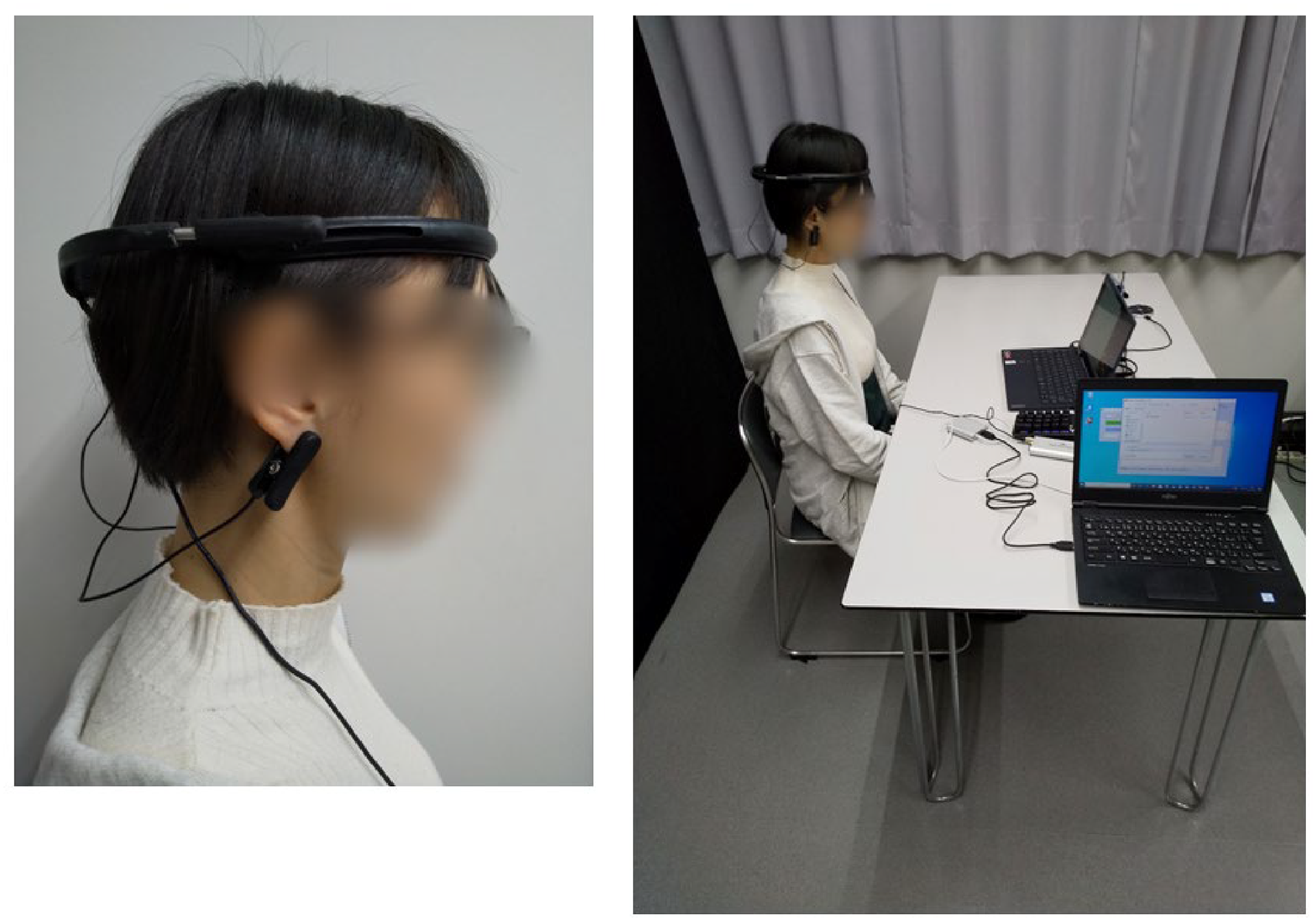
EEG measurement.
Statistical Analysis
EEG data were subjected to frequency analysis using the fast Fourier transform (FFT) at a frequency resolution of 1 Hz every second. The FFT transforms the waveform f(t), a function of time t, into a distribution function F(k) at frequency k. The window function is shown in Equation (1) using a Hanning window (Kotari, 2018).

For the statistical analysis, regression and time series analyses were conducted to examine the effects of mindfulness over time. In the regression analysis, the dependent variable was the alpha and beta wave contents of the EEG, y, and the explanatory variable was time, x, to analyze the correlation between the EEG and time. For the time series analysis, autocorrelation and partial autocorrelation analyses were used to obtain the autocorrelation coefficient ρ and partial autocorrelation coefficient Φ. Autocorrelation was analyzed using data (t), which varied with time t, a reference point t, and data shifted from t to lag k. The autocorrelation coefficient ρ and partial autocorrelation coefficient Φ of the time series data take values ranging from 1 to −1. Higher values of ρ and Φ indicate higher correlation with the reference point. In this study, the reference point was the alpha wave band content during the first 10–20 s of the task, and the time series data were analyzed with lag 16.
Results
Sample Characteristics and Research Question Results
Evaluation by Regression Analysis. Table 1 shows the coefficients of determination and probability of significance of the regression analysis of the alpha and beta wave contents of the Mi group EEG. The alpha wave content of the Mi group showed a significant trend in the CE1 and CE2 tasks in the After condition (p < .1). However, the prediction accuracy of the coefficient of determination was low. Figure 3 shows the frequency distribution of the alpha wave content in the Mi group in the After condition, where significant trends were observed in the CE1 and CE2 tasks. The horizontal axis indicates time, and the vertical axis indicates the content of alpha waves. The alpha wave content in the Mi group on the first day of the Mi task showed a negative correlation at about 55% in the After condition of CE1 and a positive correlation from about 55% to positive in the After condition of CE2.
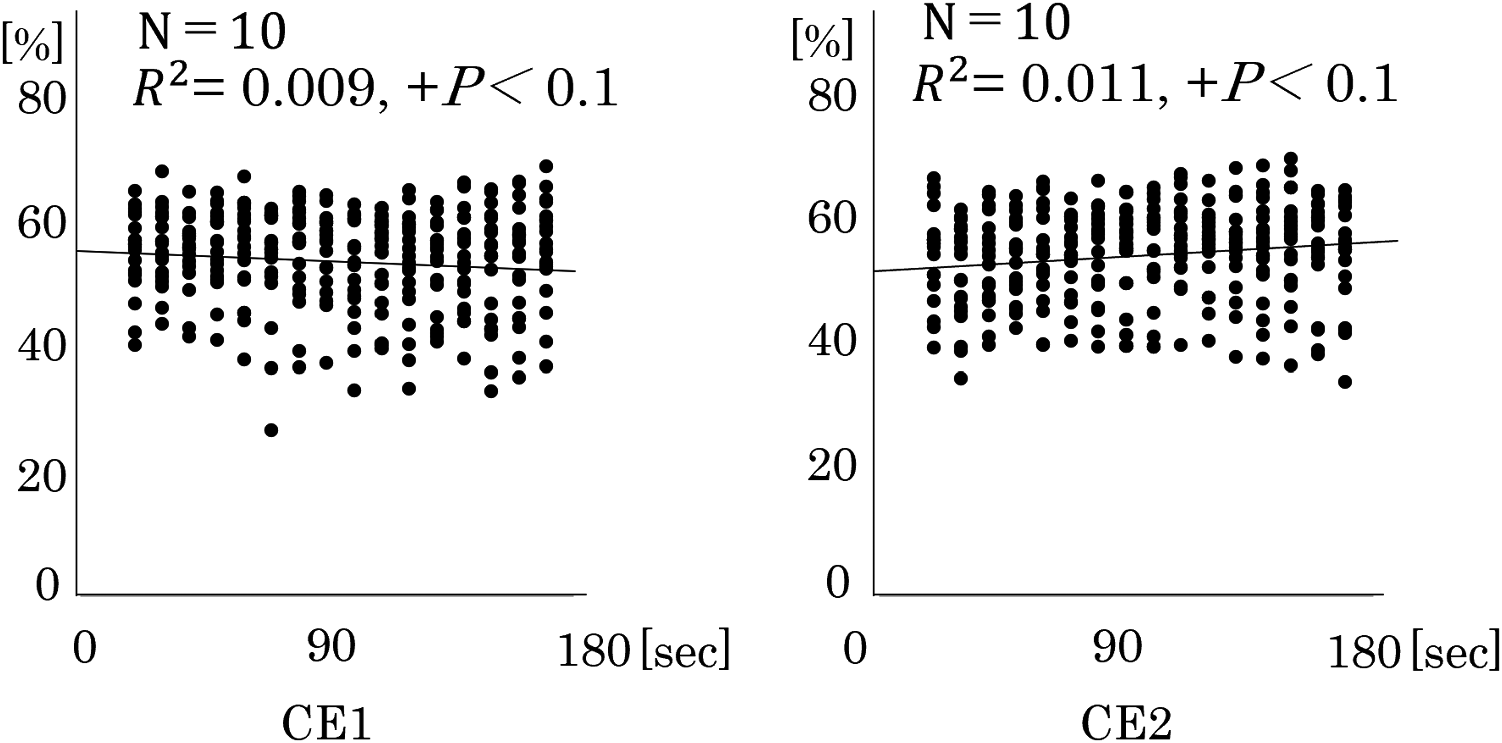
Distribution of the alpha wave content.
Regression Analysis of Mindfulness Breathing Practice Groups.

Before = before validation; After = after validation; CE1 = closed eyes 1; CE2 = closed eyes 2.
p < .1.
Table 2 shows the regression coefficients of determination and probability of significance for the alpha and beta wave contents of the EEG in the nMi group. The nMi group showed a significant difference in the Before condition of the beta wave content in the CE1 task (p < .05). However, the prediction accuracy of the coefficient of determination was low. Figure 4 shows the frequency distribution of the beta wave content in the nMi group in the Before condition, where a significant difference was found in the CE1 task. The horizontal axis indicates time, and the vertical axis indicates beta wave content. The beta wave content moved from approximately 20% to a negative correlation in the beta wave content on day 1 of the nMi group in the Before condition of CE1.

Distribution of the beta wave content.
Regression Analysis of the Mindfulness Breathing Non-practice Group.
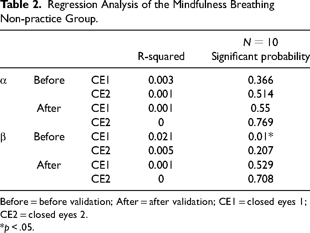
Before = before validation; After = after validation; CE1 = closed eyes 1; CE2 = closed eyes 2.
p < .05.
Evaluation by time series analysis. Figure 5(a) and (b) shows the correlograms from the alpha and beta wave band contents on day 1 for the Mi and nMi groups. The horizontal axis shows the lag at which the autocorrelation was calculated, and the vertical axis shows the autocorrelation coefficient ρ.

Autocorrelation. (a) Autocorrelation of the alpha wave content. (b) Autocorrelation of the beta wave content.
The autocorrelograms of the alpha and beta band contents of the Mi and nMi groups showed positive autocorrelations. In the alpha and beta wave band contents of both Mi and nMi groups, the ρ of the Before CE1 and CE2 showed a downward trend; in the alpha and beta wave band contents of the Mi group, the ρ of the After CE1 showed a downward trend and remained almost flat from lag 9; in the alpha and beta wave band contents of the nMi group, the ρ of the After CE1 showed a downward trend. In the alpha wave band content of the Mi group, the ρ of the After CE2 showed a downward trend overall, with an increase of about 0.1 in the ρ at three lags.
Figure 6(a) and (b) shows the partial autocorrelation plots created from the alpha and beta wave band contents of the Mi and nMi groups on day 1.The partial autocorrelation coefficients Φ in the before and after plots for the Mi and nMi groups fell to uncorrelated values at lag 2 or lag 3 and showed no correlation after lag 3.

Partial autocorrelation. (a) Partial autocorrelation of alpha wave content. (b) Partial autocorrelation of beta wave content.
Discussion
Evaluation by Regression Analysis
In the After condition after mindfulness breathing exercises in the Mi group, the content of alpha waves was positively correlated with CE2 after the task performance, and the regression equation for CE2 showed an increasing trend in the content of alpha waves. The activity in the alpha wave frequency band was suppressed by eye opening and visual stimulation, whereas it was enhanced during internal tasks such as mental arithmetic and working memory (Avram et al., 2010; Palva & Palva, 2007). This suggests that mindfulness breathing exercises affected the power spectrum values of alpha waves.
In the Before condition of the nMi group, there was a significant difference in the content of the beta wave. A previous study reported that beta wave activity was enhanced during actions with visual attention (Wróbel, 2000). Since tension may generally occur at the beginning of an experiment, it is considered that the activity of beta waves, which affects attention and concentration, was affected at the beginning of the experiment.
In the After condition after mindfulness breathing exercises in the Mi group, the alpha wave content tends to be significant, but further validation is needed because the predictive accuracy of the regression equation is low based on the results of the coefficient of determination.
Evaluation by Time Series Analysis
The results of the autocorrelation coefficients of the Mi and nMi groups showed that the autocorrelation coefficients decreased as the lag increased, and the autocorrelation coefficients showed a downward trend without large fluctuations at a specific lag. The results of the autocorrelation coefficients for the Mi and nMi groups suggest that the correlation between the EEG and time has decreased.
In the CE2 condition of the After condition of the alpha wave content in the Mi group after the validation task, the autocorrelation coefficient showed fluctuations that repeatedly rose and fell. As a simple task response, the time of beta wave activity in the cortex is said to be prolonged (Takayose et al., 2008). This suggests that the task prolonged the duration of the beta wave activity and affected the alpha waves during eye closure. In addition, previous studies have reported the possibility of a temporary functional response in the brain when emotional stimuli are large or persist for a long time (Mizuno-Matsumoto et al., 2012). In the alpha waves after mindfulness breathing exercises, it is possible that the brain activity was affected by the passage of time.
The result that the partial autocorrelation coefficient fell significantly at lag 2 or lag 3 indicates that the EEG frequency content is affected by the previous EEG only at lag 2 or lag 3. It is also suggested that the influence was small for the frequency content of the EEG after lag 3. The EEG has been shown to be affected by conditioned responses such as light stimulation (Mimura, 1955).
Partial autocorrelation coefficients are calculated by excluding the influence of values at time points prior to a given point in time. In the present experiment, it is considered that no influence between the data could be extracted in the condition of resting and closed eyes for both groups.
These results suggest that the EEG after mindfulness breathing exercises does not remain constant but rather transitions with non-linear features that showed an influence on the EEG.
Strengths and Limitations
The present study investigated the short-term effects of mindfulness breathing during an experiment. Under the conditions in which the experiment was conducted, it was possible to assess the possibility that the implementation of mindfulness breathing exercises could show an effect on the EEG rather than the EEG remaining constant. Therefore, the duration of mindfulness breathing exercises needs to be verified in the future.
Implications for Practice
Under the conditions of the present study, the EEG was shown to change over time with the implementation of mindfulness breathing exercises, as hypothesized. This could lead to a better understanding of the effects of mindfulness breathing exercises and the possibility of developing more effective interventions.
Conclusions
The results of the regression equation suggest that mindfulness breathing techniques may increase the alpha wave content over time.
The results of the autocorrelation and partial autocorrelation coefficients also suggest that mindfulness breathing techniques may make brain activity more irregular and affect the EEG after a task performance.
These results suggest that the EEG after mindfulness breathing exercises does not remain constant but rather transitions with non-linear features that showed an influence on the EEG.
Footnotes
Acknowledgements
The author would like to thank Junko Kasai for her help in photographing the experimental scenes during the preparation of this paper.
Declaration of Conflicting of interests
The author declared no potential conflicts of interest with respect to the research, authorship, and/or publication of this article.
Funding
The author disclosed receipt of the following financial support for the research, authorship, and/or publication of this article: This research was supported by JSPS Grant-in-Aid for Scientific Research 21K10566.