
Abstract
Background:
Anterior cruciate ligament (ACL) injuries are common among female soccer players and often result in long-term absence from sport. Despite extensive studies on ACL injury mechanisms, no prospective studies have investigated the distribution of dynamic plantar loading during running in players who later sustain ACL injuries.
Purpose:
To prospectively investigate the characteristics of plantar pressure during running in female soccer players who sustained ACL injuries.
Study Design:
Case-control study; Level of evidence: 3.
Methods:
A total of 121 female high school and collegiate soccer players with no history of ACL injury were enrolled. Dynamic plantar loading distribution during running was measured at baseline. Participants were monitored for 1 year and classified into an ACL injury group (ACL group) or a control group (CON group) with no injuries. Plantar pressure was analyzed across 6 regions of the foot: medial and lateral regions of the forefoot, midfoot (metatarsal), and hindfoot. Cumulative load, peak force, and the medial-to-lateral force ratio were compared between the groups.
Results:
The ACL group (11 players, 13 limbs) demonstrated significantly lower cumulative loading at the first and second medial metatarsal regions compared to the CON group (100 players, 200 limbs). The median cumulative load (N/kg) adjusted for body weight at the first metatarsal was 158.46 in the ACL group and 208.97 in the CON group (P = .01). At the second metatarsal, the values were 127.76 and 153.23, respectively (P = .04). The medial-to-lateral force ratio (%) was significantly lower in the ACL group (82.5%) than in the CON group (97.3%, P = .03).
Conclusion:
Female soccer players who sustained ACL injuries exhibited reduced medial metatarsal loading and a lower medial-to-lateral plantar force ratio during running. These findings suggest that altered plantar loading patterns may contribute to ACL injury risk.
Anterior cruciate ligament (ACL) injuries often occur under conditions of high dynamic loading on the knee joint during sports activities, with particularly high rates reported among female athletes.1,3 These injuries can be career-threatening due to prolonged absence from play, low rates of return to the preinjury performance level, and an increased risk of subsequent knee injuries after resuming sports.2,10,40,41 Female athletes are at a significantly higher risk of ACL injury than their male counterparts, with reported incidence rates 2 to 8 times greater1,3,11-13,19 and considerably lower return-to-sport rates. 18 Notably, female soccer players are at a higher risk of ACL retear after surgery than male soccer players. 21 This increased risk is attributed to various anatomic, neuromuscular, and hormonal factors, including increased ligament laxity, altered muscle activation patterns, and reduced postural stability.22,29,34
Given the growing participation of women in soccer,7,10 reducing ACL injury risk in this population is of considerable interest. Intrinsic risk factors include generalized joint laxity, 39 differences in bony anatomy,32,33,39 decreased fatigue resistance, 16 imbalanced hip abductor strength,15,17,28,31 and altered landing and cutting mechanics—often characterized by dynamic knee valgus.14,22 Furthermore, neuromuscular imbalances—such as a relatively weaker hamstring-to- quadriceps strength ratio 37 —contribute to improper force distribution during dynamic movements. Soccer demands frequent acceleration, deceleration, cutting, jumping, and landing. Several studies have reported associations between lower extremity injuries and dynamic plantar loading patterns during these agility movements.35,42 However, despite extensive research into ACL injury mechanisms and risk factors, no prospective studies have examined the characteristics of plantar loading during running in female soccer players who later sustain ACL injuries. Analyzing dynamic foot-loading patterns in female soccer players may reveal potential biomechanical differences that contribute to ACL injury risk. Therefore, this study prospectively compares the plantar force distribution between female soccer players who sustained an ACL injury and those who did not. Our hypothesis was that female soccer players who sustain an ACL injury may have an altered medial-lateral plantar loading distribution that affects lower extremity stability, compared to players who do not sustain an ACL injury.
Methods
Study Design and Participants
This prospective cohort study examined Japanese female high school and college soccer players during the 2018 to 2020 seasons. A total of 121 competitive female soccer players from 1 high school and 1 collegiate team (mean age of 17.2 ± 1.9 years) were included. Both teams were competing at the national level for their age group.No athletes participated in any other sports throughout the season; thus, there was no multisport participation among the participants. Data were collected during a preseason medical assessment each year, and participants were monitored for injuries during a single soccer season. All participants received a preseason medical assessment and completed a baseline questionnaire regarding injury history. They were then followed up for a year. Players with a history of ACL injury (n = 10) were excluded from the analysis. No participants were lost to follow-up during the injury registration period. Hence, 111 players were included in the analysis (Figure 1). Preseason measurements of players who subsequently sustained an ACL injury were compared with those of players who did not sustain an ACL injury. The latest data prior to the ACL injury were used for players who participated in the medical assessment more than once. All measurements were conducted at our institution. This study was a part of the Prospective Study of Predictors of Sports Injuries under the U Tokyo Sports Science Institute Sports Injury Prevention Project.26,27,35-37 The study protocol was approved by the institutional review board of our institution (11907-2), and all participants provided informed consent prior to enrollment.

Flow diagram of the participant selection process.
Measurements
The following measurements were performed during the preseason medical assessment.

Plantar pressure measurement while running on a flat 10-m course using the Footscan system.

Ten foot regions divided by the Footscan system.
Statistical Analysis
Players diagnosed with ACL injuries during the 1-year follow-up were assigned to the ACL group, whereas players who did not have an ACL injury were assigned to the control (CON) group.Differences in the preseason measurements between the ACL and CON groups at baseline were compared using either the unpaired t test or the Mann-Whitney U test, depending on the normality of the data (as determined by the Shapiro-Wilk test). Variables compared included age, height, BW, BMI, percentage body fat, and soccer experience. The foot–arch height ratio and plantar loading parameters during running were compared between the injured limb in the ACL group and the bilateral limbs in the CON group.For subanalysis, the correlation between age and medial-to-lateral force ratio was analyzed using Pearson’s product-moment correlation. All statistical analyses were conducted using IBM SPSS Statistics for Windows version 26.0 (SPSS) and version 11.0 (SPSS). Statistical significance was set at P < .05.
Results
During the 1-year follow-up, ACL injuries occurred in 13 knees of 11 players (Figure 1). Among the 11 injured players, 2 sustained a contralateral ACL injury after a primary ACL injury. Therefore, the measurements of both injured limbs of these players were included in the ACL group for analysis. In total, 8 ACL injuries occurred in the dominant leg and 5 in the nondominant leg.
No significant differences were found between the ACL and CON groups in age, anthropometric measurements, body fat percentage, or soccer experience (Table 1). Similarly, there were no significant differences in the foot–arch height ratio between the 2 groups (Table 2).
Comparison of Age, Anthropometric Measurements, Body Fat Percentage, and Soccer Experience Between the ACL and CON Groups a
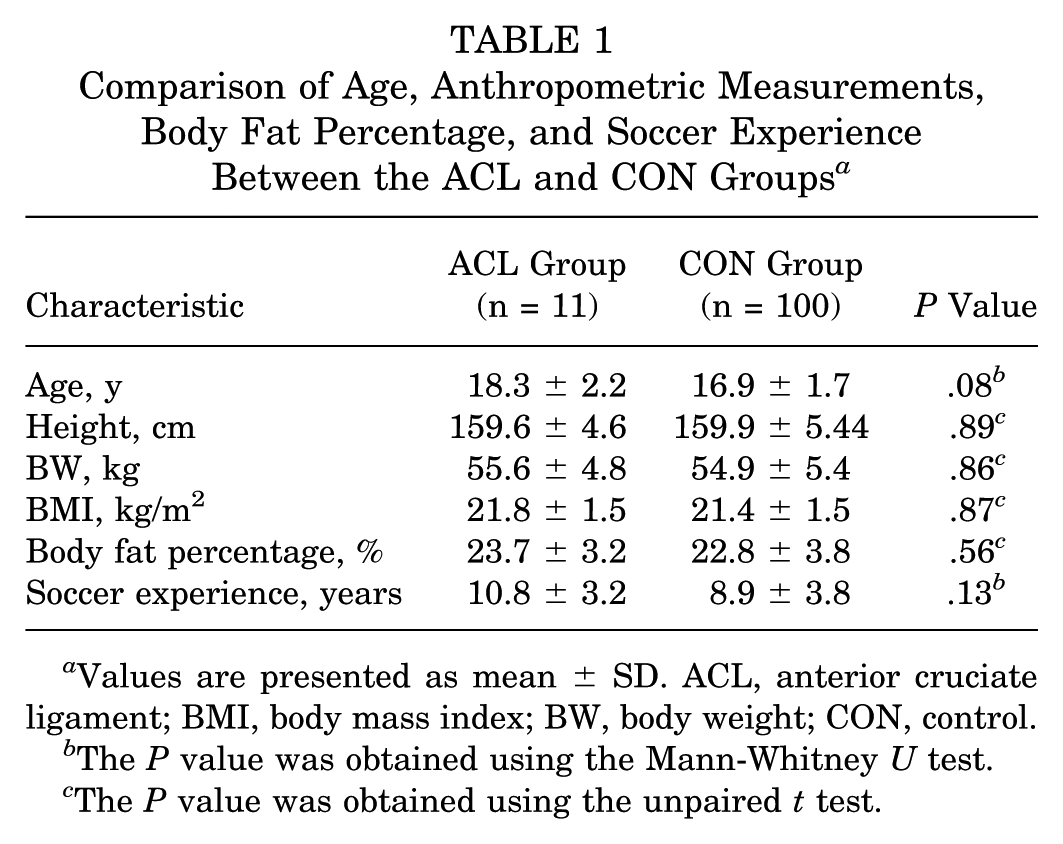
Values are presented as mean ± SD. ACL, anterior cruciate ligament; BMI, body mass index; BW, body weight; CON, control.
The P value was obtained using the Mann-Whitney U test.
The P value was obtained using the unpaired t test.
Comparison of the Foot–Arch Height Ratio Between the Injured Limb of the ACL Group and the Limb of the CON Group a
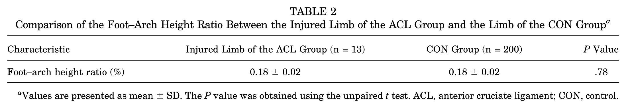
Values are presented as mean ± SD. The P value was obtained using the unpaired t test. ACL, anterior cruciate ligament; CON, control.
Comparisons of sum and peak loading values during running across 10 foot regions between the injured limb of the ACL group and the CON group are presented in Table 3. The sum of the loading value at area 3 (first metatarsal) and area 4 (second metatarsal) was significantly lower in the injured limb of the ACL group compared with the CON group (first metatarsal, P = .01; second metatarsal, P = .04). Similarly, peak loading at area 3 (first metatarsal) was significantly lower in the injured limb of the ACL group than in the CON group (P = .03).
Comparison of the Sum and Peak Loading Values Adjusted for Body Weight During Running Between the Injured Limb of the ACL Group and the Limb of the CON Group a

Values are presented as median (minimum-maximum). The P value was obtained using the Mann-Whitney U test. ACL, anterior cruciate ligament; BW, body weight; CON, control.
P < .05.
Table 4 shows a comparison of the cumulative loading force ratio in 6 localized regions and the medial-to-lateral sum loading value ratio. The sum loading ratio of the medial region to the total foot area was significantly lower in the injured limb of the ACL compared to the CON group (P = .03). Similarly, the medial-to-lateral sum loading value ratio was significantly lower in the injured limb of the ACL group than in the CON group (P = .03). While the cumulative loading value in the medial metatarsal region (areas 3 and 4) was significantly lower in the injured limb of the ACL group than in the CON group (15121.8 N, 19653.9 N; P = .01), no significant difference was found in the cumulative loading force ratio of the medial metatarsal (areas 3 and 4) relative to the total area (P = .06). Pearson’s product-moment correlation in age and medial-to-lateral force ratio was r = −0.0907 (P= .195).
Comparison of the Cumulative Loading Force Ratio in 6 Local Regions and the Medial-to-Lateral Sum Loading Value Ratio During Running Between the Injured Limb of the ACL Group and the CON Group a

Values are presented as median (minimum-maximum). The P value was obtained using the Mann-Whitney U test. ACL, anterior cruciate ligament; BW, body weight; CON, control.
P < .05.
Figure 4 illustrates the time phases of dynamic plantar pressure distribution for both the ACL and CON groups. Peak loading pressures at the medial and lateral heels occur at the beginning of the contact phase, followed by peak pressures on the metatarsal bones and toes as the foot leaves the ground with its toes. This loading pattern phase was observed in both groups. As shown in Figure 3, plantar loading on the first metatarsal area was lower than that on the second to fifth metatarsal areas in the ACL group.

Time phase of dynamic plantar pressure distribution during running. (A) Foot of the control (CON) group. (B) Ipsilateral foot of the anterior cruciate ligament (ACL) group.
Discussion
This is the first prospective study to report plantar loading distribution during running as a risk factor for ACL injuries in female soccer players. The results of this study underline a significant relationship between reduced plantar pressures in the medial foot regions—specifically the first metatarsal area—during running and an increased risk of ACL injuries in female soccer players.
Eils etal 9 and Wong etal 43 reported the patterns of plantar pressure distribution in male soccer players during soccer-specific movements. Both studies documented higher peak pressures observed in the medial region—especially at the medial forefoot (medial metatarsal)—during running, cutting, and sprinting.9,43 Increased medial loading during running may support lower extremity stability in male soccer players; however, its relationship with injury risk remains uninvestigated.
In this study, the increased lateral foot-loading pattern observed during running in players with ACL injury may reflect altered motor control related to hip rotation or ankle supination. Yang etal 45 reported that reduced hip internal rotation range of motion was associated with increased lateral plantar pressure (especially on the lateral metatarsal) during standing and walking. Therefore, reduced medial metatarsal plantar pressure and increased lateral loading during running may also indicate limited internal hip rotation. Regarding the lower limb kinetic chain, internal and external hip rotation influence ankle posture, including foot supination and pronation. 45 Although no significant differences in foot–arch height ratio (no differences in arch height) were found between the ACL and CON groups, increased plantar loading in the lateral regions of the ACL group may indicate altered loading patterns due to ankle posture during running. Previous dynamic plantar pressure studies have shown that the supinated foot is associated with reduced plantar pressure under the first metatarsal area and the medial forefoot during gait.30,38 Therefore, the increased plantar loading in the lateral regions during running in our female ACL group may be linked to a supinated foot posture at ground contact. Several studies have reported that a supinated foot is associated with lower stability during single-leg stance and jump landing.6,24 Beelen etal 6 assessed the effect of foot type on postural stability and reported that people with supinated feet exhibit less stability during a single-leg stance. McLean etal 24 investigated differences in landing strategies between male and female National Collegiate Athletic Association athletes and found that female athletes demonstrated greater initial ankle plantar flexion, ankle supination, knee abduction, and knee internal rotation compared to their male counterparts. They suggested that female tibial rotation patterns, combined with ankle supination during jump landing, may contribute to their increased risk of ACL injury, as internal tibial rotation motions and loads are known to directly affect ACL injuries. 23 In this study, increased plantar loading in lateral regions during running in the ACL group may be associated with tibial internal rotation and ankle supination posture at ground contact.
Contrary to our findings, retrospective studies by Beckett etal, 5 Loudon etal, 20 and Woodford-Rogers etal 44 reported a significant association between ACL injury history and excessive foot pronation during static posture. However, the retrospective designs in these studies limited the ability to determine whether foot posture was causative or a result of the injury. Furthermore, these studies assessed foot pronation using static measures such as the navicular drop test or prone subtalar joint position. In contrast, our study prospectively evaluated the dynamic plantar loading distribution during running, a factor previously unexplored in relation to ACL injury. Given that ACL injuries typically occur during dynamic movements, such as landing, jumping, and running, further investigation is needed to examine foot posture characteristics during these movements in female athletes who suffer ACL injuries.
Notably, a cross-sectional study investigated the relationship between plantar foot-loading patterns during normal gait and drop vertical jump biomechanics in female soccer players. 25 A previous study examined the dynamic barefoot plantar pressure distribution during walking in female soccer players, dividing participants into 2 groups based on their medial and lateral foot-loading patterns. The study reported no significant differences in sagittal or coronal plane knee joint kinematics and kinetics during drop vertical jumps between these groups. 25 Thus, the relationship between dynamic foot plantar biomechanics during running and knee landing biomechanics remains unclear. However, the reduced medial foot-loading pattern during running observed in the ACL group in this study might reflect distinctive motor control characteristics in female soccer players at risk of ACL injury.
These findings suggest that plantar pressure characteristics during running may potentially predict ACL injuries, offering a valuable benefit for prevention. Several studies reported that plantar loading and related running mechanics are modifiable, and the neuromuscular training and gait-retraining interventions can change loading mechanics.4,8 Moreover, identifying which athletes have a high-risk loading pattern is very important, which allows coaches and physicians to emphasize better neuromuscular control for these athletes at higher risk of ACL injuries. Neuromuscular training focused on improving balance mechanics and strengthening the kinetic chain during running may mitigate ACL injury risks. 34 Expanding the scope to include muscle activation patterns and joint kinematics during running—beyond gait patterns or jump-landing biomechanics—could enhance our understanding and support the development of comprehensive injury prevention strategies.
This study has several limitations. First, the number of players who sustained an ACL injury was relatively small, which limited the ability to perform multivariate analyses. Further studies with a larger sample size, including various age groups of female soccer players, are required to validate the present findings. Second, dynamic plantar pressures were measured barefoot while running, whereas ACL injury often occurs while wearing shoes. Additionally, dynamic pressure measurements were not assessed during other specific movements, such as sprinting or cutting. Third, the difference in characteristics in dynamic plantar loading between the ACL group and the CON group may be confounded by patient age, since there was a tendency for higher age in the ACL group compared to the CON group.However, there was no correlation between age and the medial-to-lateral force ratio by the subanalysis. Lastly, foot posture characteristics, such as supination or pronation, were not evaluated in our study. Regarding the relationship between foot posture and plantar loading distribution, further investigation is required to clarify how foot biomechanics and plantar loading distribution contribute to ACL injury risk.
Conclusion
This prospective study investigated the dynamic plantar loading distribution during running in young female soccer players with ACL injuries. Players with ACL injuries demonstrated reduced medial metatarsal loading and a lower medial-to-lateral plantar force ratio during running. These findings suggest that foot-loading patterns may be associated with ACL injury risk.
Footnotes
Final revision submitted November 10, 2025; accepted December 15, 2025.
One or more of the authors has declared the following potential conflict of interest or source of funding: Scholarship donations were received from East Japan Railway Company and Shimamura-Syoukai. Grants were received from the Nakatomi Foundation, Japan Sports Medicine Foundation, Japan Sport Council, Japanese Orthopaedic Society for Sports Medicine, Watanabe Memorial Foundation for the Advancement of New Technology, Japan Keirin Autorace Foundation, Japan Society for the Promotion of Science (KAKENHI 20K11358), Japan Orthopaedics Traumatology Foundation, and Japanese Orthopaedic Society of Knee, Arthroscopy and Sports Medicine. AOSSM checks author disclosures against the Open Payments Database (OPD). AOSSM has not conducted an independent investigation on the OPD and disclaims any liability or responsibility relating thereto.
Ethical approval for this study was obtained from the University of Tokyo (11907-(2)).