
Abstract
Background:
Sulcus-deepening trochleoplasty is an effective surgical treatment for high-grade trochlear dysplasia in patients with recurrent patellofemoral instability. However, concerns remain regarding the potential development of patellofemoral osteoarthritis (OA) over time and its effect on clinical outcomes.
Purpose:
To determine whether the presence of radiographic grade ≤2 OA after trochleoplasty is associated with inferior functional outcomes or patient satisfaction.
Study Design:
Cohort study; Level of evidence, 3.
Methods:
A retrospective study was conducted on 42 patients (47 knees) who underwent thick-flap sulcus-deepening trochleoplasty. After applying inclusion and exclusion criteria, 27 patients (33 knees) with a minimum follow-up of 10 years were included. Patients were categorized into OA and non-OA groups based on radiographic assessment (Iwano classification) at final follow-up. Functional outcomes were evaluated using the International Knee Documentation Committee (IKDC) and Kujala scores, while patient satisfaction was assessed on a 10-point scale. Group comparisons were performed using Mann-Whitney U and chi-square tests.
Results:
At a mean follow-up of 14.97 years (range, 10.4-19 years), 20 knees (60.6%) demonstrated radiographic OA (Iwano grades 1-2), while 13 knees (39.4%) showed no OA. Patients in the OA group were older (37.5 ± 6.7 vs 30.5 ± 3.0 years, P < .001) and had a longer follow-up duration (15.8 vs 13.0 years, P = .037). No significant differences were observed in other baseline characteristics. All patients underwent concomitant medial patellofemoral ligament reconstruction, and 14 additionally received a tibial tubercle osteotomy. Functional outcomes did not significantly differ between groups (IKDC: 79.06 ± 14.72 vs 78.04 ± 15.11, P = .726; Kujala: 80.45 ± 12.5 vs 80.07 ± 13.88, P = .462). Range of motion was comparable between groups, and overall patient satisfaction remained high (8.4 ± 1.65).
Conclusion:
Low-grade patellofemoral OA (Iwano ≤2) was observed in 60% of knees at a mean follow-up of 15 years, but its presence did not negatively affect long-term functional outcomes or patient satisfaction. Importantly, no patient developed grade 3 or 4 OA, and radiographic changes were more frequent in older patients and those with longer follow-up, supporting trochleoplasty as a reliable surgical option for patients with high-grade trochlear dysplasia.
Sulcus-deepening trochleoplasty is a well-established procedure for addressing high-grade trochlear dysplasia in patients with recurrent patellar instability by reshaping the trochlear groove, enhancing joint mechanics, and effectively reducing the risk of further dislocations.2,3,5,6,8,9,11 However, concerns persist regarding its potential long-term effect on the development of osteoarthritis (OA).1,10,17
Some authors suggest that this procedure may lead to abnormal joint loading, increased cartilage wear, and subsequent OA, potentially compromising long-term function.10,19 Others argue that radiographic OA is primarily a time-dependent finding rather than a determinant of clinical deterioration.18,21 While several studies have reported radiographic changes in the patellofemoral joint after trochleoplasty, the influence of arthritis following trochleoplasty on clinical outcomes remains unclear.5-7,17,20
This study aimed to determine whether patients who develop patellofemoral OA after trochleoplasty experience worse functional outcomes than those without OA. Additionally, we assessed differences in patient satisfaction between these groups. We hypothesized that there would be no significant differences in clinical outcomes.
Methods
Ethics
All patients provided informed consent for the use of their data for research, and the study was approved by the institutional ethics board (GCS Ramsay Sante pour l’Enseignement et la Recherche, COS-RGDS-2022-03-003-DEJOUR-D).
Study Design
The authors evaluated a retrospective consecutive series of patients who underwent thick-flap sulcus-deepening trochleoplasty by the senior surgeon (D.H.D.) between December 2003 and July 2012. Patients without follow-up radiographs at the final evaluation were excluded from the study. Additionally, patients with a history of knee surgery other than tibial tubercle osteotomy, vastus medialis plasty, or medial patellofemoral ligament (MPFL) reconstruction or those who underwent additional knee surgeries after trochleoplasty were also excluded (Figure 1).

Flowchart of the study selection and inclusion process. Patients included were categorized into OA and non-OA groups based on radiographic findings at final follow-up. OA, osteoarthritis.
During this period, the indications for trochleoplasty were objective patellar instability (≥2 documented patellar dislocations) and high-grade trochlear dysplasia (Dejour type B or D with a supratrochlear spur ≥4 mm). The contraindications included patellofemoral OA (grade ≥2 according to the Iwano classification) or skeletal immaturity (defined as an open distal femoral physis on plain radiographs).6,13,22
Surgical Technique
The surgical technique used in all cases was the previously published thick-flap trochleoplasty technique. 12
Based on preoperative measurements, a tibial tubercle osteotomy medialization was performed in cases of excessive tibial tuberosity to trochlear groove distance (>20 mm measured on computed tomography scan) or patella alta (Caton-Deschamps index >1.2 measured on lateral radiographs), aiming to reduce the tibial tuberosity to trochlear groove distance to 10 to 15 mm and the Caton-Deschamps index to 1.0.
The lateral retinaculum was released in cases of excessive lateral tightness, and the MPFL was routinely reconstructed at the end of the procedure using a doubled gracilis autograft, fixed at 20° of knee flexion, allowing a patellar mobility of 2 quadrants.
Postoperative Rehabilitation
Patients were allowed full weightbearing immediately after surgery without limitations of range of motion. However, an extension brace was used for those who underwent adjuvant tibial tuberosity osteotomy, limiting flexion to 100° for 30 days. At 45 days after surgery, the patient started performing aquatic and cycling activities, muscle reinforcement in a closed kinetic chain, and proprioceptive and postural exercises. At 3 months postoperatively, patients were prepared to return to sports with running and plyometric exercises, with a full return to sports expected at 8 months.
Postoperative Assessment
All patients were assessed at a minimum 10-year follow-up by an independent clinician (A.G.), who recorded any reoperations or patellar redislocations and collected International Knee Documentation Committee (IKDC) 14 and Kujala 16 scores.
Patients were also asked to rate their satisfaction with the procedure on a 10-point scale (1 = not satisfied, 10 = very satisfied) and whether they would undergo the surgery again (yes/no).
In bilateral cases, both knees were evaluated separately. If 1 knee met the exclusion criteria, only the contralateral knee that met the inclusion criteria was analyzed.
The radiographic assessment included weightbearing anteroposterior, sagittal (30° flexion), and axial (20° flexion) views to evaluate femorotibial and patellofemoral OA using the Iwano classification. 15 A senior expert in patellofemoral pathology performed radiographic evaluations (D.H.D.). Knees with Iwano grades 1 and 2 were classified as OA, while those with grade 0 were classified as non-OA (see Figure 2).
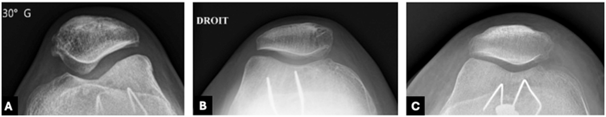
Representative radiographs of the Iwano classification: (A) no osteoarthritis, (B) grade 1, and (C) grade 2.
Statistical Analysis
Descriptive statistics were used to summarize the demographic data.
Continuous variables were expressed as means ± standard deviations, where appropriate, while the dichotomous variables were expressed as the number and percentage of patients.
Normality was assessed using the Shapiro-Wilk test to compare functional outcomes (IKDC and Kujala scores) and patient satisfaction between groups. The results indicated that while the non-OA group followed a normal distribution, the OA group did not. Consequently, nonparametric analysis was performed using the Mann-Whitney U test to determine differences between groups. For categorical variables, a chi-square test was used. SPSS (version 25; SPSS) was used to perform these statistical analyses. Significance was set at an α of P < .05.
Results
A total of 42 patients (47 knees) underwent thick-flap trochleoplasty during the study period. After applying the inclusion and exclusion criteria, 27 patients (33 knees) were included in the final analysis (see Figure 1).
Among them, 10 patients had a history of patellofemoral surgery, which included 8 cases of tibial tuberosity osteotomy and 2 cases of isolated MPFL reconstruction. None of the patients required additional knee surgery after trochleoplasty.
At the final follow-up, 20 knees (60.6%) showed some degree of patellofemoral OA, with 14 knees (70%) classified as grade 1 and 6 knees (30%) as grade 2 according to the Iwano classification, while no cases of grade 3 or 4 OA were observed. A total of 6 bilateral cases were included, and both knees were analyzed separately as independent data points.
Demographic and clinical characteristics of the OA and non-OA groups are presented in Table 1. The groups were comparable in terms of sex distribution, laterality, and the frequency of tibial tuberosity osteotomy. However, patients in the OA group were significantly older (P < .001) and had a longer follow-up period (P = .037) (see Table 1).
Patient Characteristics and Adjuvant Procedures a

Values are presented as mean ± standard deviation or number (%) unless otherwise indicated. Age and follow-up are expressed in years. Bold indicates statistical significance (P < .05). OA, osteoarthritis; TT, tibial tuberosity.
There were no significant differences between groups in IKDC scores, Kujala scores, range of motion, or patient satisfaction (see Table 2).
Postoperative Outcomes a

Values are presented as mean ± standard deviation. IKDC, International Knee Documentation Committee; OA, osteoarthritis.
When asked about their willingness to undergo surgery on the same knee again, 32 patients (97%) responded affirmatively.
We observed only 1 case (3%) of traumatic patellar dislocation in the OA group occurring 1 year after surgery. This case was managed nonoperatively, and no further dislocations were reported over the subsequent 11 years of follow-up.
Discussion
The most important finding of this study is that radiographic patellofemoral OA grade ≤2 after thick-flap sulcus-deepening trochleoplasty does not appear to be associated with inferior functional outcomes or patient satisfaction at a mean follow-up of nearly 15 years. As the development of OA remains one of the major concerns associated with this procedure, our findings indicate that patient-reported outcomes are not negatively affected by the development of mild patellofemoral OA and are comparable to those observed in patients without radiographic OA after trochleoplasty. 10
Long-term evidence on OA after trochleoplasty remains limited. In a systematic review of 1000 cases, Leclerc et al 17 found a 27% prevalence of patellofemoral OA, but the interpretation was hindered by heterogeneity in surgical techniques, imaging modalities, and the scarcity of studies with ≥10 years of follow-up. In our cohort, 60.6% of knees developed mild patellofemoral OA after nearly 15 years, higher than most previous reports, yet functional scores, range of motion, and satisfaction remained favorable.
Recent evidence has further explored the relationship between trochleoplasty on cartilage status and clinical outcomes. Frings et al 11 showed that thin-flap sulcus-deepening trochleoplasty yielded good short-term functional results without evidence of progressive cartilage deterioration, supporting the safety of the procedure in the early follow-up period. In contrast, Ackermann et al 1 found more extensive trochlear chondral changes on magnetic resonance imaging at midterm follow-up in the trochleoplasty group, whose patients were older than the controls, although these findings did not translate into worse clinical outcomes. Together, these findings highlight that structural abnormalities on imaging may not necessarily correlate with impaired function, which aligns with our observations.
Beyond cartilage integrity, patient age has also been discussed as a potential risk factor. Dan Milinkovic et al 4 demonstrated that age >30 years at the time of surgery did not compromise outcomes. We similarly found no association between age and functional results. Interestingly, the OA group in our study had a significantly longer follow-up than the non-OA group, suggesting that OA may be a time-dependent phenomenon rather than an indicator of clinical deterioration. While some studies have suggested that the procedure itself may predispose to trochlear cartilage changes, recent evidence indicates that residual postoperative incongruence may play a more important role in the long-term development of patellofemoral OA.9,19
The present study has several limitations that must be considered when interpreting the results. First, this was a retrospective long-term follow-up study with a small sample size, which limited statistical power and raised the possibility of type II error, preventing subgroup analysis or multivariable regression to identify factors predisposing to OA. Second, at the time of surgery, 10 to 20 years ago, the status of the cartilage was not routinely documented during surgery, so this information is not available, leaving uncertainty as to whether the development of OA was related to preoperative cartilage injury. Third, approximately 25% of initially eligible patients did not have final follow-up radiographs and were therefore excluded, which introduces a risk of selection bias. Fourth, no patients in our series had advanced OA (Iwano >2), limiting our ability to assess the effect of more severe disease. In addition, the small number of knees with grade 2 OA prevented any meaningful subgroup analysis. Fifth, only radiographs were used to assess OA, without magnetic resonance imaging, and most of the OA cases corresponded to grade 1, which may not be clinically meaningful. Finally, the use of the Iwano classification has intrinsic limitations, particularly in differentiating between grades 0 and 1. Although we did not evaluate inter- or intraobserver reliability in our series, a senior surgeon (D.H.D.) specializing in patellofemoral pathology performed all radiographic analyses.
Conclusion
This study provides long-term evidence that low-grade patellofemoral OA (Iwano ≤2) is common after sulcus-deepening trochleoplasty, affecting 60% of knees at a mean follow-up of nearly 15 years, yet it does not negatively affect functional outcomes or patient satisfaction. Notably, no patient developed grade 3 or 4 OA, and radiographic changes were more frequent in older patients and in those with longer follow-up. Although further prospective, multicenter studies with larger cohorts are warranted, our findings support trochleoplasty as a safe and effective surgical option for patients with high-grade trochlear dysplasia.
Footnotes
Final revision submitted September 26, 2025; accepted October 19, 2025.
The authors have declared that there are no conflicts of interest in the authorship and publication of this contribution. AOSSM checks author disclosures against the Open Payments Database (OPD). AOSSM has not conducted an independent investigation on the OPD and disclaims any liability or responsibility relating thereto.
Ethical approval for this study was obtained from GCS Ramsay Sante pour l’Enseignement et la Recherche (COS-RGDS-2022-03-003-DEJOUR-D).