
Abstract
Background:
Pathology involving the acetabular fossa is often identified during hip arthroscopy, with many of the descriptive classification systems focusing on the ligamentum teres (LT). Recently, a novel grading system for ligamentous-fossa-foveolar complex (LFFC) lesions was introduced for open hip preservation surgery that included evaluation of the cotyloid fossa and perifoveal cartilage.
Purpose:
To validate the LFFC grading system for hip arthroscopy and correlate these results with preoperative and intraoperative findings.
Study Design:
Cohort study (diagnosis); Level of evidence, 3.
Methods:
High-resolution arthroscopic images of the central compartment were obtained identifying the LT, cotyloid fossa, and perifoveal cartilage in a prospective series of patients undergoing hip arthroscopy. Each structure was graded according to increasing pathology on a scale of 0 to 4 based on a modified LFFC classification system to include the pulvinar intra-articular adipose tissue. Five surgeons graded the images, which were then randomized and regraded for intraobserver reliability. Agreement was quantified by the intraclass correlation coefficient (ICC) and kappa (κ) statistic to determine inter- and intraobserver reliability. Grading discrepancies were resolved in conference with the senior author, and final LFFC grades were compared with preoperative clinical and radiographic data as well as intraoperative findings.
Results:
A total of 93 patients were included in the study. LFFC component intraobserver reliability for 2 rounds of grading resulted in an LT ICC of 0.78 to 0.90, cotyloid fossa ICC of 0.85 to 0.93, and perifoveal cartilage ICC of 0.78 to 0.87 with an LFFC total score ICC of 0.87 to 0.95. Interobserver reliability resulted in an LT ICC of 0.73 to 0.91, cotyloid fossa ICC of 0.84 to 0.95, perifoveal cartilage ICC of 0.83 to 0.91, and LFFC total score ICC of 0.89 to 0.96. Severe central compartment pathology (LFFC total score >6) was significantly associated with age (48.2 vs 34.4; P = .0002), preoperative Tönnis grade 1 (43% vs 4%; P < .0001), preoperative Tönnis angle (8.7 vs 5.2; P = .002), and intraoperative femoral head weightbearing chondral lesions (14% vs 0%; P = .02) when compared to patients with an LFFC score <6.
Conclusion:
The modified LFFC grading system demonstrated satisfactory intraobserver and interobserver reliability for patients undergoing hip arthroscopy that compares favorably with existing arthroscopic classification systems for the acetabular fossa. The addition of a descriptive classification system for the pulvinar intra-articular adipose tissue did not decrease the reliability of the grading system. Increasing LFFC scores were found to be associated with known risk factors for inferior outcomes after hip arthroscopy providing enhanced clinical utility for this grading system.
Keywords
Pathology within the central compartment is often identified in patients undergoing hip arthroscopy for femoroacetabular impingement, hip dysplasia, trauma, and osteoarthritis.2,3,5,12,18,23 While the primary diagnostic and therapeutic focus for central compartment lesions has been the treatment of chondrolabral injury, there is frequently concomitant damage to the structures within the acetabular fossa, including the ligamentum teres (LT), pulvinar tissue within the cotyloid fossa, and perifoveal cartilage, all of which are also possible pain generators.
Pathology affecting the ligamentous-fossa-foveolar complex (LFFC) can range from mild synovitis to full-thickness defects of the involved structures, with greater levels of damage linked to the development of hip osteoarthritis and global joint deterioration.20,21 Further, intra-articular adipose tissues within the pulvinar have been shown to have a specific inflammatory phenotype compared with subcutaneous adipose tissues in the setting of hip osteoarthritis indicating that pathologic intra-articular adipose tissues signaling and interactions with the adjacent synovium may contribute to osteoarthritis progression.6-8,19 With progressive pulvinar degeneration, acetabular fossa ossification occurs with the central acetabular osteophyte (CAO), having been identified as both an early harbinger of hip osteoarthritis progression and a strong radiographic predictor of failure 10 years after hip preservation surgery.10,14
Given the biological relevance of the structures comprising the LFFC, a consistent means of analysis may help guide intraoperative treatment decisions as well as postoperative prognosis after hip arthroscopy. One of the primary limitations regarding the collective understanding of the acetabular fossa structures is the lack of a validated comprehensive arthroscopic grading system. Multiple classification systems exist describing LT pathology, but they are limited in scope as well as by reproducibility with inter- and intraobserver agreement (kappa) values ranging from 0.39 to 0.74.1,4,9,16,18 To address this, a recent study by Stetzelberger et al 21 proposed a comprehensive grading system for the LFFC that was validated for patients undergoing open hip preservation with surgical hip dislocation. The authors evaluated the LT, cotyloid fossa, and perifoveolar cartilage, grading lesions on a scale of 0 (normal) to 4 (complete defect) (Appendix Table A1) reporting excellent intraobserver and interobserver reliability.
Due to the difference in arthroscopic appearance in the acetabular fossa compared with direct visualization with open hip preservation, a slightly modified LFFC classification system was proposed with the additional goal of further describing the status of the pulvinar intra-articular adipose tissue in the cotyloid fossa. The purpose of this study was to validate this grading system for patients undergoing hip arthroscopy and correlate central compartment pathology to preoperative demographics, radiographic parameters, and intraoperative findings. We hypothesized that patients with increased central compartment pathology would have greater evidence of radiographic arthritis and intraoperative chondral damage.
Methods
Patient Recruitment
After institutional review board approval, consecutive patients undergoing hip arthroscopy at a multisurgeon quaternary referral center from August 1, 2022, to December 29, 2022, were prospectively considered for inclusion into the study. Patients undergoing primary or revision hip arthroscopy with 4 fellowship-trained hip preservation specialists (J.J.R., J.G., L.V., M.J.P.) were included. A total of 93 consecutive patients with high-resolution intraoperative images were included in the study. All patients had clinical, radiographic, and advanced imaging diagnoses consistent with intra-articular hip pathology. Preoperative demographics, radiographic parameters, and intraoperative variables were recorded through chart review and analysis of a validated prospectively collected surgical database (Table 1). Of 93 patients included in the study, 51 (55%) of the study population were female. The median age of the study population was 36 years (IQR, 25.5-47 years). Eighty patients (86%) underwent primary hip arthroscopy with a primary diagnosis of FAI, with 16 patients in this group having a secondary diagnosis of borderline dysplasia. 12 (13%) underwent revision hip arthroscopy primarily for adhesions and residual FAI. Anteroposterior pelvis radiographs for each patient were reviewed by 2 fellowship-trained orthopaedic surgeons (B.D.K., P.S.C.) for diagnosis of the sabertooth (central acetabular) osteophyte, which was not previously collected in the patient database. Preoperative outcome scores including the Western Ontario McMaster Universities Osteoarthritis Index, modified Harris Hip Score (mHHS), and Hip Outcome Score (HOS) Activities of Daily Living subscale and Sports Subscale (HOS-SS) were obtained during the initial clinic visit (Table 1).
Preoperative Patient Demographics, Radiographic Measures, and Outcome Scores (N = 93) a
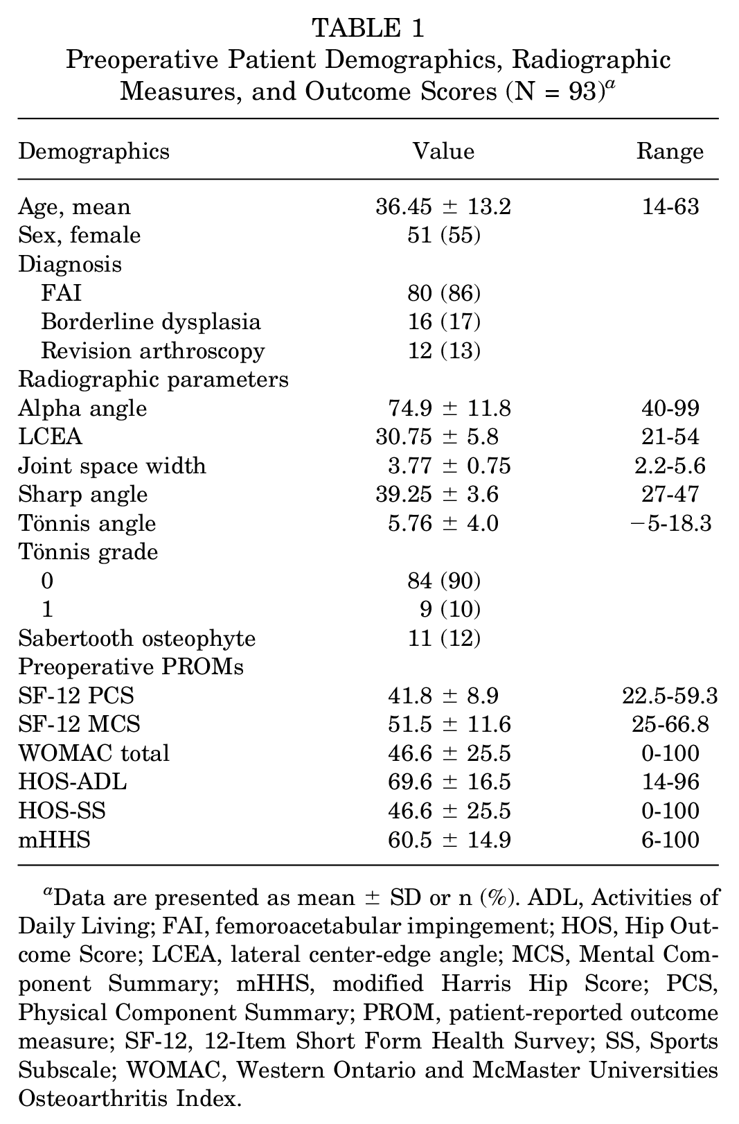
Data are presented as mean ± SD or n (%). ADL, Activities of Daily Living; FAI, femoroacetabular impingement; HOS, Hip Outcome Score; LCEA, lateral center-edge angle; MCS, Mental Component Summary; mHHS, modified Harris Hip Score; PCS, Physical Component Summary; PROM, patient-reported outcome measure; SF-12, 12-Item Short Form Health Survey; SS, Sports Subscale; WOMAC, Western Ontario and McMaster Universities Osteoarthritis Index.
Surgical Procedures and Rehabilitation
The surgical and rehabilitation techniques employed by the senior author (M.J.P.) have been described previously. Briefly, the procedure was performed in the supine position on a traction-operating table. Traction was applied with the hip in 15° of internal rotation and 10° of flexion with neutral abduction, and with joint distraction confirmed using ultrasound or fluoroscopy. Standard anterolateral and midanterior portals were established, and a diagnostic arthroscopy was performed. Central compartment work including acetabuloplasty, anterior inferior iliac spine decompression, labral repair or reconstruction, LT debridement or reconstruction, and acetabular or femoral head chondroplasty or microfracture where indicated occurred with the hip in traction. Peripheral compartment work included the femoroplasty and capsular plication or closure with the hip out of traction. After the procedure, the patients underwent previously described rehabilitation and physical therapy curated to their relevant surgical procedures.
LFFC Imaging
During the diagnostic portion of the procedure, the central compartment was visualized through a 70° arthroscope (4K; Smith & Nephew) through the traditional anterolateral and midanterior portals with the lower extremity in internal rotation. The lower extremity was then externally rotated to tension the LT allowing for improved visualization and further evaluation of related pathology. An arthroscopic probe was used to evaluate perifoveal cartilage quality and LT integrity. Viewing from the midanterior portal, 3 separate images used for grading were obtained focusing on the LT, perifoveal cartilage, and cotyloid fossa, respectively.
Imaging Analysis
Arthroscopic images were independently randomized (by J.B.) and graded by 3 senior hip preservation surgeons (J.G., J.J.R., L.V.) and 2 fellowship-trained junior reviewers (B.D.K., P.C.). The LT, cotyloid fossa, and perifoveolar cartilage were all graded according to the LFFC classification system described by Stetzelberger et al 21 with slight modification (Figure 1). Each structure was graded on a scale of 0 to 4 (0, normal; 1, inflammation; 2, degeneration; 3, partial defect; 4, full defect). Primary differences from the original classification scheme included midsubstance tearing of the LT as a grade 2 LT lesion, cotyloid fossa pulvinar punctate synovitis as a grade 1 cotyloid fossa lesion, cotyloid fossa pulvinar hyperemia as a grade 2 cotyloid fossa lesion, cotyloid fossa pulvinar atrophy as a grade 3 cotyloid fossa lesion, and cartilage softening as a grade 1 perifoveal cartilage lesion. After reviewer grading, differences were resolved in conference with the senior author to assign final grades to each structure. A single component score of >2 or total score >6 were considered markers of severe LFFC pathology.

Ligamentous-fossa-foveolar complex scoring system. Differences from the original scoring system by Stetzelberger et al 21 are in bold.
Statistical Analysis
Statistical analysis was conducted with JMP (Version 17) and MedCalc software platforms. Intraclass correlation coefficients (ICCs) were calculated using a 2-way model with absolute agreement to determine intraobserver and interobserver reliability with 95% CIs. The kappa statistic (κ) was also used to evaluate intra-and interobserver agreement. ICC values from 0.5 to 0.75 were considered moderate reliability, 0.75 to 0.9 good reliability, and >0.9 excellent reliability. 11 Kappa values from 0.41 to 0.60 were consistent with moderate agreement, 0.61 to 0.80 good agreement, and 0.81 to 1.0 very good agreement. 13 A power analysis using the Bonett method required 55 patients to achieve acceptable power with a minimal acceptable reliability of 0.75, expected reliability of 0.85, and 5 reviewers. Data were assessed for normality with the Shapiro-Wilk test. Continuous data were compared using a student t test or Mann-Whitney U test, categorical data were compared with the Pearson correlation or Fischer exact test, and categorical data were compared with continuous data using 1-way analysis of variance or Kruskal-Wallis testing where appropriate. A P value of .05 was used to determine statistical significance.
Results
Patient Demographics
Intraoperative findings and procedures as well as LFFC damage can be seen in Table 2. 12 (13%) patients had evidence of severe acetabular chondral damage and 2 (2%) patients had severe femoral head chondral damage (Outerbridge grade >2). For the definitive LFFC grades, the distribution (as also reported in Figure 2) included 12 patients (13%) having LT damage >2, 16 (17%) patients having cotyloid fossa damage >2, 14 (15%) patients having perifoveal cartilage damage >2, and 14 patients (15%) having a total LFFC score >6.
Intraoperative Findings and Procedures (N = 93) a
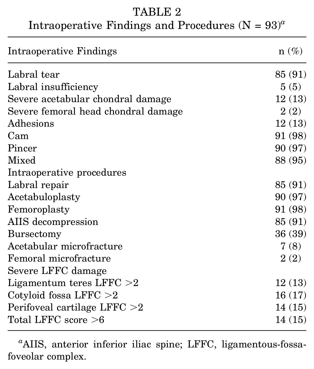
AIIS, anterior inferior iliac spine; LFFC, ligamentous-fossa-foveolar complex.

Distribution of ligamentous-fossa-foveolar complex (LFFC) component and total scores.
Intra- and Interobserver Reliability
Interobserver reliability ICC values for the LFFC grading system ranged from 0.73 to 0.96 (Table 3). LFFC component ICC values were the highest for the cotyloid fossa (range, 0.84-0.95) and lowest for the LT (range, 0.73-0.91). ICC values for the total score were consistent, ranging from 0.89 to 0.96. Kappa values for intraobserver reliability ranged from 0.64 to 0.91 (Table 4). Similarly, component kappa values were the highest for the cotyloid fossa (range, 0.81-0.91) and lowest for the LT (range, 0.64-0.86). Intraobserver reliability ICC scores for 2 rounds of LFFC grading for the component scores ranged from 0.78 to 0.93 and for the total scores ranged from 0.87 to 0.95 (Table 4). Interobserver kappa values for the component scores ranged from 0.58 to 0.82 and for the total score ranged from 0.44 to 0.53.
Interobserver Reliability and Agreement, With 95% CI a

CF, cotyloid fossa LFFC component; ICC, intraclass correlation coefficient; LFFC, ligamentous-fossa-foveolar complex; LT, ligamentum teres LFFC component; PC, perifoveal cartilage LFFC component.
Intraobserver Reliability and Agreement, With 95% CI a

CF, cotyloid fossa LFFC component; ICC, intraclass correlation coefficient; LFFC, ligamentous-fossa-foveolar complex; LT, ligamentum teres LFFC component; PC, perifoveal cartilage LFFC component.
Clinical Correlation
Severe LFFC damage (total LFFC score >6 or component score >2) was significantly associated with older age and higher rates of Tönnis grade 1 degeneration and CAOs on preoperative radiographs. Sex, revision surgery, lateral center-edge angle, and alpha angle were not associated with severe LFFC damage (Table 5). Intraoperatively, severe femoral head damage and femoral head microfracture were associated with higher LFFC component and total scores. There were no significant correlations between severe LFFC damage and preoperative outcome scores; however, patients with severe LFFC damage did have lower HOS-SS scores compared with patients without severe LFFC damage (Table 5).
Clinical Correlations With Severe LFFC Damage a

Data are presented as mean ± SD or n (%). The bold values signify significance (p < 0.05). ADL, Activities of Daily Living; HOS, Hip Outcome Score; LCEA, lateral center-edge angle; LFFC, ligamentous-fossa-foveolar complex; mHHS, modified Harris Hip Score; PCS, Physical Component Summary; SF-12, 12-Item Short Form Health Survey; SS, Sports Subscale; WOMAC, Western Ontario and McMaster Universities Osteoarthritis Index.
Discussion
The primary finding from this study was that the modified LFFC grading system was found to be both robust and reliable when evaluating the central compartment during diagnostic hip arthroscopy. The reliability and agreement statistics were comparable with the findings of the original group that validated the classification system on patients undergoing open hip preservation surgery through surgical hip dislocation with the majority of the inter- and intraobserver ICC values >0.8 and kappa agreement statistic >0.7. The addition of a descriptive classification for the pulvinar intra-articular adipose tissue did not decrease the reliability profile of the grading system and had overall higher interobserver (0.84-0.95) and intraobserver (0.85-0.93) correlation coefficients for cotyloid fossa lesions compared with the original classification (interobserver ICC, 0.74-0.95; intraobserver ICC, 0.76-0.82). Further, the clinical relevance of the grading system was strengthened by significant associations with radiographic and intraoperative parameters linked to inferior outcomes after hip arthroscopy and osteoarthritis progression such as advanced age and femoral head chondral damage. Additionally, while patients with higher LFFC damage had lower, but nonsignificant, preoperative patient-reported outcomes, the study was not powered to evaluate patient outcomes.
Existing classification systems involving the acetabular fossa have focused on LT pathology.1,9,18 Gray and Villar 9 originally published a descriptive classification system for the LT with grades 1 to 3 for complete LT rupture, partial LT rupture, and degenerative LT rupture, respectively. Subsequently, Salas and O’Donnell 18 reported a separate classification system including synovitis as well as treatment recommendations that was further modified to include hypermobility, but no existing system describes the cotyloid fossa or perifoveolar region on the femoral head. 16 Further, Devitt and colleagues 4 reported an interobserver kappa value of 0.39 and intraobserver kappa value ranging from 0.59 to 0.74 for the Grey and Villar classification and an interobserver (0.38) and intraobserver kappa (0.39-0.45) range for the descriptive LT classification system described by Botser et al. 1
Recently, Stetzelberger et al 21 proposed the LFFC grading system to include the cotyloid fossa and fovea in addition to the LT. The authors found strong reliability and agreement across their classification system that was highest for the LT (interobserver ICC, 0.91-0.92) and lowest for the acetabular fossa lesions (interobserver ICC, 0.76-0.82). The total score also had strong agreement and reliability with intraobserver ICCs ranging from 0.91 to 0.99 and interobserver ICCs ranging from 0.87 to 0.98 with interobserver kappa values ranging from 0.59 to 0.60. Modifications to the existing system that were used in the present study include atrophy of the cotyloid fossa as a grade 3 cotyloid fossa lesion, midsubstance tearing as a grade 2 LT lesion, and chondral softening as a grade 1 perifoveal cartilage lesion. Similar to Stetzelberger and colleagues, the current study found overall strong reliability and agreement statistics for each component score with marginally lower interobserver kappa agreement (0.59-0.60 vs 0.44-0.53). This may be due to slightly lower LT reliability reported in the present study, as imaging of this structure was the least consistent across the 3 components of the LFFC scale. For future use, care must be taken to thoroughly document the status of each structure and manipulate the lower extremity to maximize visualization. Despite these differences, the majority of the ICC values for both inter and intraobserver reliability reported here were in the good to excellent range with kappa scores showing moderate to very good agreement.
Clinically, LFFC lesions have been linked to osteoarthritis progression and poor outcomes after hip preservation surgery. In a retrospective study of 121 patients undergoing FAI treatment with surgical hip dislocation and 10-year follow-up, Hanke et al 10 found that the sabertooth (CAO) and perifoveal osteophytes had hazard ratios of 4.0 and 3.9, respectively, for postoperative failure defined as conversion to arthroplasty or a Harris Hip Score of <80. With regard to hip arthroscopy, a recent study by Yang et al 24 found that patients undergoing hip arthroscopy for FAI with an incidentally found and conservatively managed CAO had significantly lower rates of achieving the minimal clinically important difference and Patient Acceptable Symptom State for the mHHS. The CAO has also been associated with higher rates of LT and perifoveal cartilage damage in patients undergoing hip arthroscopy, with Lodhia et al 12 reporting 33% of patients with a CAO had severe (Outerbridge >2) femoral head chondral damage compared with 10% of patients without a CAO. Further, the CAO has been found to be more prevalent in patients with borderline dysplasia and is often present before joint space narrowing, suggesting that it is an early radiographic sign of osteoarthritis.14,22,23 These findings are consistent with the clinical correlations identified in the present study where higher LFFC scores were associated with greater Tönnis angles, as well as radiographic evidence of Tönnis grade 1 degenerative changes and the presence of a sabertooth osteophyte. In addition to the correlations identified here, preoperative measurement of cotyloid fossa and acetabular cartilage coverage indices on advanced imaging may provide additional associations with LFFC grading; however, this was not assessed in the present study.15,17
Limitations
This study has multiple limitations. While each structure was separately imaged from the midanterior portal, not all images were obtained in a uniform orientation, which may have limited some of the reliability findings from this study. The study only used the modified LFFC grading scale, and the original classification system was not separately recorded, limiting comparability between the 2 grading scales for this study. Despite this limitation, high reliability with the scoring system was reported. Additionally, a probe was used intraoperatively to assess for cartilage softening, which was not taken into account for the blinded image grading; this may explain some of the lower values for the reliability of LFFC scores for the perifoveolar cartilage. The addition of patients with revision surgery may bias the results, as there is a possibility that iatrogenic injuries were included in the description. Future studies validating the modified LFFC grading system for both primary and revision hip arthroscopy would help to clarify this. Further, the clinical correlations with LFFC scores were limited to preoperative and intraoperative data on a heterogeneous cohort of primary and revision patients, limiting the power of these associations, and no postoperative data were recorded. Application of the LFFC grading scale to postoperative outcomes will require future study. Finally, no patients undergoing combined hip arthroscopy and periacetabular osteotomy or proximal femoral osteotomy were included, so the results from the present study may not be as reliable for this cohort.
Conclusion
The modified LFFC grading system demonstrated satisfactory intraobserver and interobserver reliability for patients undergoing hip arthroscopy that compares favorably with existing arthroscopic classification systems for the acetabular fossa. The addition of a descriptive classification system for the pulvinar intra-articular adipose tissue did not decrease the reliability of the grading system. Increasing LFFC scores were found to be associated with known risk factors for inferior outcomes after hip arthroscopy providing enhanced clinical utility for this grading system.
Footnotes
Appendix
Original Ligamentous-Fossa-Foveolar-Complex (LFFC) Grading System Reported by Stetzelberger et al 21 a

| Grade | Ligamentum Teres | Cotyloid Fossa | Fovea |
|---|---|---|---|
Normal |
Pyramidal structure, homogeneous ligament structure without inflammation | Normal, homogeneous cartilage around the fossa | Pyramidal structure, homogeneous ligament structure without inflammation |
Inflammation |
Synovitis with red injection due to hyperemia of the intact synovium | Red discoloration of the central aspect of the fossa cartilage | Synovitis with red injection due to hyperemia of the intact synovium |
Degeneration |
Mucoid/fibromatous/nodular degeneration resulting in synovial folds or loss of pyramidal structure | Loss of regular contour of the fossa (excluding growth abnormalities of the triradiate cartilage) | Fissuring (striae) of the perifoveolar cartilage |
Partial defect |
Flaps of ligament avulsed from the fovea (most often) or the acetabulum | Partial-thickness cartilage defect | Partial-thickness cartilage defect |
Complete defect |
Complete disconnection of the ligament from the femoral head or the acetabulum (with or without bony avulsion) | Bony apposition with formation of a central osteophyte | Full-thickness perifoveal defect with exposure of the subchondral bone |
Component scores were graded 0-4 with a total score of 0-12.
Final revision submitted January 15, 2025; accepted February 11, 2025.
One or more of the authors has declared the following potential conflict of interest or source of funding: The study was sponsored through internal funding with the Steadman Philippon Research Institute. B.D.K. has received a grant from Arthrex; hospitality payments from Medical Device Business Services; and research support from Arthrex, Stryker Corporation, Kerecis Limited, Medical Device Business Services Inc, Medwest Associates, and Medacta USA. P.C. has received grants from Arthrex and DJO; education payments from Smith & Nephew; hospitality payments from Encore Medical; and research support from Arthrex, Smith & Nephew, Depuy Synthes, Alpha Orthopedic Systems, and Stryker Corporation. J.J.R. has received nonconsulting fees from Smith & Nephew, a grant from Arthrex, and research support from Smith & Nephew and Aesculap Biologics. J.G. has received consulting fees from Bioventus, Tornier, and DePuy Synthes Products; hospitality payments from Stryker and Medical Device Business Services; nonconsulting fees from Smith & Nephew; education payments from Gemini Mountain Medical and Arthrex; research support from Arthrex and Stryker Corporation; and stock or stock options from Nanochon and Nice Recovery Systems. L.V. has received nonconsulting fees from Smith & Nephew, research support from Smith & Nephew, and education payments from Gemini Mountain Medical and Arthrex. L.V. is a paid consultant for Stryker. M.J.P. has received research support from Smith & Nephew, Ossur, Arthrex, Siemens Medical Solutions, National Institutes of Health, National Institute of Arthritis and Musculoskeletal and Skin Diseases, National Institute on Aging, and US Department of Defense; has received consulting fees from MIS, Nice Recovery Systems, Smith & Nephew, and Olatec; is a shareholder in Arthrosurface, MJP Innovations LLC, MIS, Vail Valley Surgery Center, Vail MSO Holdings LLC, EffRx, Olatec, iBalance (Arthrex), Stryker, Trimble, Grocery Outlet, 3M, Bristol Myers Squibb, Pfizer, AbbVie, and Johnson & Johnson; has received faculty/speaker compensation from Synthes GmbH; has received education payments from ConMed Linvatec; is an investor in Manna Tree Partners; has ownership in MJP Innovations LLC, TSC Imaging, Proofpoint Biologics, and TSC DME & TSC Imaging; is co-chairman of Steadman Philippon Research Institute; has stock or stock options in 3M, AbbVie, Vail Valley Surgery Center, Bristol-Myers Squibb, Dillon Surgery Center, DocBuddy, EffRx, iBalance, Johnson & Johnson, Olatec, Pfizer, Proofpoint Biologics, Steadman Philippon Surgery Center, Stryker, Trimble, TSC DME & TSC Imaging, and Vail MSO Holdings LLC; has received IP royalties from Arthrosurface, Bledsoe, CONMED Linvatec, DJO, and Smith & Nephew; has received publishing royalties or financial or material support from SLACK Incorporated, and Elsevier; is a board or committee member of Vail Health Services, International Society of Hip Arthroscopy, ASIAM, Orthopedics Today, Steadman Philippon Research Institute, and Vail Valley Surgery Center Governing Council; is a general council member of Vail Valley Surgery Center; is an advisory board member of Orthopedics Today; and is an editorial or governing board member of the American Journal of Sports Medicine. AOSSM checks author disclosures against the Open Payments Database (OPD). AOSSM has not conducted an independent investigation on the OPD and disclaims any liability or responsibility relating thereto.
Ethical approval for this study was obtained from Vail Health (No. 2022-164).