
Abstract
Background:
Primary anterior cruciate ligament reconstruction (ACLR) using quadriceps tendon (QT) autograft may increase the risk for postoperative loss of extension, although it is unclear if this is due to QT autograft diameter.
Purpose:
To document the rate of >5° loss of knee extension after QT autograft ACLR and determine associations between loss of extension, QT autograft diameter, and notch volume.
Study Design:
Case-control study; Level of evidence, 3.
Methods:
A retrospective review of a consecutive series of patients undergoing primary ACLR with QT autograft between January 2014 and December 2021 by 7 fellowship-trained orthopaedic surgeons at a single health care institution was performed. Patients with revision ACLR, multiligamentous knee surgery, concomitant cartilage procedures, <14 years of age, unavailable preoperative magnetic resonance imaging (MRI), or <6 months of follow-up were excluded. Loss of extension was defined using the International Knee Documentation Committee criteria for abnormal knee extension (>5° loss of extension compared with the contralateral knee) 3 to 12 months after ACLR, or any subsequent surgery for loss of extension. Patients who were unable to achieve terminal knee extension (defined as 0° of extension irrespective of the contralateral knee) were also identified and analyzed. Notch volume was measured by 2 observers using preoperative MRI scans, and a ratio of QT autograft diameter to notch volume was calculated. Univariate and multivariate analyses were performed to assess factors associated with postoperative loss of extension.
Results:
A total of 500 patients were identified, of whom 333 (67%) were included (mean age, 22.8 ± 7.7 years; 151 [45%] female). The mean follow-up was 1.6 ± 1.3 years (range, 0.5-9.5 years). The rate of postoperative loss of extension was 11% (n = 37), and 70% (26/37) of those with loss of extension underwent a subsequent surgery to restore extension (mean, 1.1 ± 1.2 years postoperatively). There was no difference in QT autograft diameter (9.5 mm vs 9.6 mm; P = .81), notch volume (6.3 cm3 vs 6.5 cm3; P = .70), and the ratio between QT autograft diameter and notch volume (1.6 vs 1.6; P = .75) between patients with and without postoperative loss of extension. No differences were found in preoperative (P = .62) and postoperative (2-4 months [P = .99]; 5-8 months [P = .71]; ≥9 months [P = .95]) extension between patients with a QT autograft diameter ≥10 mm and <10 mm. Only the inability to achieve terminal extension (0°) at the initial preoperative visit was associated with postoperative loss of extension on multivariate analysis (OR, 2.23 [95% CI, 1.10-4.58]; P = .03).
Conclusion:
Eleven percent of patients undergoing QT autograft ACLR experienced a loss of >5° knee extension compared with the contralateral knee or required additional surgery to restore extension. QT autograft diameter and notch volume were not associated with postoperative loss of extension among patients who underwent primary QT autograft ACLR. The inability to achieve terminal extension (0°) at the initial preoperative presentation increased the risk of postoperative loss of extension by 2.23-fold. Surgeons may consider the lack of terminal extension preoperatively as a risk factor for postoperative loss of extension following QT autograft ACLR rather than increased QT autograft diameter or decreased notch volume.
Loss of motion after anterior cruciate ligament (ACL) reconstruction (ACLR) is a common postoperative complication that can result in severe detriments to a patient's functioning and overall physical health.10,20,21,27 Loss of extension specifically can lead to more severe consequences compared with loss of flexion and may result in the need for a reoperation if full extension is unable to be restored through nonoperative modalities. 19 Risk factors for loss of extension include female sex, concomitant meniscal repair, and poor preoperative motion.9-11,14,19 The use of a quadriceps tendon (QT) autograft has more recently been suggested as a potential risk factor for loss of extension, although the reasons for this proposed relationship are poorly understood.1,13
Previous studies have reported a positive association between larger ACL graft diameter or harvest width and loss of extension.8,24 Similarly, a narrow notch may increase the risk for loss of extension due to an association with cyclops syndrome, although this relationship is described in cohorts receiving primarily hamstring tendon autograft ACLR.4,6 In 1 retrospective series of patients undergoing all–soft tissue QT autograft ACLR, a femoral tunnel diameter >9.25 mm was associated with postoperative loss of extension among male patients. 9
Current evidence evaluating the association between loss of extension after QT autograft ACLR and QT autograft diameter is limited. Likewise, the risk for loss of extension when accounting for the ratio between QT autograft diameter and notch volume is unclear. The purpose of this study was to investigate the association between postoperative loss of extension after primary QT autograft ACLR and QT autograft diameter, notch volume, and other factors associated with postoperative loss of extension, including preoperative loss of extension. We hypothesized that patients with a large QT autograft diameter and small notch volume would have a higher rate of postoperative loss of extension compared with patients with a small QT autograft diameter and large notch volume. Additionally, we hypothesized that patients with preoperative loss of extension would be at a higher risk for postoperative loss of extension.
Methods
Study Population
This study was approved by the institutional review board (STUDY19030196). A retrospective review of a consecutive series of patients undergoing primary ACLR was performed. Patients who underwent primary QT autograft ACLR between 2014 and 2021 by 7 orthopaedic sports medicine fellowship-trained surgeons (including J.D.H. and V.M.) at a single large health care institution were eligible for study inclusion. Exclusion criteria included revision ACLR, multiligamentous knee surgery, age <14 years at the time of ACLR, undergoing a concomitant cartilage procedure, use of an ACL graft other than a QT autograft, and <6 months of follow-up. Patients with unavailable preoperative magnetic resonance imaging (MRI) scans or whose MRI scans were unable to be measured in the picture archiving and communication system for measurements of notch volume were additionally excluded. Data collection was performed via a retrospective chart review of clinical notes and operative reports.
The primary outcome was postoperative loss of extension, which was defined using the International Knee Documentation Committee (IKDC) criteria for normal range of motion. 15 Patients with >5° loss of extension compared with the contralateral knee (corresponding to an IKDC range of motion grade of C or D) between 3 and 12 months postoperatively were categorized as having loss of extension. 15 Patients who underwent a subsequent surgery for loss of extension at any postoperative time period (including >12 months) irrespective of the IKDC range of motion grading system were also categorized as having loss of extension. Patients with loss of extension due to a reinjury or reoperation unrelated to postoperative loss of extension (ie, a subsequent meniscal repair or revision ACLR) were not included when evaluating patients for loss of extension. Patients who underwent a subsequent surgery for postoperative loss of flexion (ie, >15° loss of flexion compared with the contralateral knee, corresponding to an IKDC range of motion grade of C or D) were only included with the postoperative loss of extension group if there was >5° loss of extension. Subsequent surgeries to resolve loss of extension included cyclops debridement, scar tissue debridement, and/or manipulation under anesthesia.
Knee extension was assessed by physical examination at clinical visits and documented in the electronic medical record at the initial preoperative visit and all available postoperative visits at 2 to 4 months, 5 to 8 months, and ≥9 months, including the final follow-up. Patient and surgical characteristics were collected, including concomitant meniscal procedures, timing from injury to initial presentation and ACLR, and preoperative and postoperative range of motion. The intra-articular QT autograft diameter was determined using the mean femoral and tibial tunnel diameters as documented in operative reports.
ACLR Technique and Rehabilitation
All patients underwent primary QT autograft arthroscopic ACLR using an anatomic technique. 5 QT autograft harvest techniques were left to the discretion of the operating surgeon, although most patients underwent a partial-thickness all–soft tissue harvest of the middle third of the QT. The QT defect remaining after harvest was always closed. A 30° scope was used to perform the initial diagnostic arthroscopy and to identify the need for any additional procedures (ie, meniscectomy or meniscal repair) as indicated by the operating surgeon. The femoral and tibial footprints of the native ACL were directly visualized before drilling of the bone tunnels, which were drilled in the native ACL footprints. Femur- and tibia-sided fixation were left to the discretion of the operating surgeon, but included any combination of suspensory and interference screw fixation. The knee flexion angle used for graft fixation and tension applied to the graft during fixation varied between surgeons but was generally performed at or near terminal knee extension.
Patients were scheduled to follow up at regular postoperative intervals and were initially referred to the institution's rehabilitation facilities. Specific postoperative rehabilitation protocols, including postoperative bracing, were left to the discretion of the operating surgeon; however, protocols generally consisted of a standard progression through range of motion, strength, and functional training exercises over a 9- to 12-month period. Rehabilitation exercises included passive knee extension, patellar mobilization, quadriceps sets, straight-leg raises, and active range of motion for flexion beginning within the first week after surgery. Progressive resistance exercises, including both open- and closed-chain exercises, were initiated 4 to 6 weeks postoperatively to encourage quadriceps muscle and other lower extremity muscle strengthening. Running and return-to-sports progression began 3 to 6 months postoperatively, and return to sports typically occurred ≥9 months postoperatively.
Postoperative bracing was implemented for comfort in routine cases. Early weightbearing and motion restrictions were not routinely implemented except for cases involving concomitant meniscal repairs, in which patients were kept nonweightbearing for a maximum of 4 weeks. For cases involving concomitant meniscal repairs, postoperative bracing was used to restrict deep flexion beyond 70° to 90° up to a maximum of 6 to 8 weeks. Patients with meniscal repairs would also use the brace for partial weightbearing by keeping it locked in full extension until weightbearing and motion restrictions were discontinued. Preoperative rehabilitation was prescribed for patients with risk factors for postoperative loss of extension (ie, limited preoperative range of motion or lacking terminal extension preoperatively). ACLR was delayed until swelling of the index knee was resolved. Additionally, surgeons did not perform ACLR until the range of motion of the index knee was symmetric or near symmetric to the contralateral knee.
MRI Measurements
Two observers, 1 orthopaedic surgery resident physician and 1 medical student (A.W. and J.D.G.), performed measurements of notch volume on MRI scans to assess the relationship between QT autograft diameter, notch volume, and postoperative loss of extension. Preoperative MRI scans were used to measure notch volume according to a previously described methodology. 25 Measurements were performed by measuring the serial cross-sectional area of the notch on a T2- or proton density–weighted axial view. The posterior border of the cross-sectional area was marked by the articular cartilage. The most proximal slice was determined by the slice in which the femoral condyles were first clearly visible, and the most distal slice was determined by the last slice in which the condyles remained connected (Figure 1). Notch volume was then calculated by multiplying the sum of the serial cross-sectional areas by the slice thickness, which ranged from 3.0-mm to 5.0-mm cuts (and 1 MRI scan with 1.0-mm cuts).


A random sample of 20 MRI scans were used to perform independent measurements and assess the interrater reliability between observers. The interrater reliability was excellent (mean intraclass correlation coefficient [ICC], 0.98 [95% CI, 0.95-0.99]) and comparable to previous reliability assessments. 25 Repeat measurements of the same 20 MRI scans were performed 3 months later for intraobserver reliability assessments, which were excellent for both observers (observer 1: mean ICC, 0.93 [95% CI, 0.83-0.97]; observer 2: mean ICC, 0.99 [95% CI, 0.98-1.00]).

Notch volume measurements. Example of notch volume measurement using a T2- or proton density–weighted axial view magnetic resonance imaging scan. (A) The measurements begin at the most proximal aspect of the notch, which is where the posterior border of both femoral condyles is first clearly visualized. (B) The measurements end at the most distal slice of the notch, which is defined as the last slice in which the femoral condyles remain clearly connected.
Statistical Analysis
Continuous variables are presented as means and standard deviations, whereas categorical variables are presented as counts and percentages. Normality was assessed for all continuous variables using the Shapiro-Wilk test. Variables with a parametric distribution were analyzed using the independent-samples t test, whereas variables with a nonparametric distribution were analyzed using the Mann-Whitney U test. Categorical variables were analyzed using a chi-square or Fisher exact test. The rate of postoperative loss of extension was grouped based on QT autograft diameters <9.0 mm, 9.0 to 9.9 mm, 10.0 to 10.9 mm, and ≥11.0 mm. Knee extension at various preoperative and postoperative clinical visits was compared between patients undergoing ACLR with a QT autograft diameter <10 mm and ≥10 mm. The mean notch volume and QT autograft diameter were compared in patients with and without postoperative loss of extension. A ratio of QT autograft diameter to notch volume was calculated and compared between patients with and without postoperative loss of extension. The upper and lower quartile boundaries for the QT autograft diameter to notch volume ratio were used to compare the rate of postoperative loss of extension among patients with a large QT autograft diameter in a small notch to patients with a small QT autograft diameter in a large notch.
A univariate analysis of patient and surgical factors associated with postoperative loss of extension was performed. Patients who were unable to achieve terminal knee extension (defined as the inability to achieve 0° of knee extension irrespective of the contralateral knee extension) were also identified and analyzed, although some of these patients may not have been categorized as having postoperative loss of extension using the previously described criteria. A multivariate logistic regression analysis was performed for variables demonstrating a P value <.20 on univariate analyses. QT autograft diameter, notch volume, and the ratio of QT autograft diameter to notch volume were input into regression analyses as continuous variables.
Effect sizes for QT autograft diameter, notch volume, and the ratio of QT autograft diameter to notch volume were calculated for the primary outcome of interest. A post hoc power analysis was performed using a 1-sided chi-square analysis to determine the sample size necessary to detect 5% and 10% higher rates of postoperative loss of extension in the ≥10-mm QT autograft diameter group assuming a 10% rate of postoperative loss of extension in the <10-mm QT autograft diameter group. A sample size of 157 per group would detect a 10% higher rate of postoperative loss of extension, and sample size of 540 would detect a 5% higher rate of postoperative loss of extension at a power of 80% and alpha of .05. The significance level for all tests was set at a P value <.05, and statistical analyses were performed using SPSS (Version 29.0.1.0; IBM Corp).
Results
A total of 500 patients with primary QT autograft ACLRs were initially identified, of whom 333 (67%) met the inclusion criteria (mean age, 22.8 ± 7.7 years; 151 [45%] female) (Figure 2). The mean follow-up was 1.6 ± 1.3 years (range, 0.5-9.5 years). The overall rate of postoperative loss of extension was 11% (n = 37), and 70% (26/37) of those with loss of extension underwent a subsequent surgery to resolve loss of extension at a mean of 1.1 ± 1.2 years (range, 0.2-4.9 years) postoperatively for an overall reoperation rate for postoperative loss of extension of 7.8%. Patient and surgical variables were comparable between patients with and without postoperative loss of extension, although patients in the postoperative loss of extension group had significantly longer follow-up (mean difference, 0.5 years; P = .04) (Table 1). Patients undergoing a subsequent surgery to restore extension initially presented with a mean of 6.0°± 5.3° (n = 24) of loss of extension from 0°, which improved to 2.8°± 4.9° (n = 22) immediately after surgery and −0.1°± 2.2° (n = 20) at the final follow-up.

Study inclusion flowchart. ACLR, anterior cruciate ligament reconstruction; MRI, magnetic resonance imaging; PACS, picture archiving and communication system; QT, quadriceps tendon.
Baseline Characteristics a

Data are reported as n (%) unless otherwise specified. ACL, anterior cruciate ligament; ACLR, anterior cruciate ligament reconstruction. IQR, interquartile range (25th to 75th percentiles). *Denotes statistically significant difference (P < .05).
There was no significant difference between patients with and without postoperative loss of extension with respect to QT autograft diameter (9.5 mm vs 9.6 mm; P = .81; effect size, 0.01), notch volume (6.3 cm3 vs 6.5 cm3; P = .70; effect size, 0.02), and the ratio between QT autograft diameter and notch volume (1.6 vs 1.6; P = .75; effect size, 0.02) (Table 2). The upper and lower quartiles for the ratio of QT autograft diameter to notch volume were 1.83 and 1.25, respectively, and there was no difference in rates of postoperative loss of extension between these quartiles (7.2% [6/83] vs 9.4% [8/85]; P = .61). There was no difference in the rate of postoperative loss of extension when grouping QT autograft diameters by ≤9.0 mm (10.0% [3/30]), 9.0 to 9.9 mm (10.5% [14/133]), 10.0 to 10.9 mm (13.1% [20/153]), and ≥11.0 mm (0.0% [0/14]) (P = .51) (Figure 3).
QT Autograft Diameter, Notch Volume, and Risk for Loss of Extension a

Data are reported as mean ± SD unless otherwise specified. ACL, anterior cruciate ligament; QT, quadriceps tendon.

Rate of postoperative loss of extension grouped by quadriceps tendon (QT) autograft diameter. Number of patients with postoperative loss of extension/total number of patients in each group: <9.0 mm: 3/30 (10.0%); 9.0 to 9.9 mm: 14/133 (10.5%); 10.0 to 10.9 mm: 20/153 (13.1%); ≥11.0 mm: 0/14 (0.0%). No significant difference was detected between groups with respect to the rate of postoperative loss of extension (P = .51).
There was no difference in extension values and the ability to achieve terminal extension between patients who underwent ACLR with a QT autograft diameter ≥10 mm and <10 mm at all time intervals (Table 3). Preoperatively, significantly more patients were unable to achieve terminal extension at the initial clinical presentation in the postoperative loss of extension group (59% [22/37]) than the group without postoperative loss of extension (39% [116/296]) (P = .02).
Preoperative and Postoperative Extension a
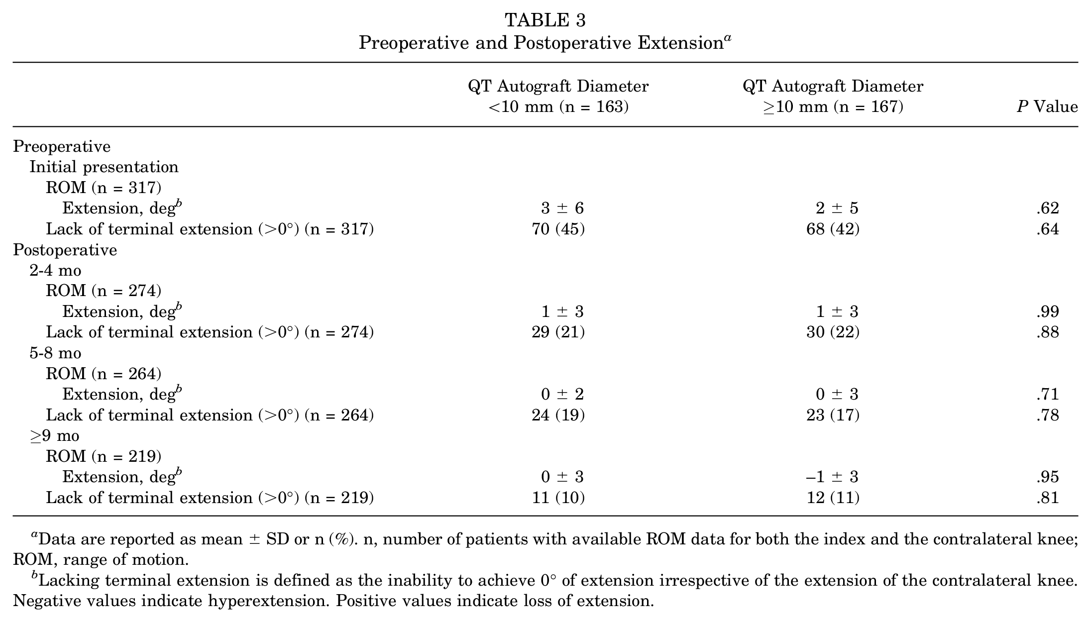
Data are reported as mean ± SD or n (%). n, number of patients with available ROM data for both the index and the contralateral knee; ROM, range of motion.
Lacking terminal extension is defined as the inability to achieve 0° of extension irrespective of the extension of the contralateral knee. Negative values indicate hyperextension. Positive values indicate loss of extension.
A univariate logistic regression analysis revealed that the inability to achieve terminal extension (defined as being unable to achieve 0° of extension irrespective of the extension of the contralateral knee) at the initial preoperative clinical presentation was significantly associated with postoperative loss of extension (OR, 2.28 [95% CI, 1.12-4.63]; P = .02). Female sex (OR, 1.68; P = .14), concomitant meniscal repair (OR, 1.16; P = .67), surgery <6 weeks from injury (OR, 1.29; P = .48), QT autograft diameter (OR, 0.92; P = .73), notch volume (OR, 0.94; P = .57), and the ratio of QT autograft diameter to notch volume (OR, 0.94; P = .88) were not significantly associated with postoperative loss of extension. Only preoperative lack of terminal extension remained significantly associated with postoperative loss of extension after performing a multivariate analysis (OR, 2.23 [95% CI, 1.10-4.58]; P = .03).
Discussion
The most important finding of this study was there was no association between QT autograft diameter, notch volume, and the ratio of QT autograft size to notch volume with postoperative loss of extension after QT autograft ACLR. There was also no difference in the preoperative or postoperative knee extension between patients who underwent ACLR with a QT autograft diameter ≥10 mm or <10 mm. Secondary findings included an independent association between preoperative lack of terminal extension at the initial clinical presentation and postoperative loss of extension after QT autograft ACLR. Additionally, the overall rate of loss of extension in this cohort was 11%, and 70% of those with loss of extension ultimately required surgical intervention to restore extension. Surgeons may use the results of the current study to identify patients who may be at risk of postoperative loss of extension after QT autograft ACLR, which primarily includes patients who have preoperative loss of extension. Undergoing ACLR with a large QT autograft diameter in a small notch is not a risk factor for loss of extension based on the results of the current cohort, although the reasons for the overall increased rate of postoperative loss of extension with QT autograft ACLR need further investigation.
QT autograft diameter was not different between patients with and without postoperative loss of extension, which was contrary to the hypothesis and results of previous studies.9,24 A femoral tunnel diameter >9.25 mm has previously been shown to be associated with postoperative loss of extension among patients receiving all–soft tissue QT autograft ACLR, although this association was only found in male patients. 9 The current study showed no difference in femoral and tibial tunnel diameters, in addition to QT autograft diameter, between patients with and without postoperative loss of extension. There was also no difference in preoperative and postoperative extension when grouping patients based on a QT autograft diameter ≥10 mm or <10 mm. Comparing postoperative loss of extension between patients in the upper and lower quartiles for the ratio of QT autograft diameter to notch volume (patients with the largest QT autografts in the smallest notches versus patients with the smallest QT autografts in the largest notches) revealed no differences.
Measurements of notch volume were similar between patients with and without postoperative loss of extension, which was also contrary to the hypothesis. A narrower notch has been found among patients who develop a cyclops lesion compared with controls.4,6 However, previous studies used 2-dimensional measurements of notch volume and included patients undergoing primarily hamstring tendon autograft ACLR. Postoperative loss of extension in the current study was also broadly defined to include all causes and not limited to cyclops lesions, which may explain differences from the results of previous literature. The lack of association between notch volume and postoperative loss of extension may also be secondary to a poor correlation between notch width and notch volume. 26
Risk factors for postoperative loss of extension in this study were limited to preoperative lack of terminal extension at the initial clinical presentation (defined as the inability to achieve 0° of extension), which corresponded with a 2.23-fold increase in the risk for postoperative loss of extension on a multivariate analysis. Such findings are expected given the close relationship between preoperative, early postoperative, and later postoperative extension that has been previously described.7,17 One previous study found a 2.79-fold increased risk of postoperative loss of extension 12 months after hamstring tendon autograft ACLR for patients who had preoperative loss of extension, which supports the findings of the present study. 28 However, there were no differences between patients with and without postoperative loss of extension with respect to sex, concomitant meniscal repair, and surgical timing. The results of the present study suggest that the preoperative extension is closely related to the postoperative extension and has a greater effect on the risk for developing postoperative loss of extension than QT autograft diameter, notch volume, or other patient and surgical characteristics.
The overall rate of postoperative loss of extension in this cohort was 11%, which is higher than that of previous studies describing a rate ranging from 1.5% to 4.5%.2,11,13,20 However, when looking specifically at QT autograft ACLR, rates are more comparable to the current study and range from 7.2% to 8.3%.9,13 Furthermore, 1 study investigating the relationship between terminal knee extension and quadriceps strength among patients undergoing QT autograft ACLR found that 7.5% (24/320) of patients underwent a subsequent surgery for loss of extension, which is closely replicated by the current study's rate of 7.8% (26/333). 14 QT autograft ACLR has also been shown to increase the risk of a subsequent manipulation under anesthesia or lysis of adhesions by 2.68-fold compared with other allograft and autograft choices, which may explain the higher rate of postoperative loss of extension compared with other graft types described in the literature. 13 QT autograft harvest may lead to increased postoperative loss of extension and/or strength deficits compared with alternative graft options and explain this higher rate of postoperative loss of extension, although evidence is mixed.3,12,14,16 While the current study did not show any increased risk for loss of extension based on the variance in QT autograft diameter, the higher rate of loss of extension in this QT autograft ACLR cohort may be due to an increased intra-articular volume that the QT autograft likely occupies compared with other graft types such as the bone–patellar tendon–bone (BPTB) autograft. It is possible that all QT autografts are “too large” compared with BPTB autografts, explaining the discrepancy in rates of loss of extension.
Donor site morbidity and/or pain at the extensor mechanism, along with quadriceps muscle inhibition 18 or quadriceps muscle weakness after QT autograft harvest, could be related to postoperative loss of extension. Arthrogenic muscle inhibition is a process that has been described in patients with ACL injuries, which is characterized by neural inhibition of quadriceps muscle activation coupled with excessive hamstring muscle contraction, and leads to loss of knee extension. 22 A QT autograft harvest and closure may affect the communication between the quadriceps muscle, QT, and neural pathways in the brain and spinal cord, resulting in a more delayed recovery of knee extension compared with other autografts. Restoring full knee extension preoperatively in all patients undergoing ACLR, but particularly in patients with arthrogenic muscle inhibition, is a critical aspect of ACL injury management that may lower the risk of postoperative loss of extension. 23 Future studies should investigate optimal perioperative management strategies to avoid postoperative loss of extension after QT autograft ACLR, which may include delaying surgery until patients demonstrate adequate quadriceps muscle activation and/or any combination of postoperative rehabilitation protocols.
Limitations
This study has a few limitations. Seven different surgeons were included in the current study, which limits standardization of postoperative rehabilitation protocols and indications for surgical procedures to address loss of extension. However, using patient data from several surgeons practicing in a large academic medical center increases the generalizability of study results. QT autograft harvest techniques were not standardized among surgeons, and such variability may influence the analysis of postoperative loss of extension. Analyses of QT autograft diameter and notch volume did not account for the volume occupied by other intra-articular structures (ie, the posterior cruciate ligament), which may alter the space available for the QT autograft and confound findings. A goniometer was not used to measure range of motion, and findings may be subject to measurement bias as a result. Patients who experienced loss of extension in the early postoperative period (ie, 3 months postoperatively) may have demonstrated improvements in extension throughout their recovery without requiring additional surgical intervention despite being categorized as having loss of extension. The post hoc power analysis indicated that the study sample size was adequately powered to detect a 10% higher rate of loss of extension in the ≥10-mm QT autograft diameter group, but not a 5% higher rate of loss of extension. The study sample size is therefore underpowered to detect smaller differences between groups. The 67% study inclusion rate also reflects the loss to follow-up in this study and may be another source of bias.
Conclusion
Eleven percent of patients undergoing QT autograft ACLR experienced a >5° loss of knee extension compared with the contralateral knee or required additional surgery to restore extension. QT autograft diameter and notch volume were not associated with postoperative loss of extension among patients who underwent primary QT autograft ACLR. The inability to achieve terminal extension (0°) at the initial preoperative presentation increased the risk of postoperative loss of extension by 2.23-fold. Surgeons may consider the lack of terminal extension preoperatively as a risk factor for postoperative loss of extension after QT autograft ACLR rather than increased QT autograft diameter or decreased notch volume.
Footnotes
Final revision submitted December 30, 2024; accepted January 31, 2025.
One or more of the authors has declared the following potential conflict of interest or source of funding: V.M. reports educational grants, consulting fees, and speaking fees from Smith & Nephew; educational grants from Arthrex and DePuy/Synthes; is a board member of the International Society of Arthroscopy, Knee Surgery and Orthopaedic Sports Medicine (ISAKOS); and deputy editor-in-chief of Knee Surgery, Sports Traumatology, Arthroscopy (KSSTA). J.J.I. is president of the board of directors for the Journal of Orthopaedic & Sports Physical Therapy (JOSPT). J.D.H. is on the editorial board of Knee Surgery, Sports Traumatology, Arthroscopy (KSSTA). AOSSM checks author disclosures against the Open Payments Database (OPD). AOSSM has not conducted an independent investigation on the OPD and disclaims any liability or responsibility relating thereto.
Ethical approval for this study was obtained from University of Pittsburgh Institutional Review Board (STUDY19030196).