
Abstract
Background:
Surgical repair of full thickness biceps tears has demonstrated adequate outcomes in short and mid-term studies. However, data on the long-term outcomes of full thickness distal biceps injuries and their treatment are currently lacking.
Purpose/Hypothesis:
The purpose of this study was to report on patient demographics, injury characteristics, and long-term outcomes for patients with full-thickness distal biceps tears. It was hypothesized that complete distal biceps tears managed operatively would demonstrate robust clinical success at long-term follow-up.
Study Design:
Case series; Level of evidence, 4.
Methods:
Patients with magnetic resonance imaging–confirmed, complete distal biceps tendon rupture sustained between 1996 and 2016 were identified. Patients were cross-referenced with a regional geographic database.
Results:
A total of 66 patients (3 female, 63 male) with a median age of 50.8 years (IQR, 41.5-60.4) and a median clinical follow-up of 14.7 years (IQR, 9.6-17.9 years) were included. Patients who sustained a full-thickness distal biceps tendon tear were likely to be in their early 50s, male, right-hand dominant, current/former smokers, and laborers with a history of traumatic injury during an intentional movement. Most of these patients had pain and supination weakness but no loss of range of motion. All included tears were treated operatively. At final follow-up, patients maintained a majority of near-normal range of motion (median total arc of flexion/extension 140° and supination/pronation 80°), excellent elbow flexion strength (91% of patients had full strength), and adequate elbow supination strength (76% of patients had full strength). The overall complication rate was 24%, with 16 out of 66 patients experiencing some type of complication between infection, rerupture, heterotopic ossification, reoperation, and nerve complications. Overall return to work was 98%, and 85% of those who returned to work did so without restrictions.
Conclusion:
Complete tears of the distal biceps were most common in patients 50 years of age, male sex, right-hand dominant, and current/former smokers. The most common profession was laborer, and injuries were primarily traumatic in nature during intentional activity. Patients managed operatively demonstrated high rates of success at long-term follow-up with respect to elbow function and clinical outcomes.
Keywords
Distal biceps tendon tears can result in significant impairment in elbow function. These injuries typically occur secondary to forceful extension of the elbow while the biceps muscles are contracted with the forearm supinated, causing sudden eccentric biceps elongation and subsequent rupture.18,25 The incidence of distal biceps tendon injury varies, with recent studies estimating an incidence of 2.6 to 3.0 per 100,000 patient years nationally,11,15,16 which represents an increase from historical estimates of 1.2 per 100,000 patient years.18,24 Injury rates appear to be rising as well, with some Nordic countries reporting a 6-fold increase in incidence over the past 20 years. 16 This increase may be a reflection of advancements in our understanding of this pathology and with improvements in the clinical history, physical examination maneuvers, and imaging necessary for injury identification. 21 Identification of distal biceps tendon tears occurs by several mechanisms. Specific physical examination maneuvers, particularly the hook test,21,32 remain important clinical tools, while magnetic resonance imaging (MRI) has emerged as the preferred modality for identifying partial- and full-thickness tears.18,21,22,32
When a full-thickness tear is identified, surgical repair is generally recommended for acute injuries in active patients to improve strength in supination and to decrease deformity while providing symptomatic relief. Surgery is ideally performed within a few weeks to reduce the risk of tendon retraction and fibrosis.18,22 Multiple techniques for repair of the distal biceps have been described in the literature. Studies have reported on the outcomes of distal biceps tendon injuries based on surgical approach 8 (single vs. dual incision) and method of fixation8,27 (anchors vs. suture vs. buttons). Several cadaveric studies have also been published.13,14 However, there is no current consensus on the optimal surgical construct, as each approach demonstrates specific advantages and pitfalls. Surgical repairs performed through a single-incision approach have demonstrated a higher rate of lateral antebrachial cutaneous nerve impairment, but this typically resolves with conservative management.1,8 For dual-incision techniques, the most commonly reported complications are related to the development of heterotopic ossification.1,2 Overall clinical outcomes tend to be quite favorable regardless of the surgical approach utilized or the method of fixation, although these investigations are largely limited to short- and mid-term follow-up.12,17
Current literature lacks substantive analyses of long-term outcomes for patients with full-thickness distal biceps tendon tears, and it is unknown if repairs are durable over long-term follow-up. Available literature is also limited in analysis of functional outcomes and impairments such as range of motion, strength, pain, and ability to return to work. Therefore, the primary goals of this study were to demonstrate patient demographics, injury characteristics, and long-term outcomes of operatively treated complete distal biceps tendon tears. We hypothesized that complete distal biceps tears managed operatively would demonstrate robust clinical success at long-term follow-up.
Methods
Study Population and Design
After Mayo Clinic institutional review board approval, patients who sustained an MRI-confirmed complete distal biceps rupture between 1996 and 2016 were identified by a musculoskeletal radiologist (A.C.J.) through a search of institutional image records. Patients from the initial search were then cross-referenced with the Rochester Epidemiology Project (REP). The REP is an electronic collection system of complete medical records of >600,000 patients, all of whom are residents of Olmstead County, Minnesota, or neighboring counties in southeast Minnesota and western Wisconsin. The methodology and generalizability of the REP has been previously described in detail.29,30 Medical charts and records were subsequently queried by 2 independent reviewers (S.E.T., A.K.R.) to confirm the diagnosis of complete distal biceps tendon rupture and to obtain demographic data.
Inclusion criteria consisted of patients with complete distal biceps tendon tear confirmed by MRI, complete medical records available for review (including imaging and operative reports), and ≥5 years of clinical follow-up. Exclusion criteria consisted of patients with polytrauma, partial-thickness distal biceps tears; unconventional biceps repairs; tears discovered incidentally without a clinical assessment; pathologic tears of the distal biceps tendon (tumor erosion); inflammatory arthritis and enthesitis (rheumatoid arthritis-related); fractures or dislocations of the elbow; patients who were treated nonoperatively; and patients who were lost to follow-up.
Medical records were then reviewed to collect patient demographic data (age at diagnosis, sex, body mass index, hand dominance, occupation), comorbidities (diagnosis of diabetes, nicotine use, etc), injury characteristics, physical examination findings, and radiographic/imaging characteristics at presentation. Particular attention in physical examination findings was given to the hook test. The hook test was conducted by actively flexing the patient's elbow to 90° while the examiner's finger attempted to hook both the medial and the lateral aspect of the biceps tendon within the antecubital fossa. In a complete biceps tendon rupture, no cordlike structure was palpated when the examiner hooked the medial or lateral aspect of the antecubital fossa. Sensitivity and specificity for the hook test have been determined to be excellent. 21 Surgical details (incision type, fixation method) as well as complications such as infection, rerupture, heterotopic ossification, and postoperative nerve dysfunction were also recorded. Outcome measures included physical examination findings at final follow-up (extension, flexion, supination, and pronation range of motion) and return-to-work status.
Surgical Technique
Surgical technique was based on surgeon preference. Most patients were treated with primary repair of the tendon directly to the bicipital tuberosity. The surgical techniques utilized in this cohort included (1) a 2-incision technique using bone tunnel-suture fixation (no implants used), (2) anterior single-incision technique using cortical button fixation alone, (3) anterior single-incision technique using cortical button and interference screw fixation, and (4) anterior single-incision technique using suture anchor fixation. For this study, the approach was classified as “2-incision” if the patient received both anterior and posterior incisions.
Statistical Analysis
Data were collected and stored in Microsoft Excel (2010; Microsoft Corp). Univariate analysis of clinical and radiographic characteristics was performed for subgroup analysis (operative vs. nonoperative). After analyzing data for parametric/nonparametric assumptions, continuous variables were compared between treatment groups utilizing Student t tests or Wilcoxon rank-sum tests, and categorical variables were similarly compared with chi-square analysis or Fisher exact tests. Missing data were omitted in data analysis. P values <.05 were considered significant. All statistical analyses were conducted in the R statistical environment (Version 4.3.1; R Foundation for Statistical Computing).
Results
Presenting Characteristics
Patient characteristics are presented with descriptive statistics using percentage counts, median with interquartile range, and mean with 95% CI as appropriate. Overall, 98 patients with MRI-confirmed, full-thickness distal biceps tendon tears treated operatively were identified. Of these, 66 individuals had ≥5-year follow-up and met all inclusion criteria for this study (Figure 1). Median follow-up time was 14.7 years (IQR, 9.6-17.9 years), median age at the time of diagnosis was 50.8 years (IQR, 41.5-60.4 years), and most patients were male (95%). Despite being majority right-hand dominant (82%), patients presented with injury to left and right extremities equally (50% and 50%, respectively). Tobacco usage was common, with 21% current and 32% former. Of total patients, 14% had diabetes, and 1 patient had chronic renal disease. Laborer was the most common occupation (56%). Complete patient demographic characteristics are found in Table 1.

Flowchart demonstrating patient inclusion and exclusion. MRI, magnetic resonance imaging.
Patient Demographics and Clinical Characteristics a

Data presented as median (IQR) or count (%). BMI, body mass index.
The median time from injury to presentation was 8 days (IQR, 2.3-20.8 days). Of the 66 patients, 76% reported a distinct “pop” at the time of injury and 56% had a visible deformity. Pain at time of consult and decreased supination strength (defined as <5/5 strength on physical examination) were the most common symptoms at presentation (98% and 89%, respectively
Presenting Characteristics of Complete Tears of the Distal Biceps Tendon a

Data presented as median (IQR), count (%) where data was available in all patients, or count/total number of patients with available data (%) where data was not available in all patients. TTP, tender to palpation.
Not performed/recorded in all patients; data reported as count/total number of patients with available data (%).
Surgical Characteristics and Outcomes at Final Follow-up
Median time from injury to surgery was 17 days (IQR, 8.25-52.5 days) (Table 2
Operative Strategies for Surgical Repair of Complete Tears of the Distal Biceps Tendon (N = 66)

Outcomes at Final Follow-up of Complete Tears of the Distal Biceps Tendon a
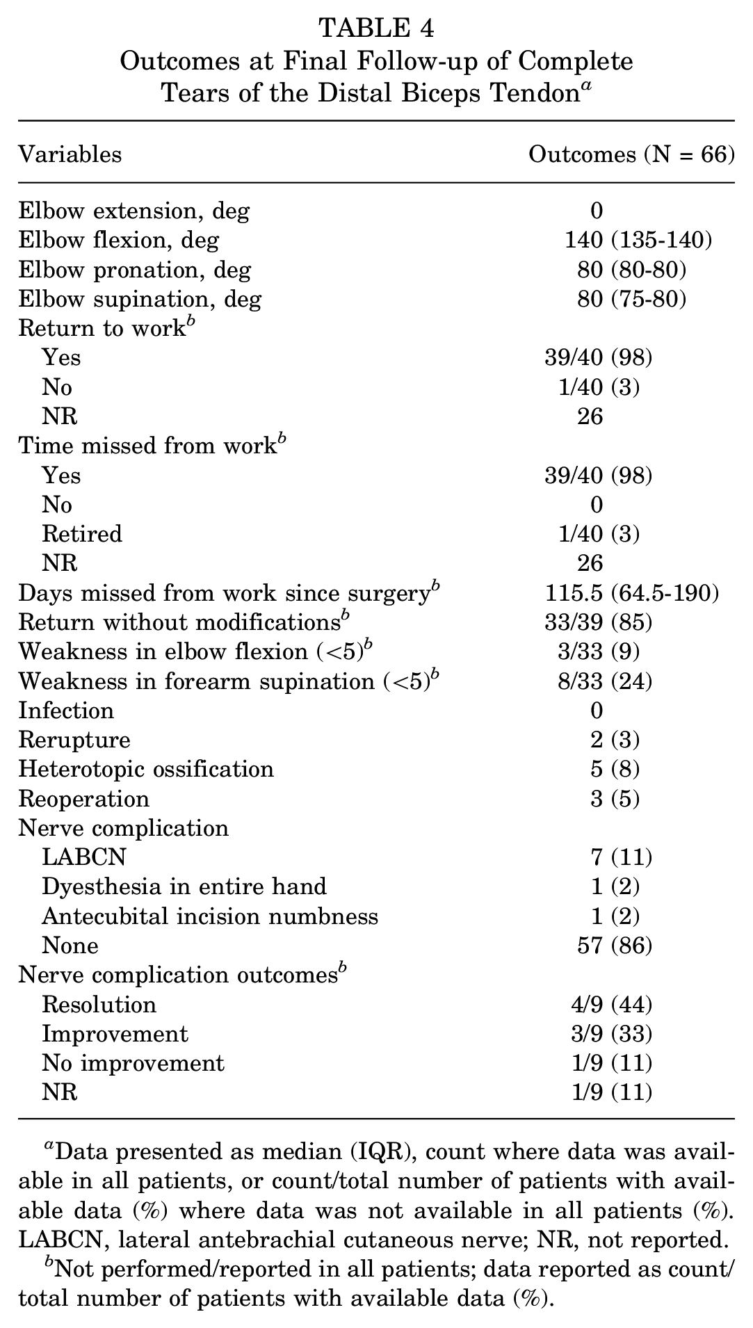
Data presented as median (IQR), count where data was available in all patients, or count/total number of patients with available data (%) where data was not available in all patients (%). LABCN, lateral antebrachial cutaneous nerve; NR, not reported.
Not performed/reported in all patients; data reported as count/total number of patients with available data (%).
On physical examination at final follow-up, the median flexion-extension arc was 140° (IQR, 135°-140°) and median supination was 80° (IQR, 75°-80°). On manual muscle testing, 3 of 33 patients (9%) demonstrated persistent elbow flexion weakness and 8 of 33 (24%) patients demonstrated supination weakness (Table 4
Discussion
In this study of long-term outcomes for complete distal biceps tendon tears, most patients were 50 years of age, male sex, right-hand dominant, current or former smokers, and laborers with a traumatic injury during an intentional movement pattern. Long-term outcomes of surgical repair were favorable and durable at a median of 14.7-year follow-up. Patients maintained majority near normal range of motion (median total arc of flexion/extension of 140° and supination/pronation of 80°), demonstrated excellent elbow flexion strength (91% had full strength), and demonstrated adequate elbow supination strength (76% had full strength) at final follow-up. Patients reported significant time missed from work due to postoperative recovery.
The demographics of patients in this present study are comparable with those of previously published, albeit limited, literature. Prior investigations have historically found that the majority of these injuries occur in male patients.5,23 Luokkala et al 19 further demonstrated this in their 2022 study, where consecutive patient sampling found a male:female case ratio of 225:1, or <0.5% female patients in their total study population. While our current investigation also demonstrated a majority male population, this difference in the ratio of sex was not as extreme in our study; female patients accounted for >5% of our population. Previously published studies have demonstrated that most patients experiencing biceps tendon ruptures are between their fourth and sixth decade of life.5,19,23,28 These findings have been closely mirrored in our study cohort. Tobacco use has been demonstrated to be a significant factor in distal biceps pathology. Prevalence of tobacco use in previous studies has been reported as between 31.2% and 43%, with active tobacco use increasing tear risk up to 7.5 times compared with that of nonsmokers.15,20,24 Similarly, a significant proportion of our study population reported significant active (21%) and former (32%) smoking status and tobacco use. Occupational risk factors have been less clearly defined, as some studies attribute elevated risk to laborer occupations, yet others are substantially less conclusive.15,31 The results from our study contribute to evidence suggesting increased association of laborer occupations with complete distal biceps tears, although no conclusive statement can be made since no direct comparison was conducted between laborers and nonlaborers.
This study demonstrated that favorable outcomes are maintained at long-term follow-up for surgically treated acute, complete tears, with acceptable rates of complication. Prior studies have demonstrated similar results in the immediate, short, and medium postoperative follow-up time points with respect to both symptomatic and functional outcomes.3,4,26,28,31,32 Previously published investigations have suggested a complication rate of around 25% without delineating operative techniques and fixation methods.6,7,15,31 We reported a comparable complication rate of 24% for all operative strategies. The majority of these were simple neurapraxias that resolved with observation alone. Numerous operative approaches and fixation techniques exist, and no single technique has demonstrated clinical superiority. Grewal et al 10 found no significant differences in outcome in a randomized clinical trial comparing single- and dual-incision repair techniques. However, they did note an increase of approximately 10% flexion strength for 2-incision repair and a 10% higher rate of transient LABCN neurapraxias in single-incision repairs. 10 A more recent investigation of 277 operatively treated patients found a substantially higher revision rate with single-incision repair, but this has not been corroborated with other studies. 9 The majority of patients in our study received a 2-incision approach and received a bone-tunnel suture construct.
Limitations
Our study is not without limitations. All data were collected retrospectively across numerous databases, which limited uniformity and consistency in data point availability. This was an unavoidable limitation of the database queried for this study. However, it was determined that the greater external validity afforded by pooling patients from numerous health systems outweighed the potential biases and limitations introduced. There were multiple operative approaches, and the postoperative treatment was not standardized across the cohort, which may have confounded postoperative outcomes depending on each patient's treatment algorithm. Additionally, a regression analysis could not be attempted with respect to surgical approach and outcome. The limited data on different approaches were not permissive of interpretable results.
Conclusion
Complete tears of the distal biceps were most common in patients 50 years of age, male sex, right-hand dominant, and current or former smokers. The most common profession was laborer, and injuries were primarily traumatic in nature during intentional activity. Patients managed operatively for complete distal biceps tears demonstrated high rates of success at long-term follow-up with respect to elbow function and clinical outcomes.
Footnotes
Acknowledgements
The authors acknowledge the support of the Foderaro-Quattrone Musculoskeletal-Orthopaedic Surgery Research Innovation Fund.
Final revision submitted April 3, 2024; accepted April 12, 2024.
One or more of the authors has declared the following potential conflict of interest or source of funding: This study was partially funded by the following: National Institute of Arthritis and Musculoskeletal and Skin Diseases for the Musculoskeletal Research Training Program (T32AR56950). This study used the resources of the Rochester Epidemiology Project medical records-linkage system, which is supported by the National Institute on Aging (AG 058738), by the Mayo Clinic Research Committee, and by fees paid annually by Rochester Epidemiology Project users. A.J.T. has received hospitality payments from Stryker and Zimmer Biomet Holdings. J.D.B. has received consulting fees from Stryker and Arthrex, education payments from Arthrex, and nonconsulting fees from Arthrex. C.L.C. has received consulting fees from Arthrex and education payments from Arthrex. AOSSM checks author disclosures against the Open Payments Database (OPD). AOSSM has not conducted an independent investigation on the OPD and disclaims any liability or responsibility relating thereto.
Ethical approval for this study was obtained from Mayo Clinic (PR16-007084-03).