
Abstract
Background:
Biocomposite screws reportedly provide equivalent graft fixation in anterior cruciate ligament reconstruction (ACLR) to metallic screws while simplifying subsequent imaging and surgery. One purported complication of biocomposite screws is paradoxical tunnel widening. Previous studies on beta-tricalcium phosphate screws have only reported outcomes at short- and midterm follow-up.
Purpose:
To radiographically assess the tibial tunnel 10 years after ACLR using hamstring tendon autografts and biocomposite interference screws in anatomic single-bundle (SB) and double-bundle (DB) methods.
Study Design:
Case series; Level of evidence, 4.
Methods:
Of the 105 initially recruited patients, 61 (58%) completed all follow-up evaluations for inclusion in this long-term study. A total of 26 patients received anatomic SB ACLR and 35 patients received DB ACLR with biocomposite interference screws containing beta-tricalcium phosphate in the tibia. Weightbearing anteroposterior and lateral radiographs of the index knee were taken in the early postoperative period and at 2, 5, and 10 years postoperatively; computed tomography (CT) imaging was performed at 10-year follow-up. Subjective and objective clinical assessments were recorded preoperatively and at 10-year follow-up.
Results:
The mean follow-up period was 122 months. In 76% of radiographs in the SB group, the width of the tibial tunnel had not increased at 10 years compared with the early postoperative period. The mean tibial tunnel volume on CT in the SB group was 2.04 cm3 (± 0.85 cm3). In the DB group, the posterolateral tunnel width had not increased in 69% of radiographs; the same was found in 63% of radiographs for the anteromedial tunnel at 10-year follow-up. The mean posterolateral tunnel volume on CT was 2.04 cm3 (±1.92 cm3) and the mean anteromedial tunnel volume was 1.38 cm3 (±0.54 cm3). There was no correlation between tunnel widths and KT-1000 arthrometer assessments. There was a moderate but statistically significant correlation between SB tibial tunnel volume on CT imaging and KT-1000 arthrometer anterior 134 N side-to-side difference (r = 0.45; P = .039).
Conclusion:
Most patients’ tibial tunnels had not increased on 1 or both radiographic views at 10-year follow-up compared with the early postoperative period after ACLR using biocomposite interference screws, with no obvious negative effect on outcomes. However, the tunnels were still visible in most patients at 10 years on standard radiographs and CT imaging.
Anterior cruciate ligament (ACL) reconstruction (ACLR) using autologous hamstring tendon grafts secured with interference screws is still a common procedure after their introduction decades ago. 13 Metallic interference screws have been used since the early 1980s with proven fixation strength and long-term biocompatibility 33 despite their disadvantages of possible graft laceration at screw insertion, artifacts on subsequent magnetic resonance imaging (MRI), and the risk of complicating eventual revision surgery.2,17,36,66
These shortcomings led to the introduction of bioabsorbable fixation screws made of poly-L-lactic acid (PLLA), polyglycolic acid (PGA) sometimes in combination with polylactic acid, polyparadioxanone, or diverse stereoisomers of lactic acid .2,7,34,43 Bioabsorbable screws have also been associated in the literature with complications such as screw breakage at insertion, bone tunnel enlargement, soft tissue reactions, cyst formation, and lack of osteoconductivity.2,3,21,32,34,37,59,61 By adding bioceramics such as hydroxyapatite or beta-tricalcium phosphate (β-TCP) to existing bioabsorbable polymers, manufacturers were able to improve osteoconductivity and accelerate graft incorporation via bone formation in the tunnel; this led to increased utilization of these biocomposite screws. 56 These screws also avoid the MRI incompatibility issues of metallic screws.6,7,40
Tunnel widening was seen as a crucial complication of bioabsorbable/biocomposite screws given their original purpose was to reduce the bony deficit after ACLR in case of the need for revision; tunnel widening with its subsequently increased bony deficit may increase the risk of revision surgery failure. 49 There is no consensus on whether any eventual tunnel widening correlates with clinical outcomes. Furthermore, we are unaware of studies that have shown enlargement to be clinically significant regarding graft laxity or increased failure rates.2,11,41
The anatomic double-bundle (DB) method reconstructs the anteromedial (AM) and posterolateral (PL) bundles of the ACL separately with the purported benefit of restoring both anteroposterior laxity and rotational laxity for which the PL bundle is primarily responsible. 31 Given that the method involves 2 tunnels, any eventual tunnel widening could theoretically affect future revision surgery more so than the single-bundle (SB) method, either directly or indirectly via tunnel communication. 29
The purpose of this prospective study was to assess and compare tibial tunnel width radiographically up to 10 years after ACLR with either anatomic SB or anatomic DB methods using hamstring tendon autografts and biocomposite interference screws. We hypothesized that no tibial tunnel enlargement would be found 10 years after ACLR in either SB or DB patients.
Methods
Ethics
The regional ethical review board at the University of Gothenburg approved the study. The participants received oral and written information about the study, after which written consent was obtained. Participants could also choose to decline further involvement at 10-year follow-up after previous participation.
Patients
Between March 2008 and September 2009, 105 patients were recruited at 2 hospitals in western Sweden (n = 31 and n = 74, respectively) and randomized to either the SB group or the DB group as part of another study 4 ; these patients were followed prospectively with radiographs assessing the tibial tunnel, up to 10 years postsurgery, for the present prospective cohort study (Figure 1). All short- and long-term clinical and subjective outcomes have previously been reported.1,4,27 The 5-year assessment of the tibial drill holes on plain radiographs in the SB group has also previously been reported. 28

Flowchart of included patients. ACL, anterior cruciate ligament; DB, double bundle; SB, single bundle.
Computed tomography (CT) images were also included at 10-year follow-up. There were 61 patients who participated in this study. The study group was unselected in regard to age, weight, height, sex, and activity level. Inclusion criteria were patients aged ≥18 years with a unilateral ACL injury at the time of index surgery. Exclusion criteria were concomitant posterior cruciate ligament injury, medial or lateral collateral ligament injury greater than grade 1, previous major knee surgery, or a contralateral ACL injury preceding the index surgery. One patient sustained a contralateral ACL injury before the date of the index operation but after preoperative clinical tests was kept in the radiographic part of the study and excluded from the side-to-side clinical assessments. The patients who fulfilled the inclusion criteria were asked to participate in the study. Surgical indication included failed nonsurgical treatment or participation in pivoting sports, whereas nonsurgical treatment was seen as an inferior treatment option.
Surgical Technique
Four senior high-volume surgeons (including J.K., M.A.) operated on the included patients, with 1 surgeon (J.K.) supervising the other 3 on how to perform both surgical techniques. These 3 surgeons were given a surgical learning curve of approximately 30 cases before the present study was initiated. The 2 surgeons at hospital 1 (n = 74) operated cases separately; surgeon 1 (J.K.) operated on 43 cases (20 DB, 23 SB) and surgeon 2 on 31 cases (19 DB, 12 SB). The other 2 surgeons (including M.A.) assisted each other with their cases, 31 in total (14 DB, 17 SB).
Standard anterolateral and AM portals were established perioperatively in both groups. Any associated intra-articular injuries, such as meniscal ruptures and chondral lesions, were addressed at the time of the index operation. Both femoral and tibial ACL footprints were identified, as well as the lateral intercondylar and bifurcate ridges on the femur; ACL remnants were then resected and any loose debris removed. Semitendinosus and gracilis tendons were harvested with an open tendon stripper via a standard longitudinal incision at the pes anserinus on the AM aspect of the proximal tibia. Femoral drilling was done using a fluted reamer through the AM portal (inside-out); tibial drilling was performed using a tibial elbow aimer at 50° and a fluted reamer. Tibial screws in both techniques were inserted until the head was just inside the cortical bone, resulting in its being behind and distal to the graft. No measurement of graft tension at the time of fixation was performed in this study.
Anatomic SB Technique
Addressing the femoral tunnel first, the femoral insertion site was marked with a Steadman awl (ConMed Linvatec) in the center of the ACL footprint with the knee at 90° of flexion. The femoral tunnel was predrilled using a 4.0-mm sharp, noncannulated drill or a guide wire before the final tunnel was drilled, its diameter determined by the size of the graft. The tibial tunnel was drilled in the center of the tibial footprint, in line with the anterior horn of the lateral meniscus. All bone tunnel diameters were approximately 0.5 mm greater than the diameters of the respective grafts. The ACL graft consisted of 4- or 5-stranded semitendinosus and gracilis tendons with a graft diameter between 7.5 and 8.5 mm (mean, 7.8 mm). Metal interference screws 7 × 25 mm (RCI; Smith & Nephew) were used on the femoral side and biocomposite screws 9 × 25 mm were used on the tibial side, made of self-reinforced 77% PLDLA and 23% β-TCP (poly-levo [96%]/dextro [4%]-lactide/β-TCP [SR-PL(96)/D(4)LA/β-TCP]) (Matryx; ConMed Linvatec) (Figure 2). Tibial fixation was performed at 10° to 20° of knee flexion. 1

Biocomposite interference screw (Matryx) used in study. Copyright Ioannis Karikis. 26
Anatomic DB Technique
Starting with the femoral tunnels, the femoral insertion sites of the AM and PL bundles were marked with a Steadman awl. The AM and PL tunnels were drilled in the middle of their respective anatomic footprints. The tibial tunnels were drilled in the center of the footprint of the AM and PL bundles, respectively. The AM tunnel was placed in line with the anterior horn of the lateral meniscus and the PL tunnel in front of the posterior cruciate ligament. On both the femoral and the tibial sides, the drill holes were 6.5 to 7 mm for the AM bundle and 6 mm for the PL bundle. All bone tunnel diameters were approximately 0.5 mm greater than the diameters of the respective grafts. Femoral fixation was achieved using metal interference screws 6 × 20 mm (RCI), while fixation on the tibial side used the same type of biocomposite screws, 7.3 × 20 mm in the AM tunnel and 7.3 × 25 mm in the PL tunnel (Figure 2).
The AM graft consisted of a doubled semitendinosus tendon, with graft diameter being 5.0 to 7.0 mm (mean, 6.3 mm), and the PL graft of a doubled or tripled gracilis tendon of 5.0 to 7.0 mm (mean, 5.9 mm) diameter. Tibial fixation was performed in 5° to 10° of knee flexion for the PL bundle and in 40° to 60° of knee flexion for the AM bundle.
Rehabilitation
All participants undertook rehabilitation in line with regional guidelines via local physical therapists, with immediate full weightbearing and full range of motion (ROM), including full extension without the use of a brace. Closed kinetic chain exercises were initiated immediately postoperatively. Running was permitted at 3 months and contact sports at 6 months after surgery at the absolute earliest, provided the patient had regained full functional stability in terms of strength, coordination, and balance compared with the contralateral leg.
Clinical Assessments
One physical therapist (N.S.) not involved in participants’ rehabilitation performed all pre- and postoperative follow-up examinations used in the study, including the laxity measurements. The physical therapist was blinded to the surgical technique to which any given patient had been randomized but not to the aim of the study at the time of the examination. Patients who sustained a contralateral ACL injury were excluded from the side-to-side clinical assessments at the 10-year follow-up but were kept in the radiographic part of the study.
During their examinations, all participants underwent multiple subjective and objective tests: ROM, single-leg hop test, 55 Lysholm knee scoring scale, 54 and Tegner activity scale. 54 Any eventual flexion or extension deficit in ROM (subsequently dichotomized to yes or no) was calculated by subtracting the respective degrees of the index knee from those of the contralateral knee.
Results of the pivot-shift test were categorized clinically with grades 0 to 3 according to International Knee Documentation Committee guidelines. 23
The instrumented KT-1000 arthrometer was used to test the anterior displacement of the tibia in relation to the femur; it was registered at 134 N and as the maximal manual test. At least 3 measurements were made on each knee, and the mean value was registered.
Patients who had sustained a contralateral ACL injury since the start of the study observation period were excluded from side-to-side clinical assessments at the 10-year follow-up.
Radiographic Assessments
Early postoperatively (SB: mean ± SD, 6 ± 0.9 months; median [range], 6 months [5-8 months]; DB: mean ± SD, 6 ± 1 months; median [range] 6 months [4-8 months]); P = .75) and at 2-, 5-, and as part of 10-year follow-up, enrolled patients underwent unilateral standard radiography with weightbearing views of the index knee in anteroposterior (AP) and lateral views, with the knee in 30° of flexion. 1 Due to technical errors at 1 examination site, 9 SB and 8 DB lateral radiographs were not recorded and are the reason for some of the missing values (see Table 5). The other missing values were due to visibility of tibial tunnels on only 1 projection of a given knee but not the other; in these cases, the tunnels were obviously not healed due to being visible on 1 projection and were thus also classified as missing values (4 DB AM tunnels visible on lateral but not AP radiographs; 1 DB PL tunnel visible on AP but not lateral radiograph).
For the standard radiographs, the width at 3 points perpendicular to the long axis of the tunnel (each end and at the middle) was measured for both AP and lateral views (Figures 3 and 4) as previously described at the 2- and 5-year follow-up of the same patients.1,27 The mean of these 3 measurements was calculated and used to define the width of the tunnel. The images were analyzed by an independent radiologist (L.R.-C.) Inter- and intrarater test-retest reliability for this method has been considered good, with interclass correlation coefficient values between 0.84 and 0.97 for tibial measurements. 35 To allow reliable tibial tunnel measurements regardless of magnification factor, the head diameter of the femoral interference RCI screw was used as a reference on both AP and lateral radiographs. Using the true diameter of the RCI screw head, the true tibial tunnel measurement was found via (1) true femoral screw head/measured femoral screw head = magnification factor and (2) tibial tunnel measurement x magnification factor = true tibial tunnel measurement.

(A, C) Anteroposterior and (B, D) lateral weightbearing digital radiographs of the right knee of a male patient showing the single-bundle tibial bone tunnels (A, B) in the early postoperative period and (C, D) at 10 years postoperatively, with (E) anteroposterior and (F) lateral computed tomography imaging at 10 years (asterisk refers to single-bundle tunnel; volume, 2.45 cm3). (A, B) Width of tunnel proximally (1), middle (2), and distally (3) and diameter of screw head (4).

(A, C) Anteroposterior and (B, D) lateral weightbearing digital radiographs of the right knee of a male patient showing the double-bundle tibial bone tunnels (A, B) in the early postoperative period and (C, D) at 10 years postoperatively. Further, (E, G) being anteroposterior and (F, H) being lateral computed tomography imaging at 10 years (blue asterisk refers to double-bundle posterolateral tunnel; volume, 1.06 cm3; yellow asterisk refers to double-bundle anteromedial tunnel; volume, 1.15 cm3). (A, B) Width of the posterolateral tunnel proximally (1), middle (2), and distally (3); screwhead diameter for posterolateral bundle (4); screwhead diameter for anteromedial bundle (5); and anteromedial tunnel width proximally (6), middle (7), and distally (8).
CT scans of the operated knee were obtained at 10-year follow-up. The images were analyzed by an independent radiologist. Tunnels were determined using sclerotic margins as landmarks. Measurements were obtained via digital measurement tools in the radiology software.
Tunnel volumes were calculated using CT imaging. Either the GE 660 (GE Healthcare) or the Toshiba Prime CT software (Toshiba Medical Systems) suites were used. Axial images with coronal and sagittal reconstructions were obtained with 1-mm sections (Figures 3 and 4). The tunnels were classified according to their appearance: either cylindrical or conical. Standard volume estimation equations were used for the truncated conical and cylindrical forms: V = 1/3πh (r1 2 +r1xr2+πr2 2 ) and V = πr 2 h, respectively; given there was a specific angle of 50° for the tibial tunnel, tunnel volume was not affected by tunnel length.
Statistical Analysis
Mean (standard deviation) and median (range) values are presented when applicable. Comparison of dichotomous variables between groups was performed with the chi-square test. In terms of comparisons of both continuous and noncontinuous variables between groups, the Mann-Whitney U test was used. The Wilcoxon signed-rank test was utilized for comparisons of the preoperative and 10-year follow-up clinical assessment data within the 2 groups.
Comparison of the tibial tunnel mean diameter means over time was performed with 1-way repeated-measures analysis of variance, whereas the Bonferroni test was used for the post hoc analyses.
The Pearson test was used for correlation analysis between the KT-1000 arthrometer laxity measurements and tibial tunnel diameter and the tibial tunnel volume on CT at 10-year follow-up.
Statistical significance was set at P < .05. To be able to detect a 1-mm decrease in tunnel width with SD of 2 mm, power of 80%, and P < .05, 33 patients per group were required.
The statistical program IBM SPSS Statistics Version 28.0.1.1 was used for the statistical analysis.
Results
Demographic information regarding the study's participants is presented in Table 1. The mean follow-up period was 122 months, with minimum 113-month follow-up. Of the initial 105 patients recruited (52 SB and 53 DB patients respectively), 26 (50%) SB and 35 (66%) DB patients had taken part in all radiographic assessments and underwent clinical examinations both preoperatively and at 10-year follow-up, though 9 SB and 8 DB patients were missing lateral radiographs due to technical issues onsite (Figure 1). Additional surgery on the index knee until the 10-year follow-up is reported in Table 1. Of note was 1 male patient in the SB group who required tibial interference screw removal due to cyst formation at the tibial screw insertion site; successful debridement and curettage was performed 42 months post–index surgery. None of the study participants was diagnosed with an ACL graft rupture during the follow-up period. One patient was excluded from the SB group because the patient received a total knee replacement in the index knee before 10-year follow-up (Figure 1).
Patient Demographics a

Significant differences could not be shown in demographics between the DB and SB groups except for the age at index operation, in bold. DB, double bundle; n.s., nonsignificant; SB, single bundle. Dashes indicate zero.
At 10-year follow-up, there was a negative pivot-shift test (grade 0) in 17 of 21 SB patients (81%) and 30 of 33 DB patients (91%; P = .15). Both groups improved significantly over time in terms of the pivot-shift test and the KT-1000 arthrometer tests as well as the Lysholm score (P < .001), with no significant change over time for both groups in Tegner activity level (Tables 2 and 3).
Knee Laxity Assessments According to the KT-1000 Arthrometer and Pivot-Shift Tests a
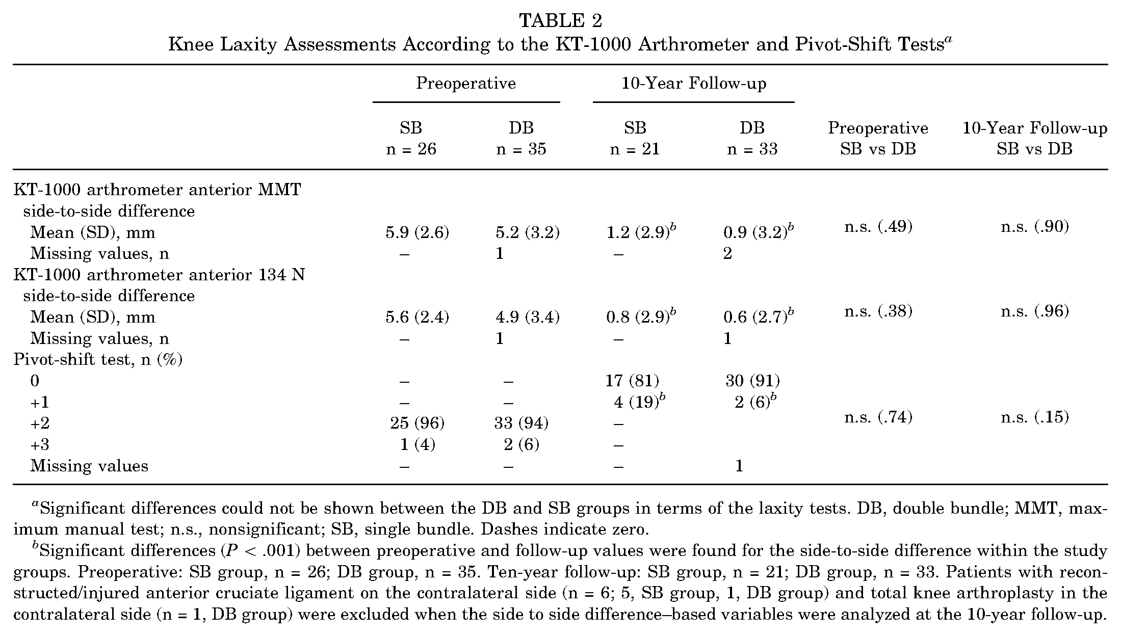
Significant differences could not be shown between the DB and SB groups in terms of the laxity tests. DB, double bundle; MMT, maximum manual test; n.s., nonsignificant; SB, single bundle. Dashes indicate zero.
Significant differences (P < .001) between preoperative and follow-up values were found for the side-to-side difference within the study groups. Preoperative: SB group, n = 26; DB group, n = 35. Ten-year follow-up: SB group, n = 21; DB group, n = 33. Patients with reconstructed/injured anterior cruciate ligament on the contralateral side (n = 6; 5, SB group, 1, DB group) and total knee arthroplasty in the contralateral side (n = 1, DB group) were excluded when the side to side difference–based variables were analyzed at the 10-year follow-up.
Objective and Subjective Results a

Significant differences could not be shown between the DB and SB groups. DB, double bundle; n.s., nonsignificant; SB, single bundle.
For side-to-side difference in flexion/extension. Preoperative: SB group, n = 26; DB group, n = 35. Ten-year follow-up: SB group, n = 21; DB group, n = 33. Patients with reconstructed/injured anterior cruciate ligament in the contralateral side (n = 6: 5, SB group; 1, DB group) and total knee arthroplasty in the contralateral side (n = 1, DB group) were excluded when the side-to-side difference-based variables were analyzed at the 10-year follow-up.
Significant improvement in terms of the Lysholm knee score in both groups (P < .001).
The minus sign indicates that the nonoperated side had better extension compared with the operated side.
The range of extension was significantly better between the preoperative and the 10-year follow-up in the DB group (P < .01).
The mean diameter of the tibial tunnels is presented in Table 4. In both groups, no significant decrease in tibial tunnel width on the AP or lateral views was found from the early postoperative period to 10-year follow-up (P = >.99) apart from the DB group's PL tibial tunnel in AP view (P = .02).
Tibial Tunnel Mean Diameters as Seen on the Radiographs a

The mean diameters of the SB tibial tunnel did not increase in both projections until the 10-year follow-up. The mean diameters of tibial AM and PL tunnels did not increase in both projections over the 10-year follow-up. AM, anteromedial; AP, anteroposterior; DB, double bundle; n.s., nonsignificant; PL, posterolateral; Postop, postoperative; SB, single bundle. Dashes indicate zero.
There was a significant decrease in PL tibial tunnel width on the AP view between the 5-year and 10-year assessments in the DB group.
In 32 of a possible 42 radiographs (76%) of 26 patients in the SB group, the width of the tibial tunnel had not increased at 10 years compared with the early postoperative period (Figure 3). In the DB group, 42 of a possible 61 PL radiographs (69%) and 35 of a possible 56 AM tunnel radiographs (63%) had no increase in tibial tunnel width at 10 years (Figure 4). The distribution of increased/decreased/unchanged tunnel widths is reported in Table 4. The mean SB tibial tunnel volume as measured on CT was 2.04 cm3 (± 0.85 cm3); DB PL tunnel volume, 2.04 cm3 (± 1.92 cm3); and DB AM tunnel volume, 1.38 cm3 (± 0.54 cm3). There was no correlation between tunnel widths and the KT-1000 arthrometer assessments, though there was a statistically significant correlation between SB tibial tunnel volume on CT imaging and KT-1000 arthrometer anterior 134 N side-to-side difference (r = 0.45; P = .039). None of the patients in this study had fully ossified tunnels on CT imaging (SB tibial tunnel volume range, 0.64-3.98 cm3; DB PL tunnel volume range, 0.53-11.66 cm3; DB AM tunnel volume range, 0.40-3.12 cm3). No tunnel communication was found in the DB group. A clarification for the missing values in 10-year plain radiographs is presented in Table 5.
Clarification of Missing Values for Plain Radiographs at 10 Years a

“True” missing values are defined as no radiographs being available. “False” missing values are defined as no tunnel being visible on the given radiographic projection while being visible in the other radiological view of the same patient. DB, double-bundle; SB, single-bundle. Dashes indicate zero.
Discussion
The key findings in this study were that the tibial tunnel mean width did not increase in 76% of SB radiographic views at 10-year follow-up after using a 77% PLDLA and 23% β-TCP biocomposite interference screws; the same was found in 69% and 63% for DB PL and AM tunnel radiographs, respectively, with no tunnel communication in the DB patients.
There is a general lack of studies comparing tunnel enlargement in SB and DB ACLR with bioresorbable or biocomposite screws, though the literature that exists suggests the degree of enlargement varies over time after ACLR. Siebold 50 found, at 1-year follow-up with 70% PLGA (poly lactic-co-glycolic acid) and 30% β-TCP screws, a 43% tibial tunnel enlargement for both PL and AM tunnels, as well as tunnel communication in 41% of patients. Järvelä et al 24 at 2-year follow-up had similar findings, with 43% PL and 39% AM tunnel enlargement using biocomposite screws of copolymers of L-lactide, D-lactide, and trimethylene carbonate. The authors claimed tibial tunnel enlargement was greater in the SB cohort despite a P value of .051 with sample sizes that risk the results being underpowered. Kiekara et al 30 stated their 5-year follow-up of DB reconstructed patients using D,L lactide, L-lactide and trimethylene carbonate screw showed tunnel enlargement at 2 years followed by narrowing at 5-year follow-up, though the tunnel diameter was still very much enlarged compared to at surgery. Similarly, Arama et al 2 found no increased tunnel widening for a PLLA-hyaluronic acid screw when compared with a titanium interference screw with SB reconstruction using a hamstring graft, though there was an increase in tunnel volume between 2 and 5 years postsurgery, negating the main supposed benefit of biocomposite screws over their titanium counterparts.
A long-term follow-up of PLLA-hyaluronic acid screws in SB reconstruction with hamstring tendon autograft demonstrated a smaller tibial tunnel volume increase with a biocomposite screw compared with a titanium interference screw between 2- and 13-year follow-up. 52 Issues regarding tunnel widening with the use of bioabsorbable screws compared to titanium raised by a previous meta-analysis do not seem applicable to all biocomposite screws according to a recent RCT.34,52 The specific polymer and any eventual isomers used seem to be key, with previous older studies highlighting higher incidence of complications such as screw migration, cyst formation, 2 foreign body tissue reactions, 12 synovitis, 20 and screw breakage during implantation 38 involving other polymers such as PGA. 52 Barbosa et al 9 examined causes for tibial cysts after ACLR and found that 16 studies revealed a relationship with bioabsorbable screws, though the vast majority were PLLA-based screws; tunnel widening in the same review was only seen in metal or PLLA-based interference screws. Other meta-analyses have mostly examined PLLA or PGA-based screws.19,34
There has been some interchangeability in the literature between the terms “bioresorbable” and “biocomposite,” which has perhaps led to surgeons discounting biocomposite screws despite their improvements on the first generation of bioresorbable screws.15,18,40,47,60 Bioabsorbable materials degrade in 5 stages: hydration, depolymerization, loss of mass integrity, absorption, and elimination. 45 Different materials lead to different degradation products with subsequently different effects on adjacent tissue 63 ; this makes it hard to claim bioresorbable or biocomposite screws in general suffer from the same complications.
Adding calcium bioceramics like β-TCP to biodegradable polymers such as PLLA or its variations with combinations of different stereoisomers creates a biocomposite material that reduces tunnel widening and increases cortical bone formation, 10 which explains their increase in popularity over time. 56 Clinical studies have confirmed that biocomposites of PLLA and β-TCP can completely degrade gradually over time with resultant osteoconductivity5-7,40; screws without a bioceramic component have not demonstrated any clear osteoconductivity. 8 The L-isomer of polylactic acid, PLLA, becomes highly hydrophobic and crystalline over a prolonged degradation period of several years; the DL-isomer is highly amorphous and less stable, making it degrade over a shorter period of time. The copolymer of these, PLDLA, is less resistant to both hydrolysis and degradation, ultimately leading to even faster degradation; it could also possibly induce fewer tissue reactions and demonstrate better biodegradability when combined with β-TCP. 47 β-TCP breaks into phosphate and calcium ions during degradation, maintaining a higher pH around the screw that may in turn buffer the acidic breakdown products of lactic acid and minimize any local reactivity or inflammatory response during reabsorption.6,26,42
Tunnels in both the SB and the DB groups in this study were, however, still visible at 10-year follow-up. Despite β-TCP or other bioceramics optimizing surrounding pH, thus fostering as osteoconductive of an environment as possible, some suggest the sheer nature of acid-based biocomposite screws in an area of low circulation limits true bone healing. 44 All patients in both groups received hamstring autographs, which have been shown to be the stiffest autograft commonly used, 46 with stiffness possibly being another factor contributing to tunnel widening. 58 Studies that have used patellar tendon autografts and biocomposite screws have been able to demonstrate high rates of screw resorption and bone replacement in addition to no increase in tunnel size over the mid- to long term.6,22,25,48 This could mean the lack of bone replacement of tunnels in this study may be more due to ACL graft choice than just the tibial graft fixation method. Despite this, 62.7% of International Society of Arthroscopy, Knee Surgery and Orthopaedic Sports Medicine surgeons (regardless of surgical volume) prefer bioabsorbable screws for tibial hamstring graft fixation, with 14.6% choosing metallic screws as the next most common alternative. 57
There was a moderate positive linear correlation in this study between KT-1000 arthrometer laxity measurements and SB patient tunnel volume. While there are studies that have investigated tunnel enlargement with biocomposite screws as the tibial fixation method, none seems to have reported any analysis of tunnel volume in relation to KT-1000 arthrometer measurements, even if both measurements were used in the study. Given the small sample size in the current study, these results should be interpreted with caution until further data are published. It is also important to note that these patients did not seem to experience this subjectively, given no changes in subjective measures as shown here and in another study on the same patient groups. 4 Kramer et al 32 highlighted the general lack of published articles on complications related to bioabsorbable/biocomposite tibial screws other than case reports or small-sample size (level 4) studies. The same study only identified low-volume surgeons as a risk factor for tibial screw–related complications; the group speculated whether this could be due to technical errors in surgical technique. 32 This does possibly relate to the study of Ayala-Mejias et al 3 that found a higher incidence of tunnel widening in tunnels with a more vertical position, which is a known risk factor for ACL graft failure and thus something high-volume surgeons may have more experience in avoiding.
Tunnel widening is suspected to be multifactorial, with possible components being biological, chemical, and mechanical factors such as more vertical tunnel position, joint fluid leakage within the bone tunnel, a longer tendinous portion of the graft within the tibial tunnel, graft-tunnel motion, the use of a bioabsorbable fixation method, and the number of tunnels drilled.16,35,37,39,51,53,64,65
Limitations
Our study had several limitations. First, the study was not randomized and there was no control group. The sample size of both groups at 10-year follow-up does lead to a risk for our study's being underpowered, partly due to the COVID-19 pandemic that prevented further clinical follow-up of the remaining participants. No subgroup analysis based on differing tunnel diameters at index surgery was performed due to too small group sizes. The use of several drill sizes, proportionate to the graft diameter, may have affected the width of the tunnel, even if the screw was the same size in all participants. Our study did not measure material density at the screw site compared with various tibial bone sites; thus, it was not possible to comment on the level of degradation of the PLDLA/β-TCP interference screws used in this study. There were technical errors at 1 examination site that led to several missing values for lateral radiographs for both groups that may have affected our study's results. CT imaging was not performed at previous follow-ups, limiting possible comparison with earlier periods; this was due to only receiving ethical approval for use of CT at the 10-year data collection. Using standard radiographs instead of CT to measure tunnel diameter does risk measurements’ being less accurate, though other groups have shown it to be an adequate alternative to detect tunnel widening; however, this method is only validated in SB grafts, which is another limitation.14,62 Using the mean of 3 tunnel diameter measurements at each end and the middle may also have been a confounder, given width decrease is more pronounced at the aperture and exit of the tibial tunnel. 48 As tunnel width measurements were defined using standard radiographs, there is also a risk for tunnel overlap affecting the results in the DB group; however, the radiologist in this study did not observe any tunnel overlap or any tunnel communication on CT in his analysis. Furthermore, the radiologist was not able to distinguish bone formation from possible screw remnants at 10 years. The earliest postoperative radiographs were a mean 6 months after surgery in this study and thus could have failed to capture any tunnel enlargement that may have occurred in the first 6 months.
Furthermore, ACL graft integrity was only examined clinically, not via MRI scans, in this study; the manual pivot-shift test has been shown to be subjective in its interpretation and thus could be questioned. Despite all these limitations, it is of interest that most of the tunnels do not appear to widen ≥10 years after insertion.
Conclusion
Most patients’ tibial tunnels had not increased on 1 or both radiographic views at 10-year follow-up compared with the early postoperative period after ACLR using biocomposite interference screws, with no obvious negative effect on outcomes. However, the tunnels were still visible in most patients at 10 years on standard radiographs and CT imaging.
Footnotes
Final revision submitted March 6, 2024; accepted April 2, 2024.
One or more of the authors has declared the following potential conflict of interest or source of funding: Research support was received from The Healthcare Board, Region Västra Götaland, Sweden. J.K. has received nonconsulting fees from ConMed Sweden. AOSSM checks author disclosures against the Open Payments Database (OPD). AOSSM has not conducted an independent investigation on the OPD and disclaims any liability or responsibility relating thereto.
Ethical approval for this study was obtained from the regional ethics committee in Gothenburg (reference No. 157-08 and T078-18).