
Abstract
Background:
Previous studies have demonstrated that medial meniscus posterior root tear (MMPRT) repair is superior to debridement in terms of patient-reported outcomes, rates of conversion to total knee arthroplasty (TKA), and long-term costs. Despite the known poor midterm outcomes, there is a paucity of long-term results of partial meniscectomy for degenerative MMPRTs.
Purpose:
To 1) evaluate long-term patient-reported and radiographic outcomes of patients who underwent partial medial meniscectomy (PMM) for MMPRTs, and 2) determine the rate of and risk factors for conversion to total knee TKA.
Study Design:
Case series; Level of evidence, 4.
Methods:
A previously identified cohort of 26 patients treated with partial meniscectomy for isolated MMPRTs between 2005 and 2013 was prospectively followed for long-term outcomes at a minimum 10-year follow-up. Patients were evaluated for International Knee Documentation Committee (IKDC) outcome score, reoperation, and conversion to TKA. Failure was defined as conversion to arthroplasty or a severely abnormal IKDC subjective score <75.4.
Results:
This study included 26 patients (10 men, 16 women; mean age, 54 ± 8.7 years [range, 38-71 years] at diagnosis; body mass index, 32.9 ± 5.5) who were followed for a mean of 14.0 ± 3.6 years (range, 10.1-19.6 years). At the final follow-up, 1 patient was deceased and 18 (72%) of the remaining 25 patients had progressed to TKA, with 1 (4%) patient undergoing repeat meniscectomy. The 6 (24%) patients who had not progressed to TKA or revision surgery reported a mean IKDC score of 57 ± 23. Nineteen patients underwent subsequent surgery and 5 demonstrated severely abnormal IKDC scores resulting in a clinical failure rate of 96% (24 of the 25 living patients) at a mean 14-year follow-up.
Conclusion:
PMM for medial meniscus posterior horn root tears demonstrated 72% progression to TKA and 96% failure according to subjective clinical outcomes at a minimum 10-year follow-up.
Keywords
Menisci are fibrocartilaginous structures that provide stability and lubrication to the knee joint. 24 Additionally, these structures allow for load bearing and shock absorption, which provides protection to the articular cartilage surface of the knee.6,7 Meniscal deficiency leads to articular cartilage degeneration and posttraumatic osteoarthritis. The mechanism of dysfunction is in the loss of hoop stress and the increased contact forces, which then leads to osteoarthritis.15,16,19 Medial meniscus posterior root tears (MMPRTs) are of particular interest as they have been shown to be biomechanically equivalent to complete meniscectomy. 1
Previous studies have demonstrated that repair of MMPRTs is superior to debridement in terms of patient-reported outcomes, rates of conversion to total knee arthroplasty (TKA), and long-term costs.8,12 Additionally, studies by Krych et al17,19 have demonstrated poor mid- and long-term outcomes with MMPRT nonoperative treatment. Yet, there remains a subset of patients who are not suitable candidates for meniscal repair because of high-grade cartilage degeneration, varus malalignment, or patient comorbidities. We reported on a cohort of patients with MMPRTs treated with partial medial meniscectomy (PMM) who demonstrated significant progression of osteoarthritis, with Kellgren-Lawrence grade 2 or higher in 91%, at a mean 5.5-year follow-up. Additionally, 54% of these patients progressed to TKA at a mean of 4.5 years after clinical presentation. 17 Despite the known poor midterm outcomes, there is a paucity of data on the long-term results of partial meniscectomy for degenerative MMPRTs. This information is crucial for surgical planning and patient counseling.
The purpose of this study was to (1) evaluate long-term patient-reported and radiographic outcomes of patients who underwent PMM for MMPRTs, and (2) determine the rate of and risk factors for conversion to TKA. We hypothesized that patients would have poor outcomes with high rates of conversion to TKA.
Methods
After institutional review board approval (IRB No. 15-000691) was obtained, a previously retrospectively identified 17 cohort of 26 patients, who were diagnosed with isolated MMPRTs between 2005 and 2013, was reviewed. These patients were consecutively enrolled and inclusion criteria included (1) a complete medial meniscus posterior root avulsion or a complete radial tear within 9 mm of the medial meniscus posterior root; (2) acute onset of clinical symptoms that correlated with magnetic resonance imaging findings; and (3) treatment with PMM, typically for perceived mechanical symptoms refractory to comprehensive nonoperative management. Exclusion criteria consisted of (1) a prior ligamentous injury, (2) meniscal repair after posterior root tear diagnosis, (3) a lateral or anterior meniscus tear, (4) a concomitant tibial plateau fracture, and (5) a minimum 10-year follow-up. Of this original cohort, 0 were lost to follow-up, resulting in 26 patients who were included in this study for a 100% long-term follow-up rate.
Electronic medical record review was performed, and patients were contacted to confirm arthroplasty status and to complete patient-reported outcomes (International Knee Documentation Committee [IKDC], visual analog scale [VAS] for pain, and Tegner activity scale scores). Failure was defined as conversion to arthroplasty or a severely abnormal IKDC subjective score <75.4.11,17
The initial radiologic reading of MMPRTs was done by a trained musculoskeletal radiologist with confirmation by a sports fellowship trained orthopaedic surgeon (M.H.). MMPRT was defined according to LaPrade et al. 21 Radiographic analysis of knee arthritis was classified using the Kellgren-Lawrence grading scale. Subsequent IKDC score and advanced imaging was evaluated by the same orthopaedic surgeon (M.H.). An intraclass correlation coefficient (ICC) analysis was performed to evaluate the validity and accuracy of the final Kellgren-Lawrence grading, demonstrating an ICC of 1, suggesting perfect interrater reliability.
Statistical Analysis
Statistical analysis was performed using BlueSky 7.4.0 software (BlueSky Statistics Inc). Continuous variables are reported as mean ± standard deviation, with nonparametric variables reported with ranges. Pearson chi-square analysis was used for categorical variables. Kaplan-Meier analysis was used to evaluate the time-dependent rate of conversion to arthroplasty. Paired t tests and Wilcoxon signed-ranked tests were used to compare baseline and final patient-reported outcomes. All statistical tests were 2-sided, with P values <.05 considered statistically significant.
Results
This study included 26 patients (10 men, 16 women; mean age, 54 ± 8.7 years [range, 38-71] at diagnosis; body mass index [BMI], 32.9 ± 5.5) who were followed for a mean of 14.0 ± 3.6 years (range, 10.1-19.6 years). At the time of initial MMPRT diagnosis, patients had a mean Kellgren-Lawrence grade of 1.3 ± 0.8 (Table 1). At the final follow-up, 18 patients had progressed to TKA, 1 went on to subsequent surgery (opening-wedge high tibial osteotomy with cancellous allografting and anterior cruciate ligament reconstruction), and 6 had not undergone additional surgical treatment. Finally, 1 patient was deceased at the time of analysis.
Kellgren-Lawrence Grade of Included Patients at Time of Initial Root Tear Diagnosis

Clinical Outcomes
The 6 patients who had not progressed to TKA or revision surgery reported a mean IKDC score of 57 ± 23 (range, 36-91), a VAS pain score of 3.2 ± 2.6 (range, 0-7), and a Tegner score of 4 ± 1.8 (range, 3-7). There was no significant change in Tegner score from the mean initial baseline of 5 ± 1.6 (range, 2-7) to the time of the final follow-up (P = .8). Notably, 5 (83%) of 6 patients reported severely abnormal IKDC scores (<75.4). Additionally, the IKDC scores at the final follow-up were significantly lower than the IKDC scores at the 5.5-year mean follow-up (57 ± 23 vs 72 ± 23; P < .037).
Radiographic Outcomes
At final follow-up, Kellgren-Lawrence grades progressed from a mean initial baseline of 1.5 ± 0.5 (range 1-2) to a final Kellgren-Lawrence grade of 2.2 ± 0.4 (range, 2-3) (P = .03) for those patients not converting to arthroplasty, resulting in a mean increase in Kellgren-Lawrence grade of 0.7 over the course of follow-up. Compared with baseline, a significant portion of these patients had progressed to Kellgren-Lawrence grade 2 arthritis at the final follow-up (100% vs 50%; P < .046). The patient undergoing revision surgery remained at Kellgren-Lawrence grade 1 before revision surgery.
Reoperation and Survivorship
At the final follow-up, 18 (72%) of the 25 patients had progressed to TKA at a mean age of 59.5 ± 7.9 years (range, 43.7-73.8 years), and 1 (4%) patient underwent a revision in the form of an opening-wedge high tibial osteotomy with cancellous allografting and anterior cruciate ligament reconstruction. The mean time to TKA or revision surgery was 5.8 ± 4.7 years (range, 0.2-16.2 years) after meniscectomy and 6.4 ± 4.5 years (range, 0.5-16.5 years) from the time of initial MMPRT diagnosis. Survival rates free of revision at 2, 5, and 10 years of follow-up were 76%, 60%, and 36%, respectively. Figure 1 demonstrates a Kaplan-Meier estimator of progression to TKA.

Kaplan-Meier estimator of progression to total knee arthroplasty (TKA). Bars demonstrate time censoring at end of follow-up for patients beyond 10 years.
Risk Factor Analysis
With 19 patients undergoing subsequent surgery and 5 demonstrating severely abnormal IKDC scores, treatment failed for 24 (96%) of the 25 patients. Age at initial diagnosis, BMI, and baseline Kellgren-Lawrence grades did not predict progression to TKA (P≥ .38) (Table 2). However, when stratified, patients with BMIs >30 progressed to TKA at a higher rate compared with patients with BMIs <30 (88% vs 50%; P = .037). Of note, the presence of meniscal extrusion >3 mm at initial diagnosis was not significantly associated with progression to TKA compared with < 3 mm extrusion (80% vs 70%; P = .60).
Risk Factor Analysis a
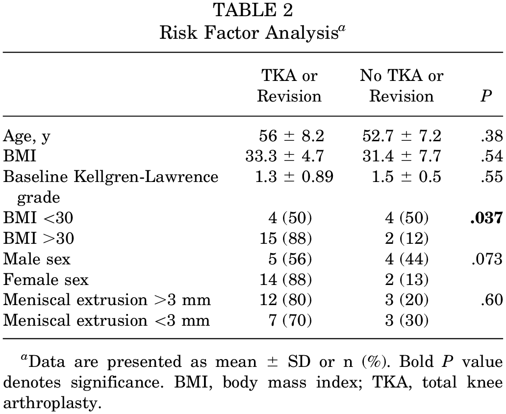
Data are presented as mean ± SD or n (%). Bold P value denotes significance. BMI, body mass index; TKA, total knee arthroplasty.
Discussion
The primary finding of this study is the high rate of failure of PMM for the treatment of degenerative MMPRTs at mean 14-year follow-up. Most patients progressed to TKA at a mean of 6 years. Of those who did not progress to TKA, 5 (83%) of 6 reported severely abnormal IKDC scores, and 100% had progression of arthritis as determined by Kellgren-Lawrence grading. The current study reports a 96% rate of failure of PMM for the treatment of MMPRTs, with 76% progressing to TKA or revision surgery.
This study demonstrated poor IKDC, Tegner, and VAS scores in patients who underwent PMM for MMPRT and had not progressed to TKA. During a previously published interim analysis at a mean follow-up of 5.5 years, this cohort reported IKDC scores of 68 ± 20. 17 The present study demonstrates further worsening in the subsequent 9 years to mean IKDC scores of 57 ± 23. Long-term data are limited; however, Chung et al 5 observed that at 8.5 ± 3.8 years, patients undergoing PMM for MMPRT had mean IKDC scores of 44 ± 19. Furthermore, a recent study by Krych et al 19 in patients managed nonoperatively demonstrated a mean IKDC score of 52 ± 22 at a mean of 14 ± 2 years. Similar studies with short-term follow-up also demonstrated that meniscectomy has no benefit over nonoperative management for MMPRTs, although it must be noted that PMM may serve to reduce pain and/or mechanical symptoms.2,17,23
At the final follow-up, radiographic progression of arthritis as determined by Kellgren-Lawrence grading was seen in 100% of patients who had not undergone revision surgery. This is similar to the midterm findings of this cohort, which demonstrated significant progression of Kellgren-Lawrence grading from a median of 1 to 2 at a mean of 3 years. 17 Similarly, Kim et al 14 observed Kellgren-Lawrence progression in 75% of patients undergoing PMM at a mean 4-year follow-up. Han et al 9 observed a lower rate, with 35% of patients progressing from Kellgren-Lawrence grades between 0 and 2 to grades between 2 and 4 at a mean 6-year follow-up. In contrast, Kellgren-Lawrence progression in patients who undergo meniscal repair has been shown to be significantly slower. 3 The cause of this difference may lie in the inability of PMM to restore hoop tension; thus, it is unsurprising to see Kellgren-Lawrence grade progression in patients undergoing PMM. 1
At a previously published 5.5-year follow-up, an overall failure rate of 77% was reported, with a 54% rate of progression to TKA. 17 Notably, the present study observed a 96% failure rate with a high rate of conversion to TKA (72%). These findings closely mirror those in a study by Krych et al, 18 which demonstrated a 96% failure rate with a 64% rate of progression to TKA in nonoperatively managed patients at a mean of 14 ± 2 years. Furthermore, the 10-year rate of revision of 64% in this study was comparable to the 10-year findings of Chung et al, 5 who demonstrated a 56% rate of conversion to TKA in PMM-treated patients. Notably, a 10-year comparative meta-analysis by Faucett et al 8 observed TKA rates of 34%, 46%, and 52% in meniscus-repaired, nonoperatively treated, and meniscectomy-treated knees with MMPRTs, respectively. Overall, these studies highlight the potential benefit of root repair and reinforce the lack of benefit observed from meniscectomy for the treatment of MMPRTs.
In the current study, BMI >30 was a statistically significant risk factor for progression to revision or TKA. The previous study on this cohort also identified this as a factor, with female sex being significantly associated with higher rates of arthroplasty and lower IKDC scores. 17 Additionally, these factors have been previously identified as associated with the development of MMPRTs.4,10 Other risk factors were not identified for conversion to TKA. Importantly, as described by Kim et al 13 and Lee et al, 22 factors that may be protective of arthritis progression and conversion to TKA after PMM for degenerative MMPRTs are appropriate alignment (<2°-3° valgus), lack of arthritis at baseline, and lower BMI. 20 Thus, patients with MMPRT, particularly those with varus knees, must be counseled carefully given the already elevated stresses on the medial compartment. Notably, the data here concern symptomatic degenerative MMPRTs, while there may be a subset of patients with asymptomatic MMPRTs.
Strengths and Limitations
The strength of this study is the long-term follow-up of a previously analyzed cohort with a 100% response rate. Study limitations include the following: 1) a high rate of conversion to TKA, which precludes subgroup analysis and identification of risk factors; 2) a small sample size, limiting the ability to make strong conclusions; 3) inconsistent full-length standing radiographs to detect varus malalignment; 4) lack of randomization with a treatment arm (meniscal repair); and 5) lack of preoperative scores, lack of additional granular intraoperative data, and inability to grade and associate progression with alignment and articular cartilage status. Last, it is important to note that not all MMPRTs are necessarily symptomatic, as well as the fact that degenerative root tears likely comprise part of the natural history of varus knee osteoarthritis, the progression of which may not be prevented by root repair.
Conclusion
PMM for medial meniscus posterior horn root tears demonstrated 72% progression to TKA and 96% failure according to subjective clinical outcomes at a minimum 10-year follow-up.
Footnotes
Final revision received February 13, 2024; accepted February 26, 2024.
One or more of the authors has declared the following potential conflict of interest or source of funding: B.A.L. has received consulting fees from Arthrex and Smith+Nephew; nonconsulting fees from Arthrex, Smith+Nephew, and Linvatec; and a royalty or license from Arthrex. M.J.S. has received a royalty or license, consulting fees, nonconsulting fees, and education payments from Arthrex; and hospitality payments from Stryker. A.J.K. has received nonconsulting fees from Arthrex; consulting fees from Arthrex and Responsive Arthroscopy; a royalty or license from Arthrex; a grant from the DJO; and honoraria from the Joint Restoration Foundation and Musculoskeletal Transplant Foundation. M.H. has received education payments from Arthrex, Foundation Medical, Medwest Associates, and Smith+Nephew; honoraria from Encore Medical; hospitality payments from Orthalign and Medical Device Business Services; and consulting fees from Vericel. AOSSM checks author disclosures against the Open Payments Database (OPD). AOSSM has not conducted an independent investigation on the OPD and disclaims any liability or responsibility relating thereto.
Ethical approval for this study was obtained from Mayo Clinic (ref No. PR 15-000601-09).