
Abstract
Background:
A steeper posterior tibial slope (PTS) is an important risk factor for anterior cruciate ligament (ACL) reinjury. The PTS may affect lower extremity biomechanics under competition-like conditions for athletes with a reconstructed ACL.
Hypothesis:
It was hypothesized that the PTS would be associated with lower extremity biomechanics, which may increase ACL strain.
Study Design:
Descriptive laboratory study.
Methods:
Included were 10 athletes (mean age, 20.9 ± 1.8 years) who had undergone ACL reconstruction. The authors recorded the 3-dimensional lower extremity biomechanics while participants performed a single-leg drop jump with the Stroop task (dual task). Kinematic and kinetic data were analyzed and compared between the involved and contralateral limbs. The medial and lateral PTSs were measured using magnetic resonance imaging scans of the involved knee. The correlation between the biomechanical data and the PTS in each knee was evaluated.
Results:
The lateral PTS was significantly correlated with the maximum hip adduction moment (r = 0.64; P < .05) and maximum internal tibial rotation angle (r = 0.71; P < .05) in the involved limb. There were no differences in kinematic and kinetic data between the involved and contralateral limbs.
Conclusion:
In athletes after ACL reconstruction, the lateral PTS was directly associated with the maximum internal tibial rotation angle during single-leg drop landing with a cognitive task.
Clinical Relevance:
The findings in this study indicate that a steeper lateral PTS may cause internal rotation of the tibia during landing, which may be associated with reinjury in athletes with a reconstructed ACL.
A steeper posterior tibial slope (PTS) is considered a risk factor for reinjury to the after anterior cruciate ligament (ACL) reconstruction. 2,3 In athletes, the rate of return to sports at the preinjury level after revision ACL reconstruction ranges from 13% to 69%, which is low and poses a significant problem. 6 Understanding the influence of the tibial slope on joint biomechanics may help account for these risk factors and aid in the development of appropriate ACL reinjury prevention strategies. ACL reinjury occurs because of a combined load being applied to the knee, which is similar to the initial ACL injury mechanism. 24,26 Studies on ACL injury that used the model-based image-matching technique, 16 and cadavers 14,15 have shown that the ACL strain increases with an increase in the knee valgus, internal tibial rotation angle, ground-reaction force, and anterior tibial shear force.
Two in vivo studies investigated the relationship between PTS and lower extremity biomechanics, and the results showed that the PTS correlated with the peak knee valgus angle, peak internal tibial rotation angle, and anterior knee joint reaction force. However, in both studies, participants with healthy knees who played sports recreationally performed only an exercise task. 18,27 Biomechanics during single-leg drop landing (SDL) and vertical drop landing have been evaluated as screening tools to assess the risk of ACL injury. 8,21 However, athletes can easily perform both tasks; therefore, the outcomes of these tests are not sufficient to investigate lower extremity biomechanics under competition-like conditions. 5,30
Biomechanical studies involving athletes need to be analyzed under more challenging conditions. The effects of PTS on lower extremity biomechanics have been assessed in healthy participants at the recreational level but not in athletes who have undergone ACL reconstruction.
To account for this, Kajiwara et al 12 used a dual task, which included the addition of a cognitive task to an exercise task, and revealed that knee biomechanics among healthy athletes were influenced under a dual-task condition.
This study aimed to investigate the relationship between the PTS and lower extremity biomechanics during an SDL combined with a cognitive task in athletes with a reconstructed ACL. It was hypothesized that the PTS would be associated with lower extremity biomechanics, which may increase ACL strain.
Methods
Participants
The study protocol was approved by the ethics committee of our institution, and informed consent was obtained from all the participants before participation. Ten athletes (5 male and 5 female individuals; mean age, 20.9 ± 1.8 years) who underwent single-bundle ACL reconstruction using a hamstring tendon and could return to their prior level of activity after completing rehabilitation were enrolled. A Tegner activity score ≥7 was an inclusion criterion. A previous study reported a correlation coefficient of 0.78 for the PTS and the peak knee internal rotation angle. 18 An a priori power analysis was performed based on an α error of .05, a 1 − β error of 0.80, and an effect size of 0.78 using G*Power software (HHU Düsseldorf). This analysis showed that at least 8 patients were required. This study had a sufficient sample size (10 knees).
Exercise Task
Each participant underwent 3-dimensional biomechanical motion analysis (Vicon MX; Vicon Motion System Inc). Kinematic data were recorded at a sampling frequency of 100 Hz using a 16-camera motion capture system (MX-T20; Vicon Motion System Inc). Before testing, each participant was equipped with 23 reflective markers of 9 mm in diameter using a point cluster technique. 1 For the cluster of points, 8 and 6 markers were attached to the thigh and shank, respectively. The bony landmarks were attached to the right and left anterior superior iliac spines; the right and left posterior superior iliac spines; and the greater trochanter, lateral epicondyles of the femur, lateral malleoli, toe (between second and third metatarsals), and heel. Kinetic data were recorded at 1000 Hz and synchronized with the motion capture system. The coordinates of the infrared reflective markers were recorded during a 40-millisecond time frame before and after the foot contact. This time frame was adapted to kinematic analyses because a previous study showed that ACL ruptures occur within 40 milliseconds of foot contact. 16
For the SDL task, the participants started with a single-leg stance on a platform (height, 30 cm) in front of the force plate (Accugait; AMTI Inc). The participants were instructed to land on the force plate with the same leg that they used for jumping off the platform and to practice this until they got used to it (maximum of 5 repetitions). The SDL task was considered successful if the participants remained still for 2 seconds after landing. After practice, all the participants performed SDL combined with a cognitive task, as described below.
Cognitive Task
The participants were required to perform the Stroop task as a cognitive task. 28 The monitor (15.6 inches; Lenovo) was connected to a foot switch on the platform (Sais) and placed 3 m in front of the platform. The monitor displayed the instruction at the same time as when the participant’s heel left the foot switch. In this task, the color indicated by the word and the color of the actual word were different (eg, the word “red” displayed in blue). The participants were instructed to respond to the color of the word rather than the color indicated by it. The participants landed on a force plate, in front of which 3 colored papers were placed (Figure 1). This task was considered successful when they landed on the appropriate spot. The SDL combined with a cognitive task was repeated until the participants successfully completed the task 3 consecutive times.
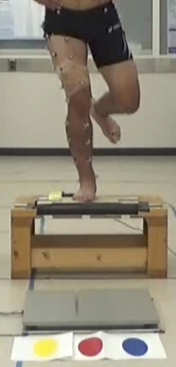
The participant jumps from a single-leg standing position on a platform (height, 30 cm) and lands on the force plate (gray square) in front of the 3 spots.
Data Analysis
Vicon Nexus Version 2.2.3 (Vicon Motion System Inc) and custom scripts in MATLAB 8.4 R2014b (MathWorks) were used for kinetic and kinematic data analysis. Based on the orientation of the distal segment coordinate system relative to that of the proximal segment coordinate system, the hip and knee angles as well as the anterior tibial translation were calculated. The inverse dynamics method was used to derive the joint kinetics data, and the data were presented as external joint moments. The external joint moments were derived via inverse dynamic calculations using the plug-in gait model of Vicon Nexus Version 2.2.3 with raw ground-reaction force data. The examined variables were joint excursion (peak-to-initial joint angle), peak angles, peak external moments of the hip and knee in all 3 planes, and anterior tibial translation. The external joint moments were normalized to body weight and height (N/BW×h). The mean values of the kinematic and kinetic variables of the 3 landing trials were analyzed.
PTS Measurement
Based on methods described by Hudek et al, 10 we measured the tibial slope of the involved knee via preoperative magnetic resonance imaging scans using software for image analysis (Osirix; Pixmeo). A 3-step procedure was applied to measure the PTS. First, the central sagittal image, in which the tibial attachment of the posterior cruciate ligament and the intercondylar eminence were visible, was obtained. Second, 1 cranial and 1 caudal circle were drawn. The cranial circle was drawn tangentially to the proximal, anterior, and posterior tibial borders, and the caudal circle was drawn in such a way that it was centered on the perimeter of the cranial circle and was tangential to the anterior and posterior tibial borders. The line connecting the centers of the cranial and caudal circles was defined as the longitudinal axis of the tibia. Finally, the angles between the line perpendicular to the longitudinal axis and the tangential lines connecting the uppermost superior-anterior and posterior cortices of the medial and lateral tibial plateaus were measured. The medial PTS (MPTS) and lateral PTS (LPTS) were measured at the centers of the medial and lateral plateau slices, respectively (Figure 2). When the contact point of the posterior plateau was inferior to that of the anterior plateau, the slope was defined as positive.

Measurement of the medial and lateral posterior tibial slope. (A) The central sagittal slice is identified. Then, 1 cranial and 1 caudal circle are drawn tangentially to the tibial border. The tibial longitudinal axis is defined by a line that connects the centers of these 2 circles. (B, C) The angles between the line perpendicular to the tibial longitudinal axis and the tangent to the medial and lateral tibial plateaus are the medial and lateral tibial posterior tibial slopes, respectively.
Statistical Analysis
All statistical analyses were performed using SPSS Statistics software (Version 26.0; IBM Corp). The threshold of significance was set at P < .05. Means and standard deviations were used to summarize the data. The Pearson correlation coefficient (r) was used to investigate the association between the MPTS or LPTS and variables of interest during the dual task. Correlation coefficients of 0.10 to 0.39, 0.40 to 0.69, 0.70 to 0.89, and 0.90 to 1.00 were regarded as weak, moderate, strong, and extremely strong, respectively. 25 The Mann-Whitney U test was used to compare the investigated variables between the involved and contralateral limbs.
Results
Correlation Between the PTS and Hip and Knee Kinetics and Kinematics in the Involved Limb
The mean MPTS and LPTS were 4.5° ± 2.5° and 5.8° ± 2.1°, respectively. The MPTS was significantly correlated with the maximum hip flexion moment (r = 0.66; P < .05), and the LPTS was significantly correlated with the maximum hip adduction moment (r = 0.64; P < .05) and the maximum internal tibial rotation angle (r = 0.71; P < .05) (Figure 3).

Association between posterior tibial slope and lower extremity biomechanics. BW, body weight; h, height; LPTS, lateral posterior tibial slope; MPTS, medial posterior tibial slope.
Kinematic Data
The results of the comparison of the kinematic data between the involved and contralateral limbs are presented in Table 1. There were no significant differences between limbs on any variable of interest. The maximum internal tibial rotation angles of the involved and contralateral limbs were 15.4° ± 7.9° and 14.4° ± 5.5°, respectively.
Comparison of Involved Versus Contralateral Limb Kinematics a

a Data are presented in degrees as mean ± SD.
Kinetic Data
The results of the comparison of the kinetic data between the involved and contralateral limbs are presented in Table 2. There were no significant differences in all variables of interest between both limbs. The maximum hip flexion moments of the involved and contralateral limbs were 6.7 ± 2.9 and 5.7 ± 2.5 N/BW×h, respectively. The maximum hip adduction moments of the involved and contralateral limbs were 4.3 ± 1.2 and 4.5 ± 1.0 N/BW×h, respectively.
Kinetics Comparing the Involved and Contralateral Limbs a

a Data were normalized to body weight and height (N/BW×h) and are presented as mean ± SD.
Discussion
This is the first study to investigate the association between the tibial slope and motion analysis using SDL with a cognitive task condition in athletes after ACL reconstruction. For the involved knee, the LPTS correlated with the internal tibial rotation angle, which could increase the ACL strain.
The tibial plateau is concave on the medial side and convex on the lateral side. When an axial load was applied, the tibia rotated internally and translated anteriorly because of the posterior tilt of the lateral tibial plateau. 19 This supports our results that the LPTS and internal tibial rotation angles were correlated. This indicates that in nonpredictive situations where the reaction time is prolonged during an actual competition, a steeper LPTS may be a risk factor for ACL reinjury.
It has been reported that there is a close relationship between ACL injury and hip biomechanics. The hip abductor strength of athletes who developed noncontact ACL injuries was significantly lower than that of noninjured individuals. 13 Furthermore, patients with ACL reconstruction had greater hip adduction during landing after a single-leg hop compared with healthy participants. 29 Regarding the relationship between tibial bone morphology and hip biomechanics, it has been reported that the tibial slope in the coronal plane correlates with hip adduction and that the PTS in the sagittal plane correlates with hip internal rotation in healthy individuals. 18 However, in this study, the PTS correlated with hip flexion and adduction. The present study included patients with ACL reconstruction whose hip abductor strength might have been different from that of healthy individuals. Further, the large PTS may have caused a large internal rotation of the tibia and an oscillation of the center of gravity, which may have caused compensatory flexion and hip adduction. In the future, research on knee biomechanics should also evaluate coronal plane alignment and hip muscle strength.
In this study, the kinematics and kinetics of the hip and knee joints of the involved limb were equivalent to those of the contralateral limb. Postural control in landing maneuvers is an important factor, as it is reportedly associated with reinjury in patients with ACL reconstruction. 23 The present results suggest that even under dual-task conditions, ACL reconstructive surgery allowed the patient to acquire a landing motion equivalent to that of the contralateral limb or similar deficits existing in both involved and contralateral limbs. Patients with ACL reconstruction have similar rates of reinjury of graft and injuring the contralateral limb, 11 and the results of this study may support this. Furthermore, although PTS on the contralateral limb was not measured in this study, considering that PTS has been reported to be similar between left and right sides in healthy individuals, 4 the fact that kinetics and kinematics on the involved and contralateral limbs were comparable suggests that PTS may be associated with a similar risk of graft rupture and contralateral ACL injury.
A survey of athletes who sustained an ACL injury during a handball game revealed that their awareness was more focused on their opponents and goals 22 and that the athletic motion directing visual attention to the external environment reduced the time taken to react with a safe and appropriate response. 7 Dual-task assessments involve a combination of motor and cognitive tasks in order to simulate multitasking situations relevant to the assessment of sports performance. 9 A dual task is one in which the participant’s attention is sensitively directed outward during the execution of the primary task. However, few studies focusing on ACL-reconstructed knees have employed dual tasks. Regarding postural control in the double-limb stance, the ACL-reconstructed knee did not show a statistical difference compared with the matched control group, even when the cognitive task was considered. 17 This may be explained by the fact that static standing is a well-learned static posture and may not be sensitive enough to evaluate the ACL-reconstructed knee. Therefore, in this study, a challenging SDL was selected as a motor task for athletes undergoing ACL reconstruction.
Limitations
This study had some limitations. First, the biomechanics of the SDL alone were not examined. Therefore, how kinematics and kinetics changed was not examined as the task became more complex. Second, although it has been reported that there is no difference in PTS between the left and right sides, 4 the PTS of the contralateral limb has not been measured. In investigating the effect of PTS on SDL after ACL reconstruction, it is necessary to measure PTS in addition to kinetics and kinematics on the contralateral limb. Third, factors that affect SDL were not evaluated, such as the neuromuscular factor, knee and hip muscle strength, knee joint instability, and foot landing position. Future research that accounts for these confounding factors is warranted. Fourth, the platform used in this study was at the same height for all participants; however, there was a substantial height difference between male and female individuals, which could have influenced the results. Finally, all participants underwent single-bundle ACL reconstruction. Although a previous study comparing single-bundle with double-bundle ACL-reconstructed knees during a drop landing and cutting reported no difference in knee biomechanics between the 2 surgical methods, 20 differences in the surgical methods could have affected our results. Further studies are warranted to address these limitations.
Conclusion
The lateral PTS was directly associated with the maximum internal tibial rotation angle during SDL with a cognitive task in athletes who had undergone ACL reconstruction. Our findings indicate that steeper lateral PTS may cause internal rotation of the tibia during landing, which may be associated with reinjury in athletes with a reconstructed ACL.
Footnotes
Final revision submitted March 27, 2022; accepted April 20, 2022.
The authors declared that they have no conflicts of interest in the authorship and publication of this contribution. AOSSM checks author disclosures against the Open Payments Database (OPD). AOSSM has not conducted an independent investigation on the OPD and disclaims any liability or responsibility relating thereto.
Ethical approval for this study was obtained from the University of Tsukuba Hospital (reference No. H28-188).