
Abstract
Background:
The windmill softball pitch is a dynamic sporting movement that places softball pitchers at high risk of injury. Unlike baseball, there is limited research into the mechanical differences between softball pitchers of varying skill levels.
Purpose/Hypothesis:
The purpose of this study was to compare pelvis and trunk kinematics between youth and collegiate softball pitchers. It was hypothesized that there would be significant differences in pelvis and trunk kinematics between these 2 groups.
Study Design:
Descriptive laboratory study.
Methods:
The pelvic and trunk kinematics of 90 softball pitchers were collected during full-effort pitching using a 3-dimensional motion capture system. Participants were grouped based on their age at the time of data collection (35 youth [mean age, 11 ± 1 years]; 55 collegiate [mean age, 20 ± 2 years]). We compared between-group differences in pelvic posterior tilt, lateral tilt, axial rotation, and axial rotation velocity as well as trunk extension, lateral flexion, axial rotation, and axial rotation velocity during the pitching phase between start of pitch and ball release (BR) using 1-dimensional statistical parametric mapping. Statistical significance was determined using Holmes-Šidák stepdown correction–adjusted P values (P ′).
Results:
Compared with youth pitchers, collegiate pitchers exhibited a more posteriorly tilted pelvis from the moment of start of pitch until 94% of the way between start of pitch and BR (P ′ = .002) and a more laterally flexed trunk toward the glove side from the moment of start of pitch until 71% of the way between start of pitch and BR (P ′ = .010).
Conclusion:
Collegiate pitchers displayed a more posteriorly tilted pelvis and more laterally flexed trunk toward the glove side during the windmill pitching motion when compared with youth pitchers.
Clinical Relevance:
These findings add to the growing body of softball research and help elucidate mechanical differences between youth and collegiate softball pitchers.
The windmill softball pitch is a dynamic, full-body movement that transfers energy from the lower extremities, pelvis, and trunk to the pitching arm to propel the ball at maximal speed toward the home plate. The sequential energy transmission from the proximal lower extremities and trunk to the distal pitching arm is commonly known as the kinetic chain. 12 Optimization of energy transmission through the kinetic chain requires coordination from the musculature surrounding the pelvis and trunk to stabilize the pitcher’s trunk as the pitching arm undergoes approximately 485° of circumduction during the windmill pitching motion. 2,10,11,17 Proximal kinetic chain deficiencies have been hypothesized to result in less proximal energy generation and transfer, placing increased demand on the pitching arm to maintain performance. 4,5,11,17 Consequently, improper pelvis or trunk mechanics may lead to suboptimal performance and place pitchers at an increased risk of injury. 4
Little research has examined the connection between windmill pitching mechanics and injury risk. Two recent studies comparing pain and pain-free National Collegiate Athletic Association Division 1 pitchers found trunk orientation at foot contact (FC) and ball release (BR) was predictive of upper extremity pain. 13,14 Specifically, increased rotation toward the pitching arm at FC and increased lateral flexion toward the nonpitching arm at BR were associated with increased incidence of pain. In baseball pitching, increased lateral flexion toward the nonpitching arm has been associated with increased pitching arm shoulder and elbow proximal forces and increased shoulder internal rotation torque and elbow varus torque. 19 These results highlight the importance of trunk orientation throughout the baseball and softball pitching motions and emphasize the need for additional research to understand the relationship between trunk orientation and injury risk in softball pitchers.
Understanding how softball pitchers’ skills and biomechanics develop with age may also help reveal potential connections between windmill pitching mechanics and injury risk. Unfortunately, relatively little research has examined the mechanics of differently aged softball pitchers despite high participation and injury rates among female youth softball athletes. 12,23,24 While several previous reports examined differences in mechanics and ground reaction force development between pitchers of varying skill levels, only 1 known study has directly compared pitching mechanics between softball pitchers of differing age groups. 12 Oliver et al 12 previously reported that older pitchers displayed improved proximal-to-distal sequencing and less reliance on the upper arm and forearm to produce pitch speed compared with less skilled pitchers. However, the aforementioned study was limited by its small sample size (approximately 6 pitchers per group) and lack of pelvis or trunk kinematic analysis.
Proximal segments, such as the pelvis and trunk, provide stability and facilitate energy transmission through the kinetic chain to the pitching arm. 1,17 Therefore, further examination of the pelvis and trunk mechanics in a larger sample of pitchers is warranted. Comparison of proximal kinetic chain mechanics between youth and collegiate softball pitchers may clarify how softball pitching mechanics progress in older, more skilled populations and help inform pitching technique improvement programs. Therefore, this study compared pelvis and trunk kinematics between youth and collegiate softball pitchers. It was hypothesized that there would be pelvis and trunk kinematic differences between youth and collegiate softball pitchers.
Methods
A total of 90 softball pitchers were chosen retrospectively from an internal participant database. Inclusion criteria were no injuries or history of surgery in the 6 months before data collection as well as participation on a competitive youth or collegiate softball team at the time of data collection. All participants in the database who met these criteria were included. The mean age, height, and weight of youth participants (n = 35) were 11 ± 1 years, 150 ± 10 cm, and 53 ± 13 kg, and the mean age, height, and weight of collegiate participants (n = 55) were 20 ± 2 years, 170 ± 7 cm, and 80 ± 12 kg, respectively. Years of participation in softball was not recorded. Testing protocols were approved by the institutional review board of Auburn University, and written informed consent was provided by all participants before data collection. In the case of underage participants, written informed assent was given in addition to parental consent.
Participants reported to the laboratory before engaging in any throwing or vigorous physical activity on the day of testing. After an overview of testing procedures, 14 six-degree-of-freedom electromagnetic sensors (Flock of Birds; Ascension Technologies Inc) were affixed to the skin over relevant body segments following previously reported methods. 17 Sensors were attached to the following locations: (1) posterior aspect of the torso at the first thoracic vertebrae (T1) spinous process; (2) posterior aspect of the pelvis at the first sacral vertebrae (S1); (3-4) flat, broad portion of the acromion on the bilateral scapula; (5-6) lateral aspect of bilateral upper arm at the deltoid tuberosity; (7-8) posterior aspect of bilateral distal forearm, centered between the radial and ulnar styloid processes; (9) posterior aspect of the throwing hand, approximately half way along the third metacarpal; (10-11) lateral aspect of bilateral upper leg, centered between the greater trochanter and the lateral condyle of the knee; (12-13) lateral aspect of bilateral lower leg, centered between the head of the fibula and lateral malleolus; and (14) superior aspect of the nonthrowing foot, approximately halfway along the third metatarsal. A 15th movable sensor was then used to digitize bony landmarks to develop a full-body linked-segment model consistent with International Society of Biomechanics standards. 25,26
After segment digitization, participants were allotted an unlimited amount of time to prepare for full-effort pitching. Individual warm-up routines were allowed to ensure that participants could most closely mimic in-game effort levels. 22 Once pitchers indicated they were ready, participants performed a minimum of 3 full-effort fastball pitches for strikes to a catcher located at an age-appropriate regulation distance (youth: 40 ft/12.2 m; collegiate: 43 ft/13.1 m). Sensor positions and orientations were collected at 100 Hz using an electromagnetic tracking device (trakSTAR; Ascension Technologies Inc) synchronized with The MotionMonitor software (Innovative Sports Training). Pitch speed was recorded to the nearest mile per hour using a calibrated radar gun (StalkerPro II; Stalker Radar).
The global reference frame was set up such that the x-axis represented the anteroposterior (AP) direction with positive pointing toward home plate. The global y-axis represented the vertical axis with positive pointing upward. Orthogonal to the global x- and y-axes and pointing toward third base was the positive z-axis. Global sensor position and orientation data were filtered using a fourth-order Butterworth low-pass filter with a cutoff frequency of 6.8 Hz 27 and then transformed into local coordinate systems of the pelvis and trunk (the digitized space between C7 and T12). Pelvis and trunk orientation were described using a z-x′-y″ rotation sequence with the pelvis motion defined relative to the laboratory coordinate system and the trunk motion defined relative to the pelvis coordinate system. 25 Trunk motion was described relative to the pelvis instead of the laboratory to provide a more clinically relevant joint angle interpretation. The first rotation about the reference frame z-axis represented trunk flexion/extension and pelvis AP tilt. The second rotation about the reference frame x′-axis represented trunk and pelvis lateral flexion/tilt. The third rotation about the reference frame y″-axis represented trunk and pelvis longitudinal (axial) rotation. Pelvis and trunk rotation velocities were defined as the time derivatives of the respective joint angle. Frontal and transverse plane rotations for left-handed pitchers were scaled by −1 to match right-hand rule sign conventions.
Before analysis, 4 key pitching events were identified (Figure 1). The first event was termed “start” and was defined as the frame in which the humerus was parallel to the ground in the positive x direction. Top of pitch (TOP) was defined as the frame in which the humerus was oriented vertically, perpendicular to the ground. FC was identified using an inground nonconductive force plate (Bertec 4060 NC; Bertec Corp) and was defined as the first frame in which the lead foot made contact with the force plate, producing a nonzero ground-reaction force. BR was defined to be coincident with peak angular hand velocity in the laboratory reference frame. 24 Data between the start of pitch and BR were extracted and temporally normalized to 100 data points to represent 0% to 100% of the pitching motion.

Depiction of the events during the windmill softball pitch. BR, ball release; FC, foot contact; Start, start of pitch; TOP, top of pitch.
Statistical Analysis
Time-normalized kinematic data were compared using 1-dimensional statistical parametric mapping (SPM) in the open-source software package spm1d. 7,20,21 Six independent-samples SPM{t} tests were conducted to compare pelvis posterior tilt, lateral tilt, and axial rotation as well as trunk extension, lateral tilt, and axial rotation between start of pitch and BR. Additionally, 2 independent-samples SPM{t} tests were conducted to compare pelvis and trunk axial rotation velocity over the same time period. Tests were conducted using an α threshold of .05 in MATLAB 2020A (MathWorks). The iterative Holmes-Šidák stepdown correction was applied to each observed P value to account for multiple comparisons. 6,9 This resulted in adjusted P values (P ′) for each comparison, which were then used for hypothesis testing (Table 1).
Hypothesis Testing Results a
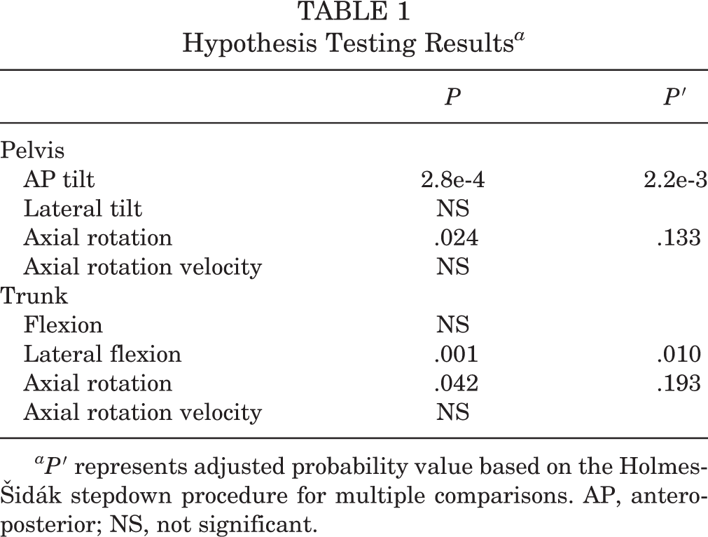
aP ′ represents adjusted probability value based on the Holmes-Šidák stepdown procedure for multiple comparisons. AP, anteroposterior; NS, not significant.
Results
The average pitch speed for youth and collegiate pitchers was 41 mph/65 kmh (range, 31-52 mph/50-84 kmh) and 57 mph/92 kmh (range, 50-61 mph/80-98 kmh), respectively. Compared with youth pitchers, collegiate pitchers exhibited a more posteriorly tilted pelvis from the moment of start of pitch until 94% of the way between start of pitch and BR (P ′ = .002) and a more laterally flexed trunk toward the glove side from the moment of start of pitch until 71% of the way between start of pitch and BR (P ′ = .010). No differences in pelvis lateral tilt, pelvis axial rotation, trunk extension, or trunk axial rotation were observed. No differences in pelvis or trunk axial rotation velocity were observed. Time-normalized comparisons for significant variables are presented visually in Figures 2 and 3. No correlation was seen between ball speed and either trunk or pelvic rotation.

(A) Comparison of pelvis posterior tilt angle between collegiate (dark) and youth (light) pitchers. Thick lines represent the mean value at each instant in time. Shaded cloud represents ±1 SD of the mean. Dashed vertical lines represent the mean timing of pitching events. (B) SPM plot. Black line represents SPM{t} test statistic at each point in time. Horizontal dashed lines represent the test statistic critical threshold. BR, ball release; FC, foot contact; SPM, statistical parametric mapping; TOP, top of pitch.
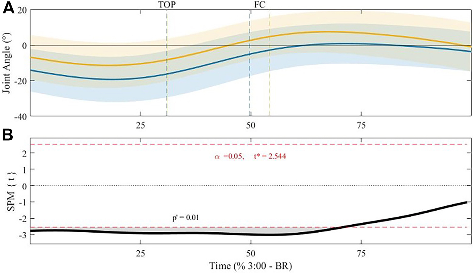
(A) Comparison of lateral trunk flexion angle between collegiate (dark) and youth (light) pitchers. Thick lines represent the mean value at each instant in time. Shaded cloud represents ±1 SD of the mean. Dashed vertical lines represent the mean timing of pitching events. (B) SPM plot. Black line represents SPM{t} test statistic at each point in time. Horizontal dashed lines represent the test statistic critical threshold. BR, ball release; FC, foot contact; SPM, statistical parametric mapping; TOP, top of pitch.
Discussion
This study compared the pelvis and trunk kinematics of youth and collegiate windmill softball pitchers. The results indicate that, compared with youth pitchers, collegiate pitchers tended to exhibit a more posteriorly tilted pelvis and a more laterally flexed trunk toward the glove side for much of the windup and delivery phases of the windmill pitch. The magnitude of differences for both angles was generally between 5° and 10° during earlier portions of the pitch and decreased as pitchers approached BR (Figures 2 and 3). These differences may indicate greater core and lumbopelvic-hip complex stability that provides a more stable base of support from which the upper and lower extremity musculature can contract. In softball, decreased lumbopelvic-hip complex stability has previously been associated with altered pelvis, trunk, and upper arm mechanics. 3 Likewise, less stable baseball pitchers have displayed increased pitching arm kinetics. 8
Increased posterior pelvic tilt in collegiate compared with youth pitchers was prominent throughout most of the pitch (Figure 2). In addition to extending and externally rotating the hip, the drive leg gluteal muscles also have leverage to posteriorly tilt the pelvis. Based on this, we hypothesized that the observed differences in pelvic tilt could be the result of increased gluteal muscle activity and result in a stronger push-off by the collegiate pitchers. This hypothesis aligns with previous reports showing high activation of the gluteal muscles between start of pitch and BR 16 and a positive association between hip external rotation strength and distal energy outflow from the trunk and pitching arm in youth softball pitchers. 18 We believe youth pitchers should strengthen the gluteal and abdominal musculature to improve pelvic stability and their ability to push off the pitching rubber.
Increased lateral trunk flexion toward the glove side was also observed in collegiate pitchers (Figure 3). Because the trunk was rotated away from home plate for much of the pitch, lateral flexion toward the glove side rotated the trunk toward home plate when viewed globally. Therefore, increased lateral trunk flexion may allow the collegiate pitchers to keep their trunk upright and momentum heading toward the target. However, a previous report demonstrated increased lateral trunk flexion to the glove arm side and increased shoulder distraction force at BR in pitchers who had experienced upper extremity pain compared with pain-free pitchers of similar skill levels. 15 Therefore, while pitchers may improve performance with increased glove-side lateral trunk flexion, they may also be placing their body in a more injury-prone position. The association between lateral trunk flexion and increased pitching arm kinetics has also been observed in baseball pitchers. Oyama et al 19 demonstrated increased pitching arm kinetics in pitchers displaying excessive lateral trunk flexion compared with those who did not.
After FC, softball pitchers must transmit energy up the kinetic chain and into the pitching arm. Weakness in the musculature surrounding the pelvis and trunk can interfere with this transmission and reduce pitching performance. 14 We hypothesize that a failure to efficiently transfer energy up the kinetic chain may present altered trunk kinematics. 4,5,11,17 Coaches regularly describe a trunk that is not upright as one that has “collapsed” or “caved” and work to coach pitchers out of this position and to “stay tall.” It is hypothesized that the increased posterior pelvic tilt and lateral trunk flexion toward the glove side demonstrated by collegiate pitchers may alter the length-tension relationships of the abdominal and lumbopelvic musculature and improve stability through the middle of the body. Stability through the trunk and lumbopelvic-hip complex is critical for kinetic chain function during upper extremity open-chain athletic tasks such as baseball and softball pitching. 4,11 Thus, it should be emphasized in daily conditioning protocols.
Limitations
Although the present study provides novel insight into the mechanical differences between youth and collegiate softball pitchers, it is not without limitations. While the controlled laboratory setting did allow for precise measurement of pelvis and trunk kinematics, participants may not have been able to fully replicate in-game effort levels. This could have, in turn, affected their pitching mechanics. Additionally, whether a difference of 5° to 10° is meaningful for clinical practice remains unclear. Future studies should focus on technique interventions to elucidate what constitutes a meaningful change in pelvis and trunk kinematics. The analysis was limited to the only the fastball pitch. Other pitches may require different pitching techniques that could present different trunk and pelvis mechanics than those observed in this study. Additionally, because participants were required to abstain from strenuous physical activity before data collection and threw a small number of pitches compared with normal game circumstances, we did not investigate the possible role of fatigue on pelvic and trunk kinematics. Additionally, the study’s cross-sectional nature limited the ability to draw inferences about how mechanics change over time within individual pitchers. Future studies should evaluate pitchers longitudinally to elucidate how pitching mechanics mature over time and examine how kinematic differences influence joint forces and moments during pitching.
Another limitation is the overlap in pitch velocity ranges between groups. Multiple advanced youth pitchers threw as fast as some collegiate pitchers. This overlap raises the question of whether age is a sufficient differentiator for examining mechanical differences between pitchers. Future research should examine mechanics, performance, and injury considering other factors such as pitch velocity, years of experience, earned run average, and so forth, to elucidate the best way to categorize pitching performance levels. Finally, the hypothesis of trunk mechanics indicating improved stability is, at this point, speculative. Additional research combining clinical measures of trunk and lumbopelvic-hip complex function is needed to investigate this hypothesis.
Conclusion
The current study found that collegiate pitchers tended to display a more posteriorly tilted pelvis and more laterally flexed trunk toward their glove side compared with youth pitchers. We believe that youth pitchers should incorporate exercises that improve their pelvic and core stability to enhance their pitching performance.
Footnotes
Final revision submitted February 2, 2021; accepted February 25, 2021.
The authors declared that there are no conflicts of interest in the authorship and publication of this contribution. AOSSM checks author disclosures against the Open Payments Database (OPD). AOSSM has not conducted an independent investigation on the OPD and disclaims any liability or responsibility relating thereto.
Ethical approval for this study was obtained from Auburn University (protocol No. 15-474 EP 1512).