



Abstract

Development of posttraumatic subchondral bone cysts without arthritis is infrequent. Subchondral cysts themselves are common, as they are classically a degenerative finding associated with osteoarthritis. However, subchondral cysts associated with microtrauma and no arthritis are uncommon, and there is little literature to guide surgical management of posttraumatic subchondral bone cysts, as they are often managed conservatively. 10,11 Development of posttraumatic bone cysts in young patients within the weightbearing area of the femoral head presents a unique challenge, particularly in the absence of arthritis, and the best surgical treatment for isolated lesions remains controversial. 15
A majority of the available evidence deals with osteochondral lesions of the knee and ankle. 8,9,13,14 Symptoms vary but can include pain with weightbearing activity and range of motion, as well as mechanical symptoms. Progression of disease and joint surface collapse are also concerns, given the nature of high contact forces through the hip joint and potential surface incongruity that may result in the early development of osteoarthritis. Hip arthroscopy has been increasing in popularity for several decades, and new areas of application continue to emerge. 1 –3,5,17 –19
This article reports the case of a young patient with the development of an acute postmicrotraumatic subchondral femoral head cyst and no evidence of osteoarthritis, which was successfully managed with arthroscopic-assisted femoral head drilling and filling the defect with a combination of local autograft and cadaveric bone graft. The evolution of the injury and subsequent cyst formation is clearly documented with advanced imaging and, to our knowledge, has not been described before.
Case Report
A 31-year-old otherwise healthy Indian American man was seen initially at an outside facility with an 8-month history of vague right hip pain. His initial injury occurred while playing tennis, where he had an acute onset of right hip pain while hitting a backhand (the patient is right-handed) and thus was weightbearing on the right lower extremity and pivoting from external rotation to internal rotation. The pain was initially localized to the lateral hip. His symptoms improved with activity modification, namely cessation of impact exercise, but did not completely resolve. While visiting family in Asia several months after the injury, he underwent a magnetic resonance imaging (MRI) study and was diagnosed with an insufficiency fracture of the femoral head and instructed by his treating physician in India to avoid impact sports (Figure 1).

Coronal T2 magnetic resonance imaging obtained 3 months after injury. The image demonstrates edema within the superior portion of the right femoral head. No significant subchondral collapse is evident. There is subtle loss of sphericity without collapse.
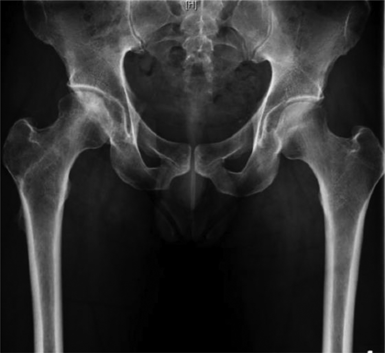
Eight months after injury, he was seen in the United States, where initial management was physical therapy and progressive increased activity with an Anti-Gravity Treadmill (AlterG). An anteroposterior pelvis radiograph from that visit demonstrated a mild crossover sign and cam deformity on the right but not the left (Figure 2).

Anteroposterior pelvic radiograph 8 months after injury. The image demonstrates a mild crossover sign and cam deformity on the right, which is not present on the left. The lateral center-edge angle measures 30° on the right and 21° on the left, and the Tönnis angle is 12° on the right and 15° on the left.
Six weeks later (9.5 months after injury), his hip pain was not improved, and a magnetic resonance arthrogram (MRA) was obtained to rule out a labral injury. The MRA demonstrated a healing osteochondral lesion of the superior weightbearing portion of the femoral head and a posterior labral tear (Figure 3).

Magnetic resonance arthrogram 9.5 months after injury demonstrates healing osteochondral lesion of the superior weightbearing portion of the femoral head.
The recommendation by his physician was to continue with physical therapy and anti-inflammatory medications. The patient was seen again in follow-up at 2, 4, and 6 months, with minimal symptomatic improvement. Follow-up MRI and radiographs of the hip were obtained 18 months after injury (Figures 4 and 5), which demonstrated an increase in size of the osteochondral lesion, subsequent cyst formation, and concern for avascular necrosis or pigmented villonodular synovitis (PVNS).

Magnetic resonance imaging 18 months after injury demonstrates an increase in size of the osteochondral lesion, subsequent cyst formation, and edema within the femoral head and subchondral zone.

Anteroposterior pelvic radiograph 18 months after injury demonstrates decreased density and cyst formation within the superior aspect of the femoral head.
The patient and his treating physician sought a second opinion, and he was referred to the senior author (M.R.S.) 20 months after initially presenting for treatment and more than 2 years since his initial injury. By this point, his symptoms had worsened and now included deep anterior hip/groin pain with activity. MRI with gradient echo sequences was performed, ruling out PVNS as a potential cause of the subchondral cyst (Figure 6).

Magnetic resonance imaging 19 months after injury demonstrates no intra-articular pathology, successfully ruling out pigmented villonodular synovitis. Redemonstration of an enlarging subchondral cyst without collapse of the subchondral plate.
A computed tomography scan of the right hip was obtained to assess for incongruity of the articular surface, and none was identified (Figure 7).

Computed tomography scan 20 months after injury demonstrates a subchondral cyst with preservation of the joint space.
Surgery was discussed with the patient, with options including surgical dislocation and osteochondral allograft versus arthroscopic drilling with or without bone grafting. He wished to proceed with the arthroscopic approach with bone grafting and elected not to have the cam deformity addressed.
Surgical Technique
The patient underwent routine hip arthroscopy in the supine position. Anterolateral, modified midanterior, and posterolateral portals were used during arthroscopy. The patient was found to have a 2 × 1.5–cm chondral flap with complete delamination of the superior pole of the femoral head. This chondral flap was debrided with curettes, and microfracture was performed of the subchondral bone. Figure 8 shows the femoral head after debridement and microfracture.

Femoral head lesion after debridement and microfracture.
The cystic lesion was then accessed by drilling from the lateral femur, just distal to the greater trochanter under fluoroscopic image guidance. The guide wire was placed with an anterior cruciate ligament (ACL) tibial guide, with the tip on the center of the femoral head intra-articularly and with the other part of the guide through a separate lateral incision to allow the guide wire to be passed to the femoral head lesion. The guide wire was advanced under arthroscopic and fluoroscopic visualization to ensure that it did not penetrate the subchondral bone of the femoral head but entered the cystic lesion. A cannulated 8-mm acorn ACL drill was then used to enlarge the size of the channel to the joint (Figure 9).

Intraoperative fluoroscopy verifies the location of the anterior cruciate ligament drill within the femoral head subchondral cyst.
Straight and curved long curettes were used to debride the subchondral cyst. Once the cyst was adequately decompressed, local cancellous autograft was combined with allograft bone chips and passed up the channel to fill the cyst.
Postoperative treatment consisted of limited weightbearing (20 lb, flat foot) while allowing full range of motion, followed by progressive weightbearing under the guidance of a physical therapist. The patient used continuous passive motion for 8 hours a day for the 6 weeks that he was limited in weightbearing. At the 9-month follow-up, the patient had minimal hip discomfort without the use of anti-inflammatory medications (Figure 10).
Anteroposterior pelvic radiograph 9 months after surgery. Interval healing and consolidation of subchondral cyst.
At a 28-month postoperative visit, the patient reported that his hip felt 95% recovered. He was extremely happy, having returned to bicycling, tennis, swimming, and running several miles per day with only mild morning tightness. Manual motor testing was 5 out of 5 in all muscle groups, and his passive range of motion of both hips was flexion to 150°, external rotation to 30° on the right and 70° on the left, and internal rotation of 10° bilaterally. The most recent imaging demonstrated remodeling of the cyst with preservation of joint space (Figure 11).

(A) Anteroposterior pelvic and (B) cross-table radiographs 28 months after surgery demonstrate remodeling of the cyst with preservation of the joint space.
Discussion
Subchondral cysts are a rare phenomenon in young patients with no evidence of osteoarthritis. A review by Schajowicz et al 18 provides a detailed description of the symptoms, sites, clinical features, and radiographic characteristics of juxta-articular cysts. Briefly, they are defined as benign cystic and often multiloculated lesions made up of fibrous tissue located in the subchondral bone adjacent to a joint. The pathogenesis of cyst formation in nonarthritic patients is almost exclusively an initial injury and cartilage defect. Mechanical stress and repeated microtrauma subsequently lead to vascular disruption. Microtrauma and local necrosis that is due to disruption of the subchondral blood supply in turn lead to local resorption of bone and subsequent cyst formation. 18 Other rare causes of cyst formation with a joint can be lesions such as PVNS, intra-articular ganglia, geode, or chondroblastoma. 19 Often these cysts involve the carpal bones, ankle, knee, and hip (usually acetabulum). In adults, the natural history of osteochondral lesions is typically one of poor healing with gradual progression to osteoarthritis. 12 MRI is usually the study of choice to evaluate and characterize cyst architecture. 16
It is our suspicion that the patient in this report sustained an acute osteochondral injury, as he was symptom-free prior to a specific injury that he remembers incurring while hitting a tennis shot. Interestingly, our patient had repeated MRI prior to seeing the senior surgeon (M.R.S.), allowing for documentation of the evolution of the initial osteochondral injury to a subchondral cyst. This case nicely demonstrates the natural history and progression of a chondral injury to cyst formation over the course of approximately 3 years. Specifically, it highlights the importance of identifying lesions in the femoral head that may require surgical intervention. Fortunately, this patient had not yet begun to develop radiographic evidence of joint space narrowing. This made arthroscopic hip surgery a reasonable surgical option given his persistent symptoms.
Additional evidence exists for the use of arthroscopy in posttraumatic hip injuries in athletes. Byrd and Jones 6,7 reported a median improvement of 45 points on the modified Harris Hip Score for athletes who underwent hip arthroscopy for intra-articular pathology. Interestingly, 5 athletes who had radiographic evidence of arthritis all went on to require total hip arthroplasty. The patient in this report did not have signs of arthritis at the time of surgery and has not developed joint space narrowing in the 2-year period following surgery.
Byrd 4 published a case series of 4 patients who sustained a lateral impaction injury to the hip joint after falling directly onto the greater trochanter. The hallmark of these patients was femoral head edema (or positive bone scan) on the weightbearing surface of the femoral head as seen on MRI, without any other obvious cause of pain. However, at arthroscopy, femoral head chondral damage overlying the area of the edema was seen. The question would be whether these patients would have developed subchondral cystic change if left untreated, as the patient in this report did, having similar initial MRI findings as patients in the Byrd study. Byrd’s patients had symptoms 4 to 15 months before surgery. The case series by Byrd also comments on the importance of preserving the subchondral plate if possible. Without the vascular supply of the plate, cell death occurs in overlying osteocytes and chondrocytes, which ultimately leads to osteochondral fragment demise. As the patient in the current report developed the subchondral cyst between the MRA at 9.5 months and the MRI at 18 months postinjury, one wonders if earlier intervention would have prevented the subchondral cyst, as Byrd did not report subchondral cyst formation in his patients prior to surgery. It is also not known if the patients in the Byrd report subsequently developed subchondral cystic change.
Furthermore, the Byrd 4 study reported that patients with traumatic onset of symptoms had the largest degree of improvement. The case presented here is consistent with these findings. In our patient’s case, he initially presented to a nonoperative sports medicine clinic and was not evaluated by a surgeon or hip specialist until 20 months after injury. Upon referral to a hip specialist, the patient was diagnosed with a chondral injury and promptly taken for surgery. A clear benefit is emerging in patients who undergo hip arthroscopy to address intra-articular pathology prior to radiographic evidence of osteoarthritis. Although no comparative studies exist, the evolution of this patient’s edema to cyst formation would suggest that early intervention to prevent cyst formation and the potential for joint surface collapse would be the recommended approach for patients with a similar presentation to those reported by Byrd. 2,4
Conclusion
Symptomatic juxta-articular posttraumatic cyst formation is a relatively uncommon occurrence, particularly in patients without osteoarthritis. Careful history should be taken and early advanced imaging obtained to identify those at risk of progression. Arthroscopic management prior to radiographic evidence of arthritis is ideal, and the case reported here demonstrates the evolution of a posttraumatic subchondral femoral head cyst successfully treated with arthroscopically aided drilling, debridement, and bone grafting.
Footnotes
One or more of the authors has declared the following potential conflict of interest or source of funding: M.R.S. has received fellowship support from Breg, ConMed, Ossur, and Smith & Nephew; research support from Ferring and Smith & Nephew; educational support from Biomet; consulting fees from Medacta and Smith & Nephew; and royalties from DJO, Elsevier, Smith & Nephew, Stryker, and Williams & Wilkins. AOSSM checks author disclosures against the Open Payments Database (OPD). AOSSM has not conducted an independent investigation on the OPD and disclaims any liability or responsibility relating thereto.