
Abstract
Osteogenesis imperfecta (OI) is a rare genetic disorder characterized by bone fragility. Its association with dentinogenesis imperfecta (DI) is well documented, but the concurrent presentation with nephrocalcinosis is uncommon and poorly understood. We documented the case of an 18-year-old male presenting with a triad of OI, DI, and nephrocalcinosis. The patient exhibited characteristic features including blue sclera, multiple fractures, dental abnormalities, bowing of long bones, a short stature, and biochemical evidence of altered calcium metabolism. Genetic testing revealed mutations in COL1A1, confirming the diagnosis of OI Type I. This case highlights the importance of comprehensive evaluation in OI patients, emphasizing the need for dental and renal assessment. The presence of nephrocalcinosis in OI demands further investigation into the mechanisms of calcium dysregulation in disorders of these kinds.
Summary
This is a case of an 18-year-old male with dentinogenesis imperfecta type I (DI-I) associated with osteogenesis imperfecta (OI) who visited the outpatient department (OPD) of Periodontology Department with a chief complaint of swollen gums that were easy to bleed. The patient exhibited characteristic dental abnormalities, including discolored teeth and enamel chipping, alongside systemic manifestations such as blue sclera, short stature, and multiple bone fractures. Notably, the case was complicated by nephrocalcinosis and chronic metabolic acidosis, underscoring the multisystem impact of OI. This report highlights the importance of comprehensive evaluation and multidisciplinary management in patients presenting with dental anomalies suggestive of genetic disorders.
Background
Dentinogenesis imperfecta (DI) is a rare hereditary disorder affecting tooth development, primarily involving defective dentin formation. DI-I, specifically associated with OI, presents a unique challenge in diagnosis and management due to its systemic implications. OI, characterized by bone fragility and collagen defects, exhibits varying severity across its 8 recognized types. The genetic basis of these conditions involves mutations in COL1A1 and COL1A2 genes, crucial for type I collagen synthesis.
The incidence of OI is approximately 1 in 30 000 to 50 000 births, with DI-I occurring in about 50% of OI cases. This case report aims to elucidate the clinical presentation, diagnostic approach, and management considerations in a patient with DI-I and OI, emphasizing the rare complication of nephrocalcinosis.
Introduction
OI is a heterogeneous group of genetic disorders primarily affecting type I collagen, the most abundant protein in the bone and teeth. The incidence of OI is estimated at 1 in 10 000 to 20 000 births. OI is characterized by bone fragility, recurrent fractures, and various extra-skeletal manifestations including blue sclera, hearing loss, and defective dentin formation leading to discolored teeth with aberrant structure of the dentin component of the teeth, known as DI.1,2
DI-I is a genetic disorder of tooth development characterized by opalescent dentin and abnormal dentin structure. It occurs in about 50% of individuals with OI. The dental manifestations include discolored teeth that appear opalescent gray or brownish, bulbous crowns, and obliterated pulp chambers.3,4
Nephrocalcinosis, the deposition of calcium in kidney tissue, is not typically associated with OI. However, recent studies have suggested a potential link between OI and altered calcium metabolism. The co-occurrence of OI, DI, and nephrocalcinosis presents a unique clinical scenario that merits further investigation.5,6
This case report describes an 18-year-old male presenting with this rare triad, providing insights into the potential interplay between these conditions and highlighting the importance of comprehensive evaluation in patients with OI.
Case Presentation
An 18-year-old male presented to the Outpatient Department of Khyber College of Dentistry with chief complaints of swollen, bleeding gums, and concerns about the appearance of his teeth. The patient reported rough tooth surfaces and abnormal coloration but minimal tooth sensitivity.
History
The patient had a history of multiple fractures since early childhood, primarily affecting the long bones. He reported delayed motor milestones and recurrent bone pain. A family history revealed a deceased brother with similar symptoms, but no other family members were affected.
Physical Examination
On general physical examination, the patient presented with a short stature (height below the third percentile for the age) with evidences of long bone deformities, particularly bowing of the long bones (Figures 1 and 2). A surgical scar on the right leg indicates interventions for orthopedic reasons, particularly due to recurrent fractures. A blue-tinted sclera was observed, which was more pronounced under direct light (Figure 3).

Bowing of the legs, a classic sign of osteogensis imperfecta and surgical scars on the shins indicating previous orthopedic interventions due to recurrent fractures.

Bowing of the long bones, an orthopedic anomaly of dentinogenesis imperfecta type 1.

Blue sclera, a classical sign of patients with dentinogensis imperfecta type 1.
Extra-oral examination showed normal facial features and symmetry with no swellings, scars, or palpable lymphadenopathy.
Intra-oral examination revealed a mixed dentition, having multiple retained deciduous resulting poor oral hygiene. The teeth also showed signs of generalized enamel chipping and brownish discoloration.
Investigations
Comprehensive investigations were undertaken to elucidate the patient’s condition. A panoramic radiograph revealed multiple dental abnormalities characteristic of DI associated with OI. These included multiple unerupted teeth, retained deciduous teeth, bulbous crowns with marked cervical constriction, obliterated pulp chambers, and short, constricted roots. These findings are consistent with the delayed dental development often observed in OI patients and confirm the presence of DI, as shown in Figure 4.

Panoramic radiographs of the oral cavity showing retained primary teeth, unerupted permanent teeth and a narrow mandible bone.
Serum biochemistry yielded notable results. The patient’s serum calcium was elevated at 11.0 mg/dL (normal range: 8.6-10.2 mg/dL), indicating hypercalcemia. Biochemical investigations further elucidated the cause of hypercalcemia. The patient’s fasting serum parathyroid hormone (PTH) level was within the normal range (10-65 pg/mL). However, the 25-hydroxyvitamin D (25-OHD) level was measured at 18 ng/mL (normal range: 30-100 ng/mL), indicating vitamin D deficiency. This deficiency likely contributes to the patient’s bone deformities and elevated serum alkaline phosphatase levels. Vitamin D plays a crucial role in calcium and phosphate metabolism, and its deficiency can impair bone mineralization, exacerbating the metabolic disturbances associated with OI. Additionally, the calcitriol (1,25-dihydroxyvitamin D) level was slightly elevated (75 pg/mL, normal range: 20-70 pg/mL), reflecting increased vitamin D activity. This finding aligns with the metabolic disturbances in OI, which is characterized by increased bone turnover and altered calcium metabolism. Alkaline phosphatase levels were at the higher end of normal at 269 IU/L (range : 54-369 IU/L), potentially suggesting increased bone turnover. These findings prompted further investigation of the patient’s calcium metabolism (Figure 5).
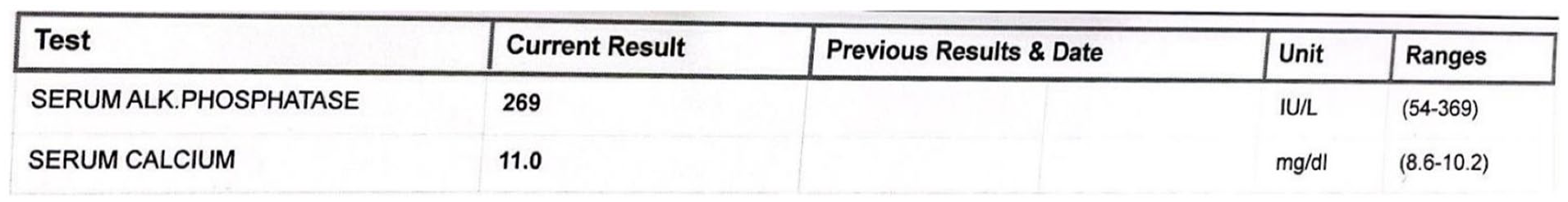
Elevated serum calcium levels indicating osteoporosis and potentially nephrocalcinosis.
The patient’s blood was later analyzed for metabolic profile including Cr, electrolytes, Ca, P, Mg, PTH, and vitamin D level at 4 different occasions over the course of 4 weeks to establish the diagnosis of nephrocalcinosis. A detailed table of the 4 different profiles were given in Table 1. The patient’s serum calcium levels are consistently elevated (10.8-11.2 mg/dL), which is a key indicator of hypercalcemia and a risk factor for nephrocalcinosis. The patient’s 25-OHD levels are low (18-19 ng/mL), indicating vitamin D deficiency, which can affect calcium metabolism. The patient’s 1,25-dihydroxyvitamin D levels are slightly elevated (74-76 pg/mL), which can increase calcium absorption and contribute to hypercalcemia.
A detailed report of the pateint’s metabolic profile on four different occasions.

Abbreviations: 25-OHD, 25-hydroxyvitamin D; PTH, parathyroid hormone.
Urinalysis provided additional insights into the patient’s mineral homeostasis. The 24-hour urinary calcium excretion was within normal limits at 154.23 mg/24 hour (range: 100-300 mg/24 hour), an intriguing finding given the elevated serum calcium. Notably, 24-hour urinary phosphate was low at 268.71 mg/24 hour (range: 400-1300 mg/24 hour), suggesting altered renal phosphate handling. The urinary citrate level is slightly lower than the normal range (315 mg/24 hour), which can increase the risk of calcium stone formation as shown in Table 2. Urine pH has been normal at 6.5 (range: 5.0-8.0; Figures 6 and 7).
A 24-hour urinalysis report of the patient showing low urinary phosphate suggesting altered renal phosphate handling.


Elevated urine phosphorous levels, a result of bone dissolution in dentinogenesis imperfecta type 1.

Detailed urine analysis report.
Renal ultrasonography confirmed nephrocalcinosis, an un-common finding in OI patients. This discovery, in conjunction with the biochemical abnormalities, underscores the complexity of this case and highlights the importance of comprehensive renal evaluation in OI patients.
During the diagnostic workup, nephrocalcinosis was incidentally identified. This finding, in conjunction with hypercalcemia, normal PTH levels, and elevated calcitriol, suggests a potential dysregulation in calcium metabolism that requires further evaluation. The exact mechanism linking OI to nephrocalcinosis remains unclear and requires additional investigation.
Genetic testing revealed a pathogenic variant in the COL1A1 gene, confirming the diagnosis of OI Type I. This finding provides a molecular basis for the patient’s clinical presentation and supports the observed phenotype of mild OI with associated DI.
Collectively, these investigations paint a picture of a rare and complex case of OI Type I with concurrent DI and nephrocalcinosis. The biochemical profile, particularly the juxtaposition of hypercalcemia with normal urinary calcium excretion and low urinary phosphorus, suggests a nuanced disturbance in mineral metabolism. This case underscores the need for thorough investigation of calcium-phosphate homeostasis in OI patients, especially when renal complications are present.
Discussion
This case shows a rare combination of OI, DI, and nephrocalcinosis in an 18-year-old male. The diagnosis of OI was based on the clinical presentation of multiple fractures, blue sclera, and bone deformities, supported by the genetic finding of a COL1A1 mutation. 7
The presence of DI-I, occurring in approximately 50% of OI cases. This is evident from the dental manifestations and radiographic findings. 8 The clinical manifestation included grayish brown discoloration of the teeth, over-retained primary teeth, and un-erupted permanent teeth, while the panoramic radiograph demonstrated classic features of DI, including bulbous crowns, obliterated pulp chambers, and short roots. 9
The most intriguing aspect of this case is the concurrent nephrocalcinosis which is an uncommon finding in OI. The patient’s elevated serum calcium and alkaline phosphatase levels, with the low levels of 25-OHD, affecting calcium metabolism coupled with increased levels of 1,25-OHD can contribute to hypercalcemia, an increased risk factor, suggestive of nephrocalcinosis. While the exact mechanism linking OI and nephrocalcinosis remains unclear, it may relate to altered calcium metabolism associated with abnormal collagen formation. 10
The biochemical profile in this case, particularly the elevated serum calcium and low urinary phosphorus is suggestive of a complex disturbance in mineral metabolism. This finding underscores the importance of regular monitoring of calcium homeostasis in OI patients, even in the absence of any obvious renal symptoms. 11
From a dental perspective, the management of DI-I in OI patients requires a multidisciplinary approach. Early dental intervention is crucial to prevent rapid wear of dentition and to maintain oral function and aesthetics. 12 In this case, the mixed dentition and multiple unerupted teeth present additional challenges in dental management, like swollen gums, tending to bleed upon probing.
The familial aspect of this case, with a similarly affected deceased sibling, highlights the importance of genetic counseling in OI. The autosomal dominant inheritance pattern of most OI types necessitates family screening and long-term follow-up. 13
Conclusion
This case report presents a rare disease of the bones and teeth, OI Type I with concurrent DI-I and nephrocalcinosis. It signifies the importance of a comprehensive, multisystem approach in the evaluation and management of patients with OI. The presence of nephrocalcinosis in this case raises questions about the broader implications of collagen disorders on calcium metabolism and renal function.
Future research should focus on elucidating the mechanisms linking abnormal collagen formation with calcium homeostasis and renal calcification in OI. Long-term follow-up studies of OI patients may provide insights into the prevalence and progression of renal complications in this disorder.
In clinical practice, this case emphasizes the need for:
Comprehensive initial evaluation of OI patients, including detailed dental and renal assessment.
Follow-up regrading dental health care.
Regular monitoring of calcium metabolism and renal function in OI patients.
Early dental intervention and ongoing dental care in patients with OI and DI.
Genetic counseling and family screening in cases of OI.
By sharing this unique case, we hope to alert clinicians to the possibility of renal complications in OI and to stimulate further research into the complex interplay between bone, teeth, and kidney health in collagen disorders.
Footnotes
Acknowledgements
We would like to acknowledge the support we got from all our colleagues, especially Mr. Aizaz Ali, a student of final-year MBBS at Khyber Medical College Peshawar.
Declaration of Conflicting Interests
The author(s) declared no potential conflicts of interest with respect to the research, authorship, and/or publication of this article.
Funding
The author(s) received no financial support for the research, authorship, and/or publication of this article.
Ethical Approval
Our institution does not require ethical approval for reporting individual cases or case series.
Informed Consent
Written informed consent was obtained from the patient for the publication of their case along with all the images.