
Abstract
Globally, health care providers have been challenged to provide adequate care during the coronavirus disease-2019 (COVID-19) pandemic. Due to the ever changing and rapidly evolving nature of the novel coronavirus, there is increased public anxiety and knowledge gaps that have created major dilemmas in health care delivery. In this environment, there is tremendous pressure on clinicians to diagnose each and every case of COVID-19. This has led to a situation in which clinicians are primed to suspect all respiratory illness is due to COVID-19 infection until proven otherwise. Because of this, providers may misdiagnose patients who have illnesses that are distinct from COVID-19 but present in a similar manner. In the current article, we present the case of e-cigarette- and vaping-associated acute lung injury (EVALI) mimicking pneumonia secondary to the novel coronavirus. It is unknown if vaping puts patients at higher risk of respiratory failure if coinfected with COVID-19. Therefore, exposure history in patients presenting with pneumonia-like syndrome is important. Physicians should be aware of the overlap between these conditions and should pay particular attention during history taking to distinguish EVALI from COVID-19 pneumonia.
Case Presentation
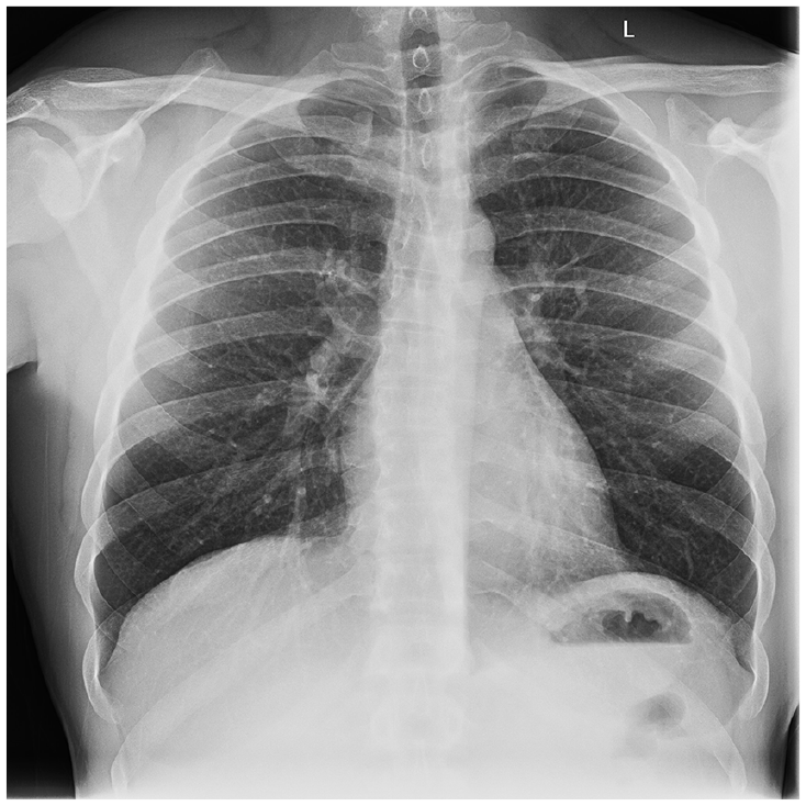
A 31-year-old male with past medical history of Crohn’s disease presented with 4 days of fever, fatigue, dry cough, and dyspnea. His medications included infliximab, taken every 6 weeks with the last dose being 1 month prior to admission. On arrival, the patient had a heart rate of 110 bpm, temperature of 102 °F, and SpO2 of 86% on room air. Auscultation revealed crepitations at the lung bases bilaterally. Arterial blood gas revealed a pO2 of 48 mm Hg, a widened A-a gradient, and respiratory alkalosis. Chest radiograph (Figure 1) demonstrated faint bilateral pulmonary interstitial opacities. Transthoracic echocardiogram was performed and was normal. Contrasted computed tomography (CT) of the chest (Figure 2a and b) showed diffuse ground-glass opacities. Based on initial presentation, the patient was suspected to have coronavirus disease-2019 (COVID-19) pneumonia and was placed in negative-pressure isolation. Respiratory viral panel including testing for influenza A and B, Legionella pneumophila, Streptococcus pneumoniae, and Mycoplasma pneumoniae were negative. Furthermore, sputum culture, blood cultures, urine legionella antigen, as well as serum histoplasma and cryptococcal antigens were ordered and were negative. Reverse transcriptase–polymerase chain reaction testing for COVID-19 was negative on days 1 and 3. During initial hospitalization, he remained dependent on high-flow oxygen to maintain saturations >90%. Because of the lack of improvement in clinical status, further workup was initiated. Serum β-D-glucan was negative. Serum ACE (angiotensin-converting enzyme) was 35 U/L (normal = 16-85 U/L) and serum BNP (brain natriuretic peptide) was 30 pg/mL (normal = 6-100 pg/mL). Serum LDH (lactate dehydrogenase) was also performed, and was elevated at 366 U/L (normal = 140-271 U/L). HIV 1 and 2 antibody screening was negative. Rheumatoid factor, anti-dsDNA, anti-Jo-1, anti-Scl-70, anti-Smith, SSA-52 and SSA-60, SSB, and anti-chromatin antibodies were negative. Procalcitonin was <0.10 ng/mL (normal ≤0.15 ng/mL).

Chest X-ray showing faint bilateral pulmonary interstitial opacities.

(a) Coronal computed tomography (CT) scan of the chest demonstrating diffuse pattern of ground-glass opacities without consolidation or interlobular septal thickening. No pleural effusions are noted. (b) Axial CT scan of the chest demonstrating diffuse pattern of ground-glass opacities without consolidation or interlobular septal thickening. No pleural effusions are noted.
On further questioning, the patient reported frequently binge smoking disposable E-cigarette pods, which included vaping nicotine (40-50 hits/day), tetrahydrocannabinol (140-150 hits/day), and cannabidiol. He attributed this to anxiety secondary to the stay-at-home orders. A diagnosis of e-cigarette- and vaping-associated acute lung injury (EVALI) was made. The patient developed significant clinical and radiographic improvement (Figure 3) over the next week after cessation of vaping and completion of a 1-week course of oral steroids.
Chest X-ray showing improvement of hazy ground-glass opacities in both lungs.
Discussion
COVID-19 pneumonia and EVALI both lead to aerosol-induced acute lung injury with similar clinical presentation. Both processes include respiratory symptoms and similar radiographic findings. EVALI is thought to be due to inhalation of the aerosolized content of e-cigarettes, including vitamin E acetate, coconut oil, and limonene, while COVID-19 is transmitted through respiratory droplets and aerosols.1,2 The difference between these 2 states lies in the etiologic nature of the lung disease, as COVID pneumonia is due an infectious disease (SARS-CoV-2 [severe acute respiratory syndrome coronavirus 2] infection), whereas damage secondary to EVALI is due to noninfectious inhaled agents that can cause acute respiratory failure and lung injury. Table 1 provides a comparison between these 2 disorders. Electronic cigarettes and electronic nicotine delivery systems have rapidly gained popularity over the past several years, especially among young adults. By 2018-2019, the number of young adults using these products have reached epidemic levels. 3 A 2019 survey conducted by the Centers for Disease Control and Prevention evaluating the current use of electronic cigarettes in middle school and high school students found that in the past 30 days from the time of the survey, approximately 10.5% of middle school students and 27.5% of high school students had used electronic cigarettes. 3 Since February 2020, the Centers for Disease Control and Prevention has reported a total of 2807 hospitalizations/deaths secondary to EVALI.4,5 In comparison, as of August 20, 2020, the COVID-19 pandemic has infected 5 506 929 people in the United States with 172 416 deaths. 6 There have been reports of increased incidence of COVID pneumonia in smokers, but there is a paucity of data regarding an association between vaping and COVID-19 rates. 7 Although increased public awareness has led to a decline in the incidence of EVALI, further patient and public education is needed in order to eradicate this disease process.
Comparison Between EVALI and COVID-19.

Abbreviations: EVALI, e-cigarette- and vaping-associated acute lung injury; COVID-19, coronavirus disease-2019; E-CIGs, electronic cigarettes; ENDS, electronic nicotine delivery systems; ESR, erythrocyte sedimentation rate; SARS-CoV-2, severe acute respiratory syndrome coronavirus 2; RT-PCR, reverse transcriptase-polymerase chain reaction.
In EVALI, patients often present with respiratory symptoms, including dyspnea, cough, and chest pain. 8 In addition, gastrointestinal symptoms such as nausea, vomiting, and diarrhea, as well as constitutional symptoms such as fever >100.4 °F are common. 8 Laboratory findings include nonspecific leukocytosis, elevated inflammatory markers such as erythrocyte sedimentation rate and procalcitonin, and elevated liver enzymes. 9 Elevated liver enzymes and inflammatory markers can also be seen in COVID-19; however, patients with COVID-19 often exhibit lymphopenia as opposed to lymphocytosis. 2 In the current case, our patient also presented with lactic acidosis. While this finding is rather nonspecific for EVALI, it is likely that the patient developed this finding due to the profound inflammation and hypoxia associated with acute lung injury.
With regard to imaging, EVALI typically presents with patterns of basilar-predominant consolidation and ground-glass opacity, with areas of lobular or subpleural sparing on CT scan. 1 In some patients, there may be findings suggestive of rapidly developing acute lung injury, including acute eosinophilic pneumonia and diffuse alveolar damage. 1 In contrast, the imaging findings in COVID-19 vary. In early disease, inflammatory infiltrates may be found in subpleural or peribronchovascular regions. With advanced illness, ground-glass opacities, crazy-paving pattern, and consolidation in bilateral lobes are the most common findings.1,10 Histopathologic findings in EVALI may include foamy macrophages, acute fibrinous pneumonitis, diffuse alveolar injury, or organizing pneumonia. 11 Histopathologic characteristics in COVID-19 disease are nonspecific and include inflammatory changes, vascular congestion, proteinaceous exudates, and findings of diffuse alveolar damage. 12 Microscopic findings may include viral infectious changes, such as multinucleated-enlarged pneumocytes with large nuclei, amphophilic cytoplasm, and prominent nucleoli in alveolar spaces. 13
EVALI is a diagnosis of exclusion, and as such, there is no single confirmatory test. The diagnosis of COVID-19 is made by reverse transcriptase-polymerase chain reaction of respiratory tract specimens. 2 As such, appropriate history taking and early testing are critical to establishing the appropriate diagnosis.
Conclusion
There is significant overlap in the presentation of acute lung injury due to COVID-19 and EVALI, with syndromes ranging from completely asymptomatic to severe respiratory failure and multiorgan dysfunction. EVALI is a diagnosis of exclusion. High index of suspicion and particular attention to detail during history taking is crucial to prompt recognition and the initiation of proper management.
Footnotes
Declaration of Conflicting Interests
The author(s) declared no potential conflicts of interest with respect to the research, authorship, and/or publication of this article.
Funding
The author(s) received no financial support for the research, authorship, and/or publication of this article.
Ethics Approval
Our institution does not require ethical approval for reporting individual cases or case series.
Informed Consent
Verbal informed consent was obtained from the patient(s) for their anonymized information.