
Abstract
Background:
Deviated nasal septum (DNS) is an influential variant of the sinonasal region due to its possible association with varied sinonasal pathologies, mid-facial (skeletal or occlusal) disharmonies and orofacial pain disorders. The aim of the study is to document the prevalence of DNS and to observe and assess the radiographic attributes of its types in adults.
Materials and Methods:
It is a descriptive cross-sectional study in which, after getting ethical approval, computed tomography paranasal sinus volumes of 100 adult subjects are collected from the medical college on the basis of convenience sampling. Paediatric subjects and patients with a previous history of mid-facial trauma, malignancy or surgery are excluded from the study. DICOM data thus collected are retrospectively analysed by RadiAnt DICOM viewer to determine the prevalence of DNS to study its association across gender and with increased (>1 mm) mucosal thickening of the maxillary sinus. DNS is recorded by Mladina’s classification and by septal deviation angle, and association is studied by the Chi-square test and Mann–Whitney U test.
Results:
The overall prevalence of DNS is 82%. Association of deviated septum with gender and mucosal thickening of the sinus lining is not statistically significant. Overall type V (spur) deviation is most common; however, in subjects with thickened mucosal lining type III (“C” or reverse “C”), deviation is found to be most prevalent. All the cases of type VI deviation have a tilted nasal floor, a finding of dental surgeon’s interest. “Bridging Spur,” an uncommon form of spur deformity, is also recorded in one case.
Conclusions:
Deviated septum is far more common than straight septum in the adult population. Types III, II and IV are more frequent in subjects with increased mucosal thickening. Association of type VI with tilted floor and rare variants like “Bridging spur” is also reported.
Keywords
List of Abbreviations
3D: Three dimensional
CT: Computed tomography
Df: Degree of freedom
DICOM: Digital Imaging and Communications in Medicine
DNS: Deviated nasal septum
FOV: Field of view
MDCT: Multidetector computed tomography
NVA: Nasal valve angle
OSA: Obstructive sleep apnoea
PNS: Paranasal sinus
SDA: Septal deviation angle
SPSS: Statistical Package of Social Sciences
TN: Trigeminal neuralgia.
Introduction
Deviated nasal septum (DNS) is being studied as an etiologic factor for several pathologic conditions for which patients often consult dental surgeons. Pain of maxillary sinusitis often radiates to the maxillary teeth region and is perceived as odontogenic pain or facial pain.1–3 Deviation near the Cottle’s/nasal valve region is often linked with snoring/obstructive sleep apnoea (OSA).4,5 Deviation also plays a credible role in the development of sagittal occlusal disharmonies and in facial retrusion with cleft lip and palate.3,6,7 Deviation in the deepest part of the nose in the form of a spur often results in the impaction of its tip in the region of the sphenopalatine foramen and may cause unilateral hemi cranial headaches of Sluder’s type, an important differential diagnosis of trigeminal neuralgia (TN).8,9 Furthermore, Mladina et al. 10 advocated that horizontal deformities are dominantly inherited deformities (type V and type VII) and are not related to trauma and thus have a role in forensics.
CT imaging with a large field of view (FOV) is often indicated by dental surgeons for oral and maxillofacial pathologies such as osteomyelitis, benign cystic lesions, tumours and so on. It is also indicated for presurgical implant planning, sinus lift procedures or orthognathic surgeries. It is a non-invasive modality and provides a permanent three-dimensional (3D) record of the bone as well as the soft tissue of the entire length of the nasal septum and accurately assesses paranasal sinus pathology.11,12
This study aims to observe the common and rare variants of septal deviation in a CT data set which forms the basis for the interpretation of the tomographic image as a thorough understanding of radioanatomy of this utile landmark is essential for an oral and maxillofacial radiologist and stomatologist for accurate diagnosis and treatment planning. The objectives of the study were (1) to study the prevalence (overall and type-wise) and association of DNS with gender, (2) to compare septal deviation angle (SDA) across gender, (3) to study the prevalence (overall and type-wise) and association of DNS with increased mucosal thickening of maxillary sinus lining, a characteristic radiographic feature of sinusitis and (4) to compare SDA across the categories of absence or presence of increased mucosal thickening of maxillary sinus lining.
Materials and Method
Setting and Design
It is a descriptive cross-sectional hospital-based study that aims to evaluate the variants of nasal septum from CT data. The study is conducted at the Gujarat University affiliated dental college and hospital, and permission for the procurement of the CT data for the retrospective analysis is taken from the medical college after getting approval from the Institutional Review Board (AMC Dental College, Khokhra-Ahmedabad. Reg. No.: ECR/236/Indt/GJ/2015/RR-18).
Data from non-contrast-enhanced computed tomography paranasal sinus (CT PNS) scans are collected from the medical college over a period of 3 years starting from 2018 to 2020. Radiographic data of all the adult patients (>18 years) who have undergone CT PNS analysis are collected irrespective of gender and ethnicity. Paediatric patients and subjects with a previous history of trauma to the mid-facial region, malignancy or surgery are excluded. A total of 100 subjects were included in the study on the basis of convenience sampling.
Specifications and Data Capturing
Volumetric data of the sinonasal region are collected from the archives of the MX 16 Slice machine (Philips Health Care, the Netherlands) where exposure parameters for PNS are set at 120 kV, 250 mA s, with 1–3 mm slice thickness. The first scan observed is a coronal scan, and later axial and sagittal scans are further analysed with a RadiAnt DICOM (Digital Imaging and Communication in Medicine) viewer. The nasal septum and its variants are then evaluated by an experienced oral and maxillofacial radiologist twice with an interval of more than two weeks. Intra-examiner variation showed excellent agreement (kappa statistics value 0.9).
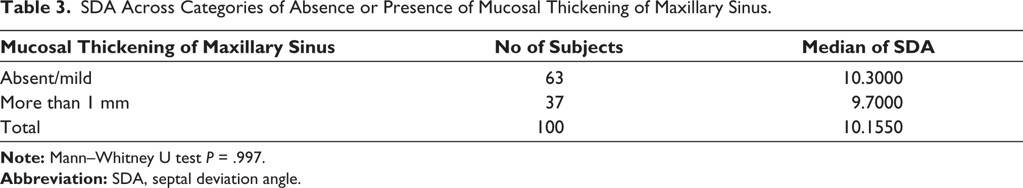
As thickened sinus lining (>1 mm) is one of the characteristic radiographic features of sinusitis, subjects are divided into two groups based on the absence or presence of thickened sinus mucosal lining. 13
Observational Parameters
Subjects are grouped according to gender and then on the basis of the thickened mucosal lining of the maxillary sinus. DNS is then studied for the prevalence and association with gender as well as across categories of absence/presence of increased mucosal lining of the maxillary sinus.
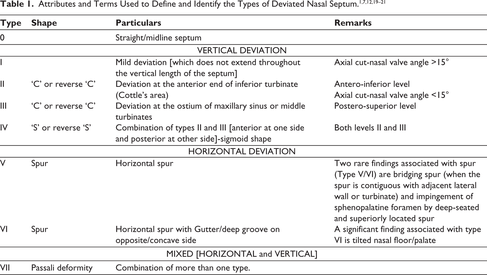
To study the nasal septum, various classification systems are in practice, and among them, Mladina’s classification system14–18 has been cited in more than 40 clinical research works. Deviation is thus noted and classified using Mladina’s classification and its user-friendly modification (in types I and II) proposed by Rao 1 so that a thorough examination of the coronal section suffices to document the type on the basis of the shape and location of deviation. The attributes of the different types of deviation observed in this study are shown in tabular and pictorial forms. It also includes the terms used to describe different types of deviations and the relation of DNS with the significant landmarks of the lateral wall of the nose (Table 1 and Figures 1 and 2).1,7,12,19–21
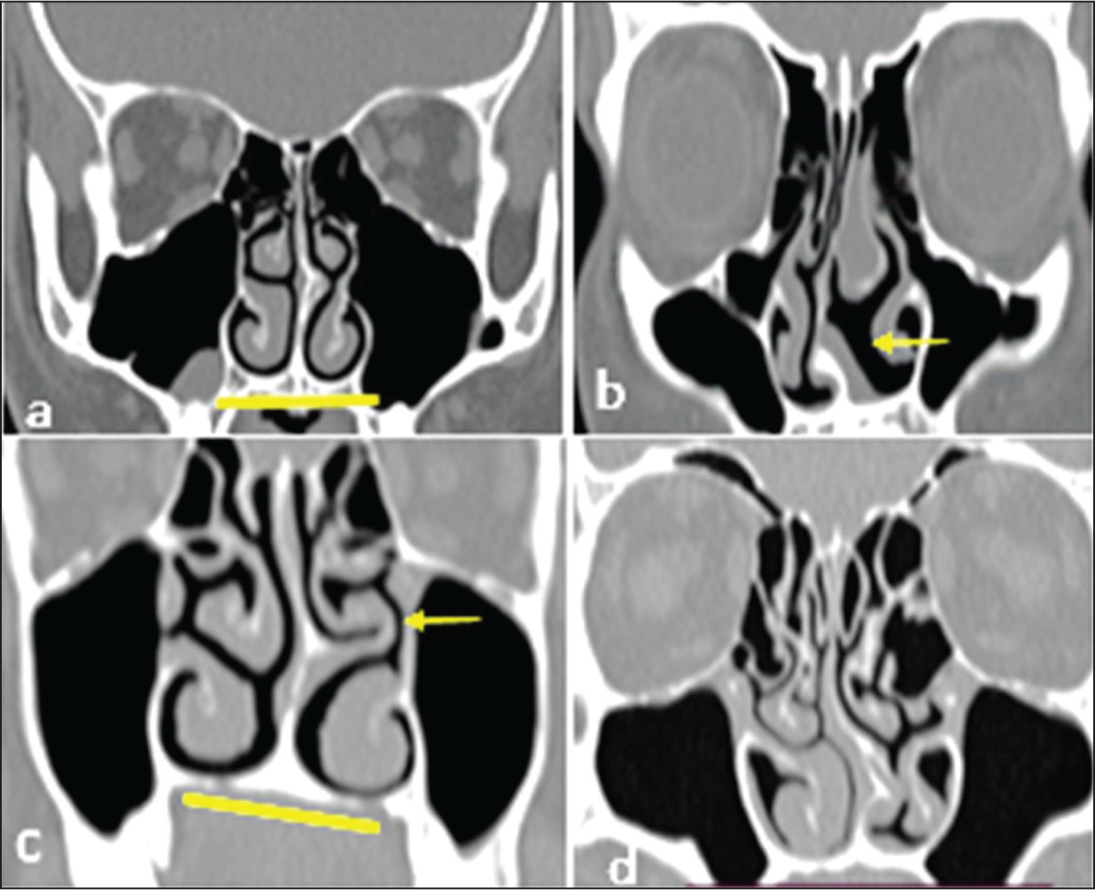
Midline Septum and Vertical Deviations of Septum in Coronal Section: Type 0 to Type IV. (a) Type 0 – No Deviation, (b) Type I – Mild Deviation, (c, d) Type II – Severe Deviation in Cottle’s Area, Coronal and Axial Sections, (e) Type III – ‘c’ or Reverse ‘c’ Shape and (f) Type IV – ‘s’ or Reverse ‘s’ Shape. Arrow Indicates the Site of Deviation, and Vertical Line Indicates Reference Midline.
In order to measure the degree/angle of deviation (SDA), a midline is drawn between the crista galli (upper reference point) and the nasal crest (lower reference point), a deviation line is drawn between the crista galli and the most prominent point of the deviation, and then the angle is recorded at the upper point/crista galli.22,23
Statistical Analysis
Recorded data are analysed by IBM SPSS Statistics 20 (IBM Corp., Armonk, NY, USA). To study the association between nominal/categorical data, the Chi-square test is applied. Shapiro-Wilk normality test is performed for the continuous data and p < .05, data are not normally distributed. Thus, a non-parametric test (Mann-Whitney test) is used to compare the median of SDA across genders and across categories of absence or presence of thickened mucosal lining of the maxillary sinus. The level of significance is set at P-value < .05.
Result
Demographic Data
CT data of a total of 100 adult subjects are examined with ages ranging from 18 to 80 years. Out of these, 66 subjects are males and 34 are females. When the mucosal lining of the maxillary sinus is observed for thickening (>1 mm), 37 subjects are identified with thickened maxillary sinus lining, while 63 subjects have minimal/no thickness.
Nasal Septum Deviation
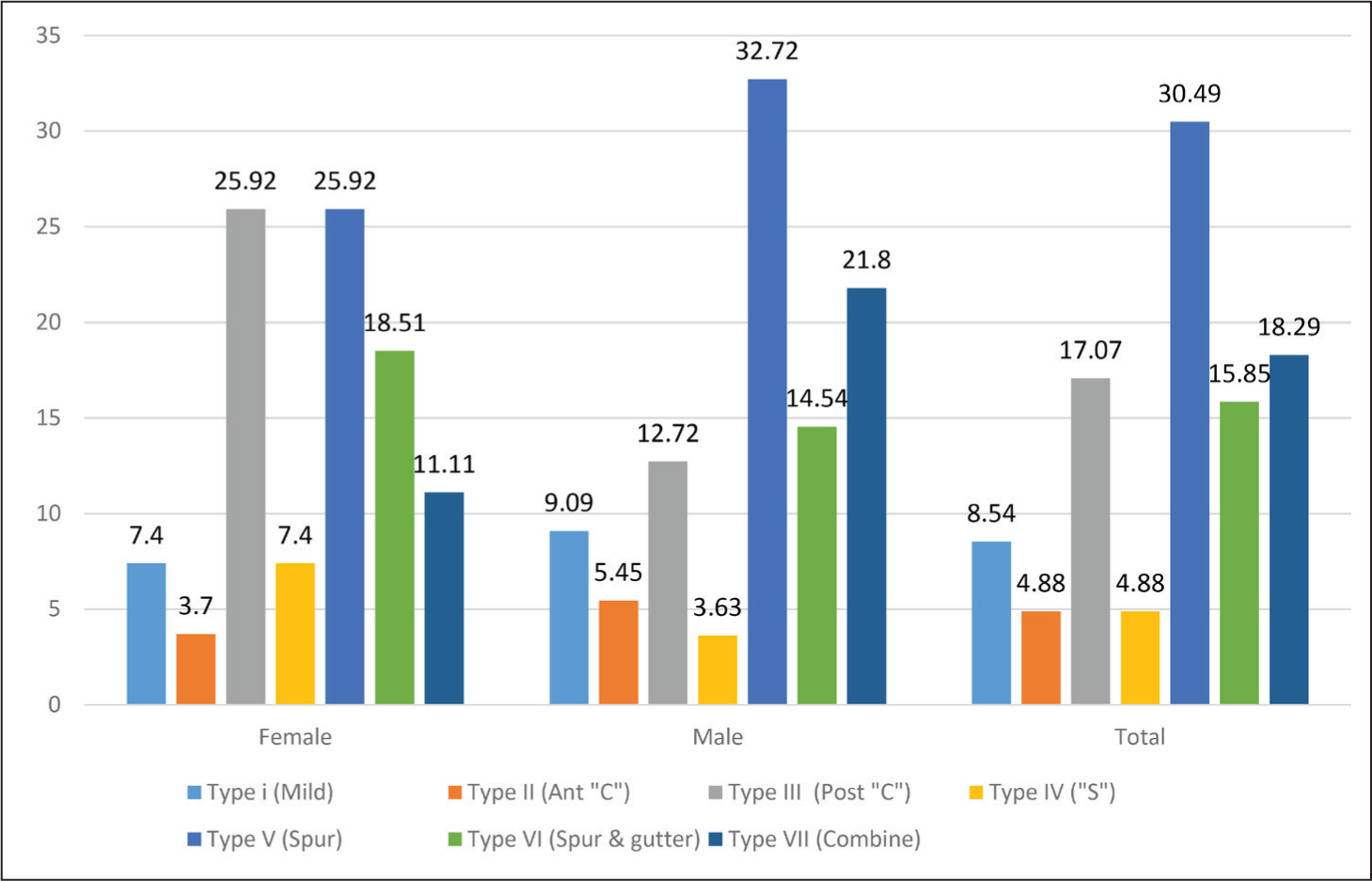
The prevalence of DNS recorded in this study is 82% [82/100]. It is 79.41% [27/34] in females and 83.33% [55/66] in males. Association of DNS with gender is statistically not significant (P = 0.629, df = 1). The most prevalent types documented in the study are type V (30.49%, n = 25/82), type VII (18.29%, n = 15/82) and type III (17.07%, n = 14/82). In male subjects, the order of prevalence remains the same [type V (32.73%, n = 18/55), type VII (21.82%, n = 12/55) and type III (12.73%, n = 07/55)], and in female subjects, the order of prevalence is type V and type III (25.93% each, n = 0/27) and type VI (22.22%, n = 06/27) (Graph 1).
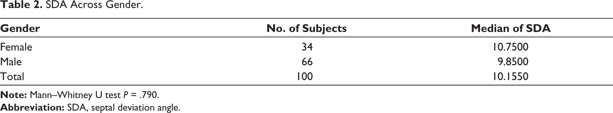
Comparison of the median of the SDA across gender by Mann–Whitney U test (P = .790) shows statistically non-significant results (Table 2).
SDA Across Gender.
The increased mucosal thickening of the maxillary sinus lining is present in 37% [37/100] and absent in 63% [63/100] of the total study group. The presence of DNS in both groups is 83.78% [31/37] and 80.95% [51/63], respectively. The association of DNS with the presence or absence of thickening of the maxillary sinus lining is statistically not significant (P = .722, df = 1).
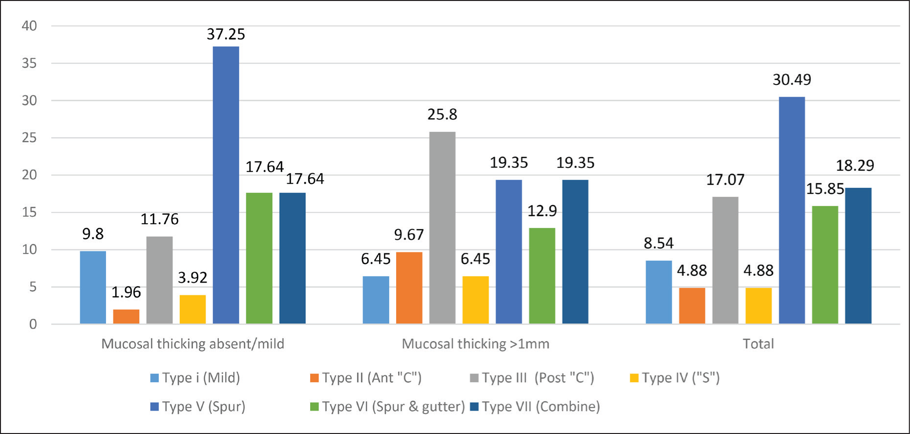
The correlation of types of DNS and absence or presence of increased mucosal thickening is reported as under. Type V (37.25%, n = 19/51), type VII, VI (17.65% each, n = 09/51) and type III (11.76%, n = 06/51) are most prevalent with the absence of mucosal thickening, and type III (25.80%, n = 08/31), type VII, V (19.35% each, n = 06/31) and type VI (12.90%, n = 04/31) are most prevalent with the presence of mucosal thickening (Graph 2).
Comparison of the median of the SDA across categories of absence/presence of mucosal thickening is carried out by the Mann–Whitney U test (P = .997) which shows statistically non-significant results (Table 3).
SDA Across Categories of Absence or Presence of Mucosal Thickening of Maxillary Sinus.
Discussion
Nasal septum is one of the most frequent landmarks that have been appreciated even in the intraoral radiographs of maxillary teeth. Deviated septum is studied by many authors due to its possible association with obstructive pathologies of the sinonasal region. Thus, this study is carried out to study the prevalence, radiographic attributes of the DNS (shape, location and severity) and its probable association with the thickened mucosal lining of the maxillary sinus.
The deviation recorded (82%, n = 82) in this study is four times more than that of straight septum/midline septum (18%, n = 18). It is reported equally in both genders. Our findings are equivalent to that of Tiwari et al. 24 and Periyasamy et al. 12 who reported the prevalence of DNS as 88.2% and 73.40% which supports the fact the septal deformities are far more common than a non-deviation and its attributes should be defined precisely while recording the variant.25–27
In this study, we observed that the most prevalent types of deviated septum are type V, type VII and type III which is consistent with that of the study of Janovic et al. 28 In our study, we found that type III deviation is most prevalent in subjects with increased mucosal thickening. Our results match with that of Mladina et al. 7 who recorded the occurrence of type III absolute and type III within type VII and concluded it to be the most frequently found type in rhino sinusitis patients. Moorthi et al. 3 reported type IV is most commonly associated with sinusitis. This difference could be attributed to the difference in the type of study. Their criteria for sinusitis were based on clinical or symptomatic presentation. Grossly ‘C’ shaped defect in the posterior aspect of the nasal septum at the ostium of the maxillary sinus or at the middle turbinate region (seen in type III and type IV as well) is significant and possibly plays a role in obstructive sinusitis.
Close proximity of DNS (mostly in the form of spur) with the lateral wall of the nose could be associated with contact point headache or neuralgia-like pain and thus when present these remarkable findings are to be noted while interpreting the radiograph. A rare but significant finding reported in this study is a variant called ‘Bridging Spur’ (Figure 2), where a spur from the septum is contiguous with the middle turbinate but not forming the bridge of bone. 20
Horizontal Deviations of Septum and Passali Deformity in Coronal Section: Type V to Type VII. (a) Type V-Spur With Horizontal Nasal Floor/Palate (Untitled Reference line), (b) Type VI-Spur With a Gutter/Indentation (Arrow) on the Opposite Side and Tilted Nasal Floor/Palate (Tilted Horizontal Reference Line), (c) Type VI-Bridging Spur Is Contiguous With the Middle Turbinate (Arrow) and (d) Type VII-Passali Deformity (Combination of One Horizontal and One Vertical Form of Deviation).
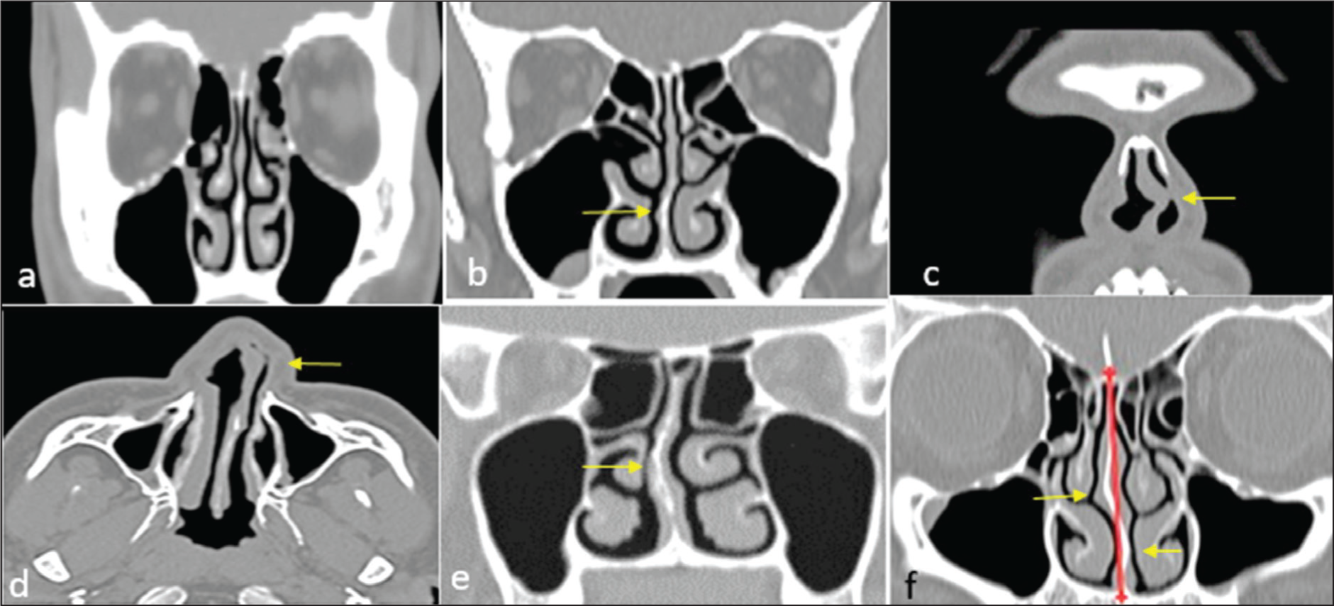
Along with that, we have also noted that type VI deformity is always associated with a tilted nasal floor/palate, which is an important finding from the dental perspective. 7
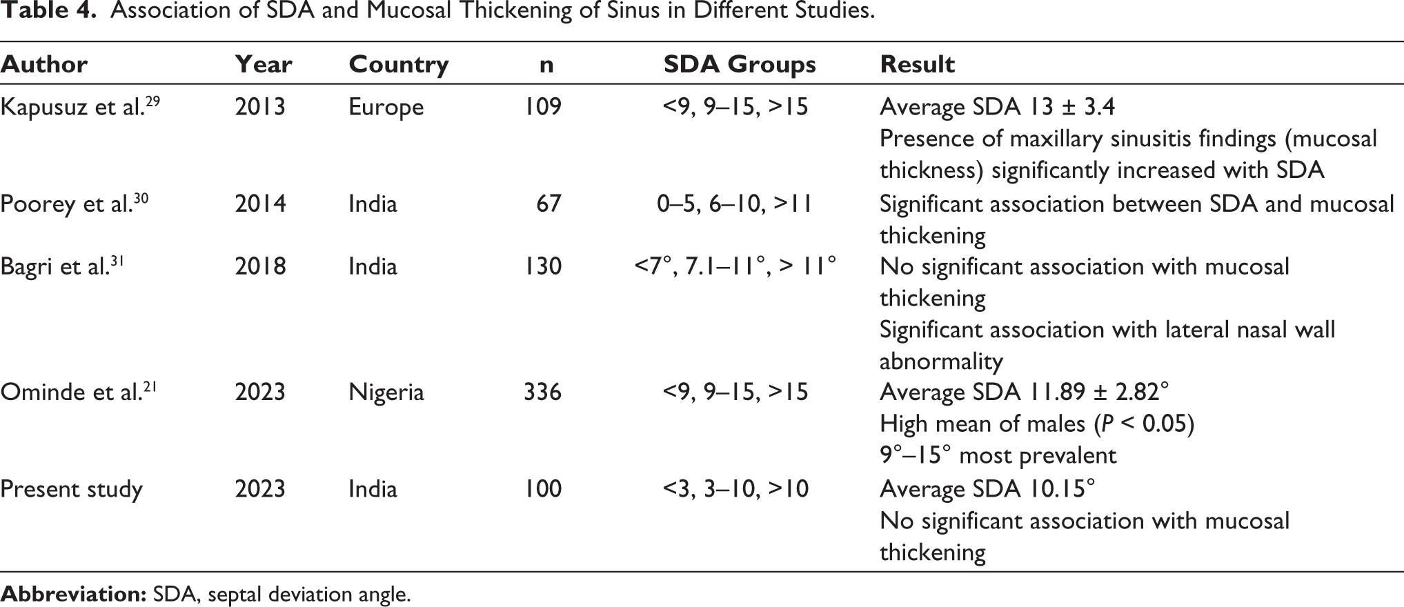
While comparing the SDA across groups, on average, we have noted marginally high SDA in females and in absent thickening of the mucosal lining of the maxillary sinus as compared with their normal counterparts. The average SDA reported is 10.15. We have also noted that measuring the deviation angle to characterize the severity gave similar results for moderate deviation at the inferior turbinate region and mild deviation at the middle turbinate region which supports the statement of Tomblinson et al. 25 suggested measuring the severity of the deviation along with the height of the deviation from the nasal floor. The association of SDA and the presence of mucosal thickening of sinus in different studies are compared with our study results (Table 4).21,29–31 It is found that there are many different classification schemes, but the average SDA is almost above 10°.21,29 Ominde et al. 21 conducted a similar study outside India and have reported high average SDA in males (p < .05) which is not matching with the study results. These data may aid for forensic studies, if baseline data are established by carrying out the study in large sample. Studies conducted in India by Poorey et al. 30 and Bagri 31 showed significant association with the findings of maxillary sinusitis (mucosal thickening) and with lateral nasal wall abnormalities, respectively, which makes the landmark remarkable while interpreting the CT data volume.
Association of SDA and Mucosal Thickening of Sinus in Different Studies.
Recording DNS with its type, relation with the turbinates or lateral wall (anteroposterior location), height (superoinferior location) and severity yields useful information for diagnosis and management of obstructive pathologies of the sinonasal region.
Limitation of the Study
It is a retrospective radiographic study wherein the sinusitis group is formulated based on increased thickening of the lining of the maxillary sinus as seen on a CT scan. Future studies with clinical correlation can confirm the findings reported in this study and also its application for the assessment of patients with orofacial pain in dental OPD. The gender-wise prevalence of variants of DNS reported in this study can be used for gender and ethnicity determination in forensic odontology. 32
Conclusion
DNS and its variants are best viewed in coronal scans. Axial scan is viewed to determine nasal valve angle for the confirmation of type II DNS and as an adjunct to appreciate the proximity of the deviation with the lateral nasal wall or sphenopalatine foramen.
Type III deviation [C-shaped] is more frequent in the sinusitis group.
Rare entity like “Bridging spur” when present in close contact with the lateral wall must be recorded in the interpretation of the sinonasal region.
The presence of a tilted nasal floor/palate is a remarkable radiographic finding of type VI deviation, which is often seen in patients with cleft palate and vice versa. The finding is also invaluable in sinus lift or implant planning procedures of the maxillary posterior region.
In conclusion, deviation of nasal septum is more frequent than straight septum. A precise description of its shape, location, severity and association with the lateral wall of the nose is recommended during the reporting of CT scans of the maxillofacial region.
Footnotes
Acknowledgements
The authors are thankful to the AMC MET medical college and hospital for providing permission for the procurement of CT data volumes for the observational retrospective study and to the Department of Radiology and the staff for their instrumental support and guidance.
Author Contributions
All authors contributed to the study conception and design. Material preparation, data collection and analysis were performed by KT. The first draft of the manuscript was written by KT, and all authors commented on previous versions of the manuscript. All authors read and approved the final manuscript.
Data Availability Statement
The data used to support the findings of these studies are included within the article.
Declaration of Conflicting Interests
The authors declared no potential conflicts of interest with respect to the research, authorship and/or publication of this article.
Ethical Approval and Informed Consent
This study was conducted retrospective from the radiographic data obtained for clinical purposes after getting the ethical approval from the IRB of AMC MET dental college where the study got an exemption from obtaining consent from the patients (Reg. No.: ECR/236/Indt/GJ/2015/RR-18).
Funding
The authors received no financial support for the research, authorship and/or publication of this article.