
Abstract
Aim:
To evaluate the bond strengths of pulp capping materials (Dycal, ProRoot MTA, Biodentine, TheraCal LC, Calcimol LC, and ApaCal ART) and different adhesive systems (Gluma 2 Bond, Clearfil SE Protect, Gluma Self Etch, Clearfil S 3 Bond Plus, Gluma Bond Universal, Clearfil S 3 Bond Universal).
Materials and Methods:
Two hundred fifty-two acrylic blocks were prepared in which cylindrical cavities of 4 × 2 mm 3 were formed. Pulp capping materials were placed in the cavities. Different adhesive systems were applied to each pulp capping material group. After applying the composite resin, the shear bond strength (SBS) values of the specimens were determined in the Instron test device. Fracture types were evaluated using a stereomicroscope and a scanning electron microscope. Data were analyzed by Shapiro–Wilk’s and Kruskal–Wallis H test.
Results:
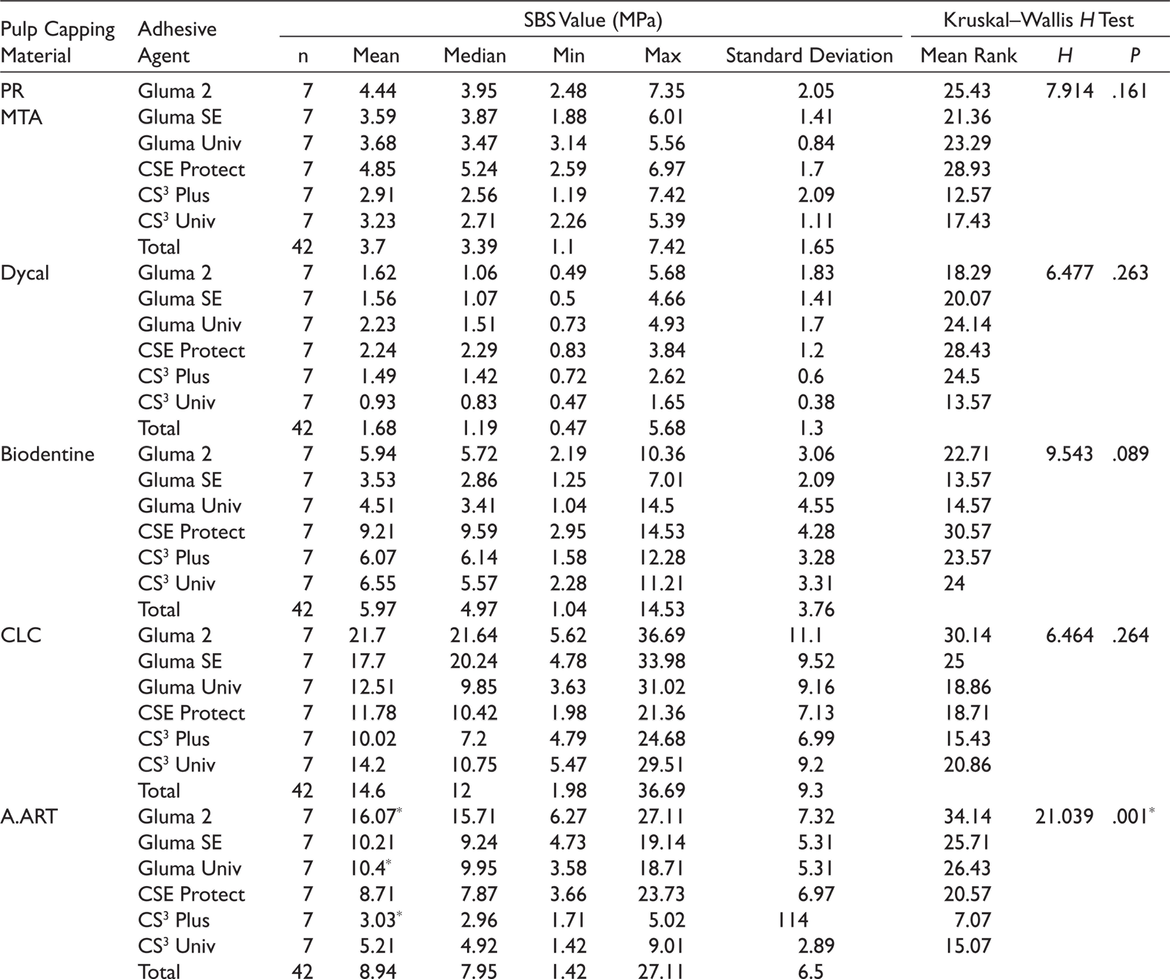
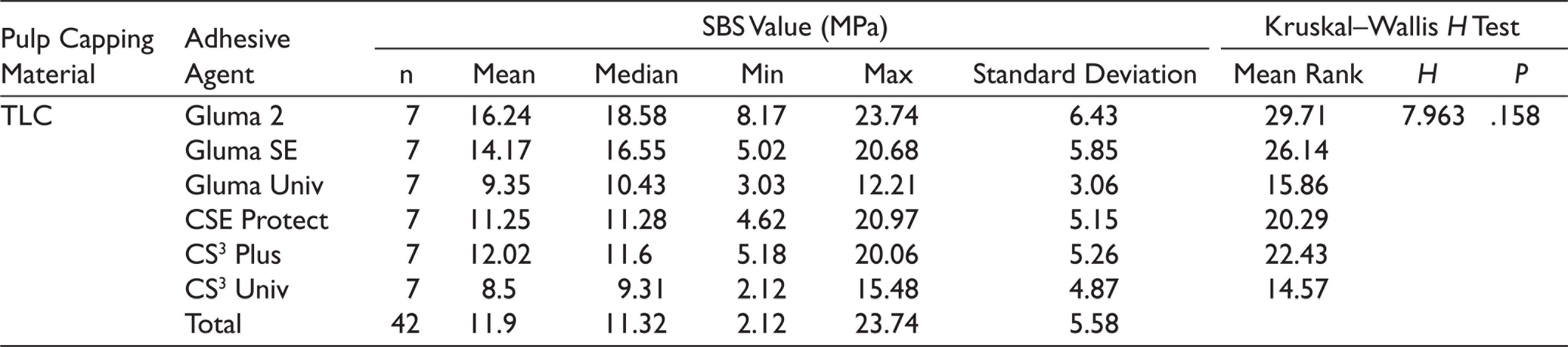
There is a statistically significant difference between pulp capping materials in terms of SBS values (P < .05). Dycal’s SBS was found significantly lower than other materials, and the highest bond strength was observed in Calcimol LC material. Although there is no statistically significant difference (P > .05) between the adhesive agent groups in terms of SBS, Gluma 2 Bond showed the highest bond strength value.
Conclusion:
In traditional pulp capping materials such as Dycal, MTA, and Biodentine, using a two-step self-etch adhesive system can result in higher bond strength values. In resin-based TheraCal LC,, ApaCal ART, and Calcimol LC materials, it may be recommended to use a two-step etch and rinse adhesive system.
Introduction
The main purpose of restorative dentistry is to protect the vitality of the pulp of vital teeth. Vital pulp can be exposed mechanically during dental preparation or in traumatic injuries and caries; treatment options include pulp capping, pulpotomy, and pulpectomy. Pulp capping treatment, which is the most applied vital pulp therapy procedure, is a form of treatment in which the tissue is covered with therapeutic materials in order to protect the exposed vital pulp tissue against thermal, chemical, and harmful stimuli. 1
Calcium hydroxide (CH) is a material that has been used in pulp capping treatment for years and is considered the gold standard. CH has biological properties, such as neutralizing the acidic environment because of its alkaline pH, stimulating tertiary dentinogenesis, forming dentin bridge, and showing antibacterial properties. 2 However, it has high solubility, low elastic modulus and compressive strength, and poor adhesion to dentin and resin-based materials. 3 Light-curing CH paste has been put on the market to aid eliminate these weak properties of traditional CH. Light-curing CH contains visible light-activated initiators and accelerators, with CH and barium sulphate dispersed in a urethane dimethacrylate resin. 3 It can be used as a base material under restorative materials in indirect pulp capping.
Mineral trioxide aggregate (MTA) is the first bioceramic material in dentistry, which was introduced in 1993 by Dr Torabinejad. 2 It has properties of hermetic sealing, low solubility, antibacterial and anti-inflammatory effect, is biocompatible, and also has a long hardening time, is difficult to apply to the cavity, can cause tooth discoloration, and is an expensive material.4–6 In order to improve the clinical properties of MTA, Biodentine, a calcium silicate-based material, was launched in 2009. It has been reported that the mechanism of action in the direct pulp capping is similar to that of MTA. It does not require a two-step restoration procedure as setting is fast. 7
TheraCal LC (TLC) is a visible light-cured resin-modified calcium silicate-based pulp capping material. Calcium ions released from TLC play a role in the proliferation and differentiation of dental pulp cells and the formation of new mineralized hard tissues. 8 It has low solubility and good sealing properties. Its ability to bond to dentin and resin-based restorative materials is good. On the other hand, it has been reported that it shows more irregular dentin bridge formation compared to other pulp capping materials that do not contain resin. 9
ApaCal ART (A.ART) is a pulp capping material with light-cured resin-modified tricalcium phosphate and added hydroxyapatite. It is said to have advantages such as accelerated calcium ion release and dentin bridge formation. It is claimed that A.ART can be used as a pulp protector after hemostasis is achieved in direct and indirect pulp capping for deep cavities. 10
One of the factors that maintain the vitality and function of the tooth is the good bonding between the pulp capping material and the restoration, and this is achieved with adhesive agents. Adhesive systems that we generally use in these treatments are etch and rinse (ER), self-etch (SE), and universal systems. Bond strength tests are frequently used to evaluate the adhesion properties of restorative materials in vitro. The most preferred bond strength measurement method is shear bond test. 11
Materials and Methods
This in vitro study was completed in approximately five days in Faculty of Dentistry Research Laboratory, Erciyes University and Erciyes University Technology Research and Application Center.
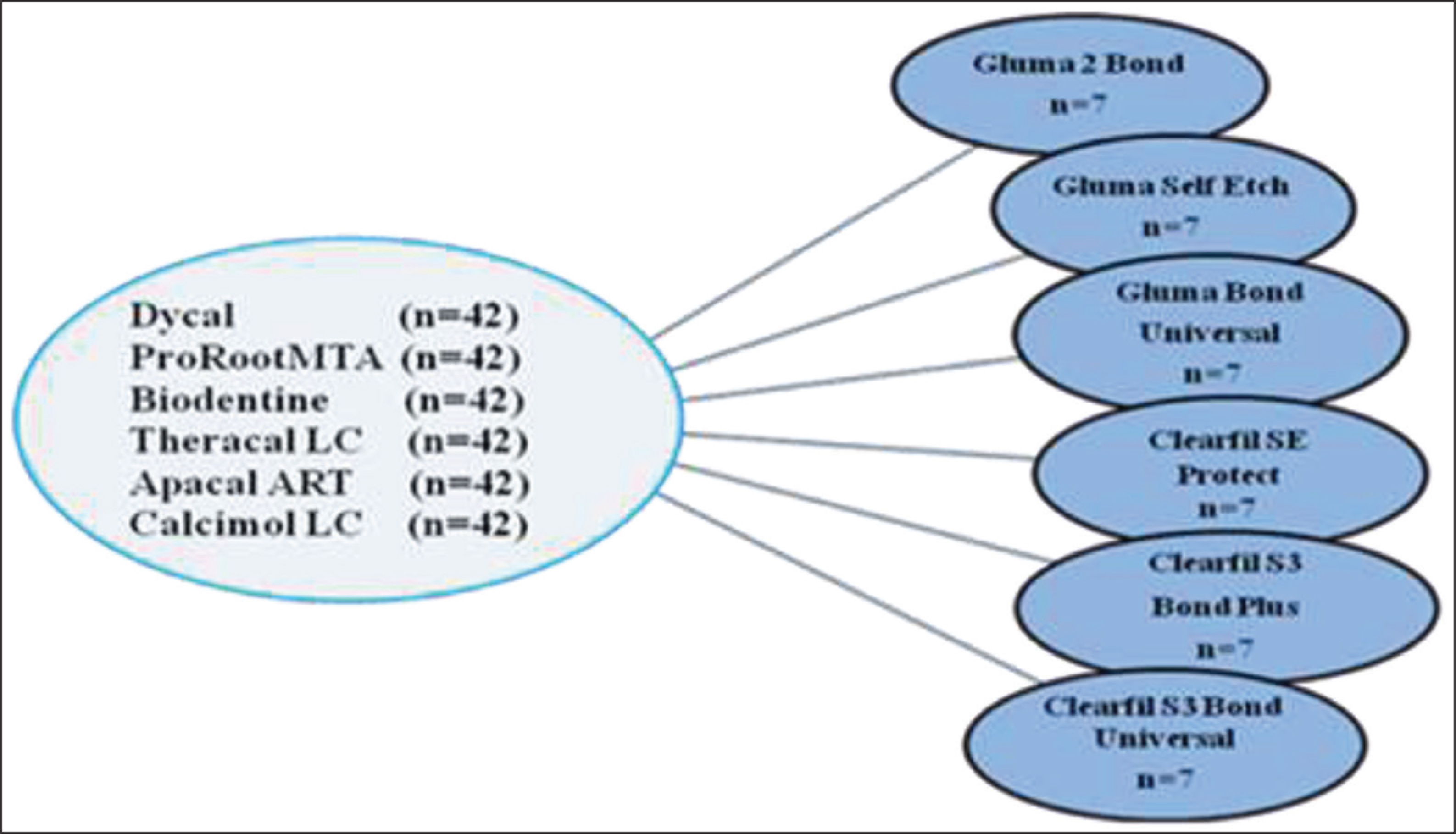
The number of specimens to be studied was estimated on the basis of a power analysis and a significance level of 0.05. The specimen size was determined as n = 42. All specimens with no fracture detected were included in the bond strength test.
Study steps:
Preparation of acrylic blocks. Placement of pulp capping materials in the cavity created in acrylic blocks. Application of adhesive agents to the surfaces of pulp capping materials. Application of a composite resin on the surfaces of the adhesive applied specimens. Shear bond strength (SBS) testing. Examination of all specimens with a stereomicroscope. Examination of specimens under scanning electron microscopy (SEM). It was carried out as a statistical analysis of the obtained data.
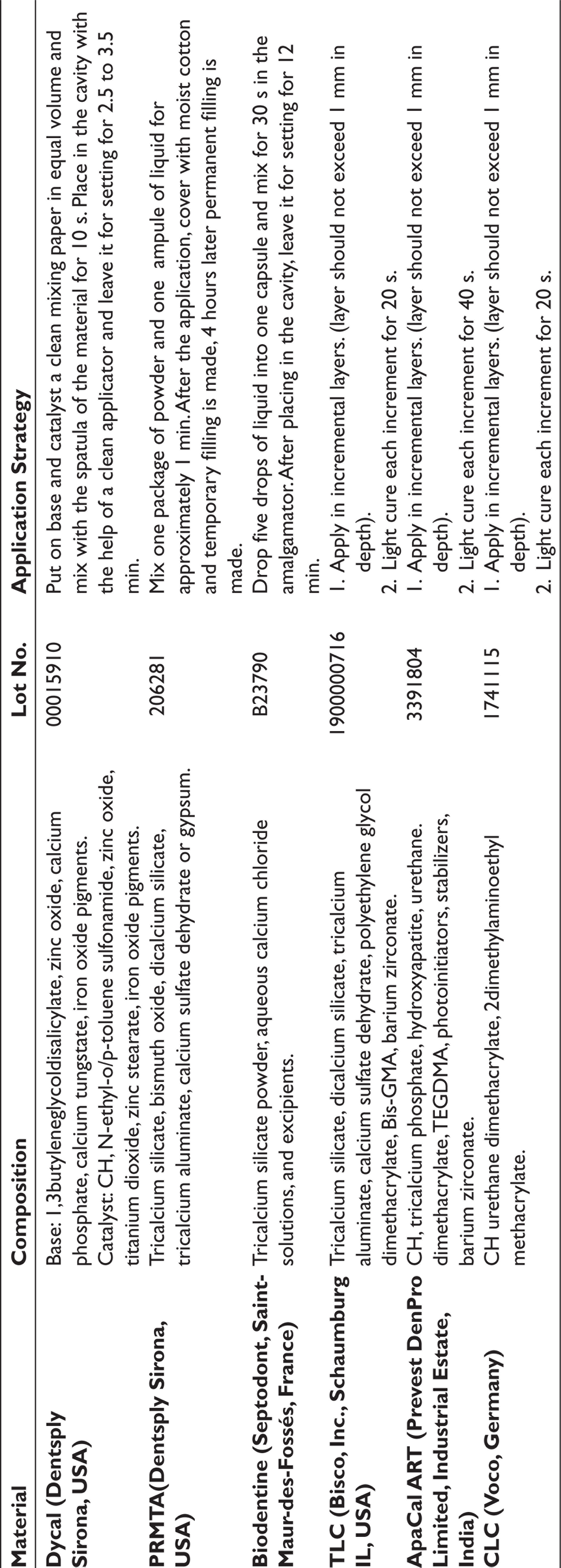
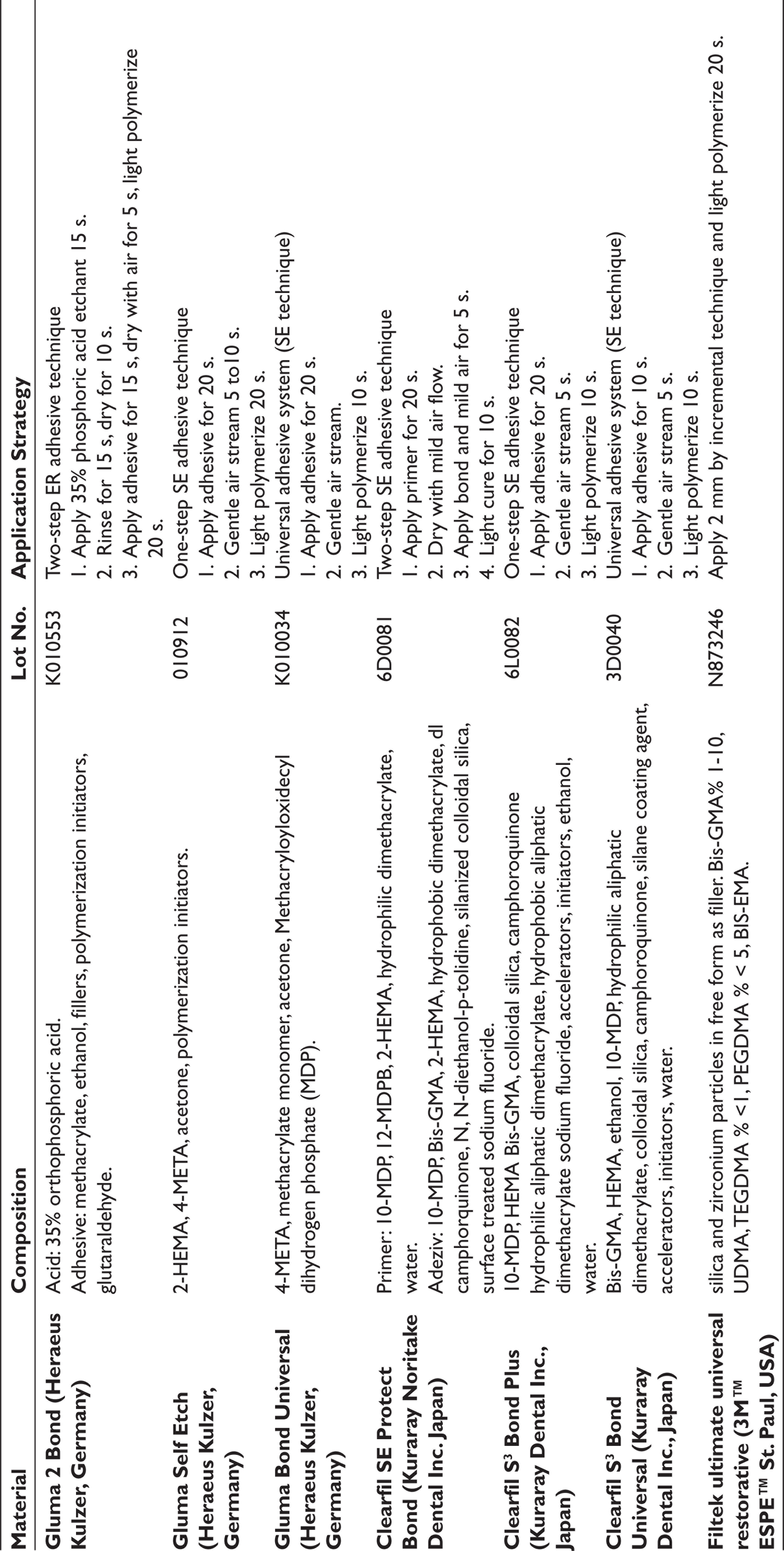
Manufacturer company information, contents, and application methods of the materials used in the study are shown in Tables 1 and 2.
Preparation of Specimens
In our study, 252 acrylic blocks with 2 × 2 cm dimensions were obtained. Cylindrical cavities of 4 × 2 mm were created in the middle of the acrylic blocks. Forty-two acrylic blocks were separated for each pulp capping material and were placed in cylindrical cavities in line with the manufacturer’s recommendations (Table 1). Adhesive agents were applied after the polymerization of each pulp capping material. Six different adhesive systems, Gluma 2 Bond (G2B) two-step ER technique, Gluma Self Etch (GSE) and Clearfil S 3 Bond Plus (CS 3 P) one-step SE technique, Clearfil SE Protect (CSEP) two-step SE technique, Gluma Bond Universal (GBU) and Clearfil S 3 Bond Universal (CS 3 U) with one-step SE technique, seven of each sample was applied, in line with the manufacturer’s instructions (Table 2) and adhesives were polymerized with a light-emitting diode light cure (Woodpecker Led-G, China). Groups and subgroups are given in Figure 1.
Pulp Capping Materials Used in the Study
Adhesive Systems and Composite Resin Used in the Study
Groups and Subgroups
Composite Resin Application
A composite resin [Filtek ultimate universal restorative (3M ESPE St. Paul, USA)] was applied with the help of a 2 × 2 mm cylindrical plastic tube over the pulp capping materials, and polymerized for 20 s with an LED light device. The composite resin was polymerized for another 20 s after the plastic tube was cut and removed with a scalpel tip. Before the bond strength test, all specimens were kept in the oven (100% humid environment at 37°C) for 24 h in order to create conditions similar to the mouth environment, taking reference from other studies.
SBS Testing
Universal test device (Instron, Lyoyd Instruments, England) was used for SBS measurements and a separating force was applied at a speed of 1 mm per minute. The force value of the composite resin sample at the moment of separation from the surface of the capping material was obtained in Newton and converted to “MPa” (megapascal) by dividing the surface area in the rupture area.
Examination of Fracture Surfaces
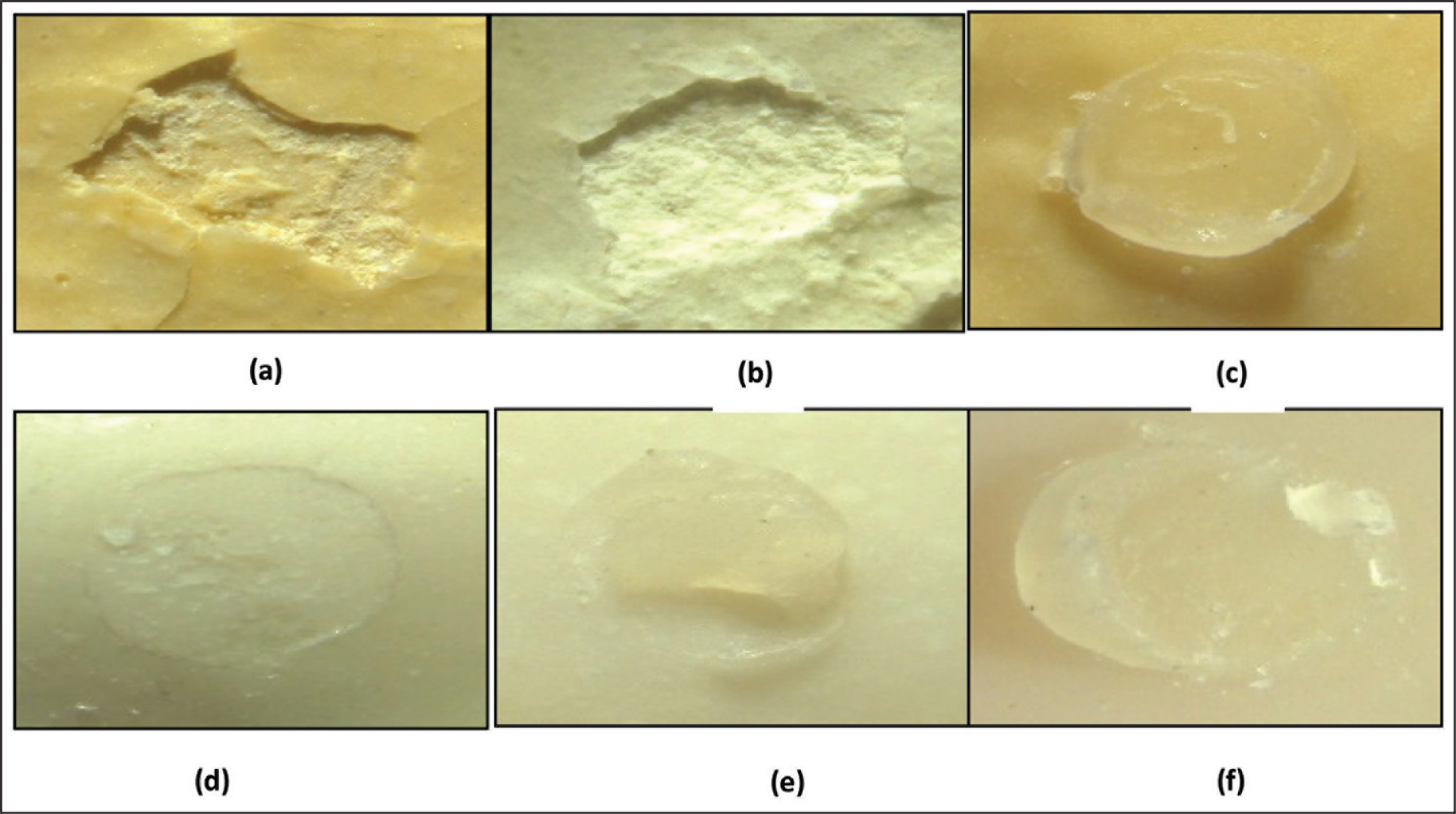
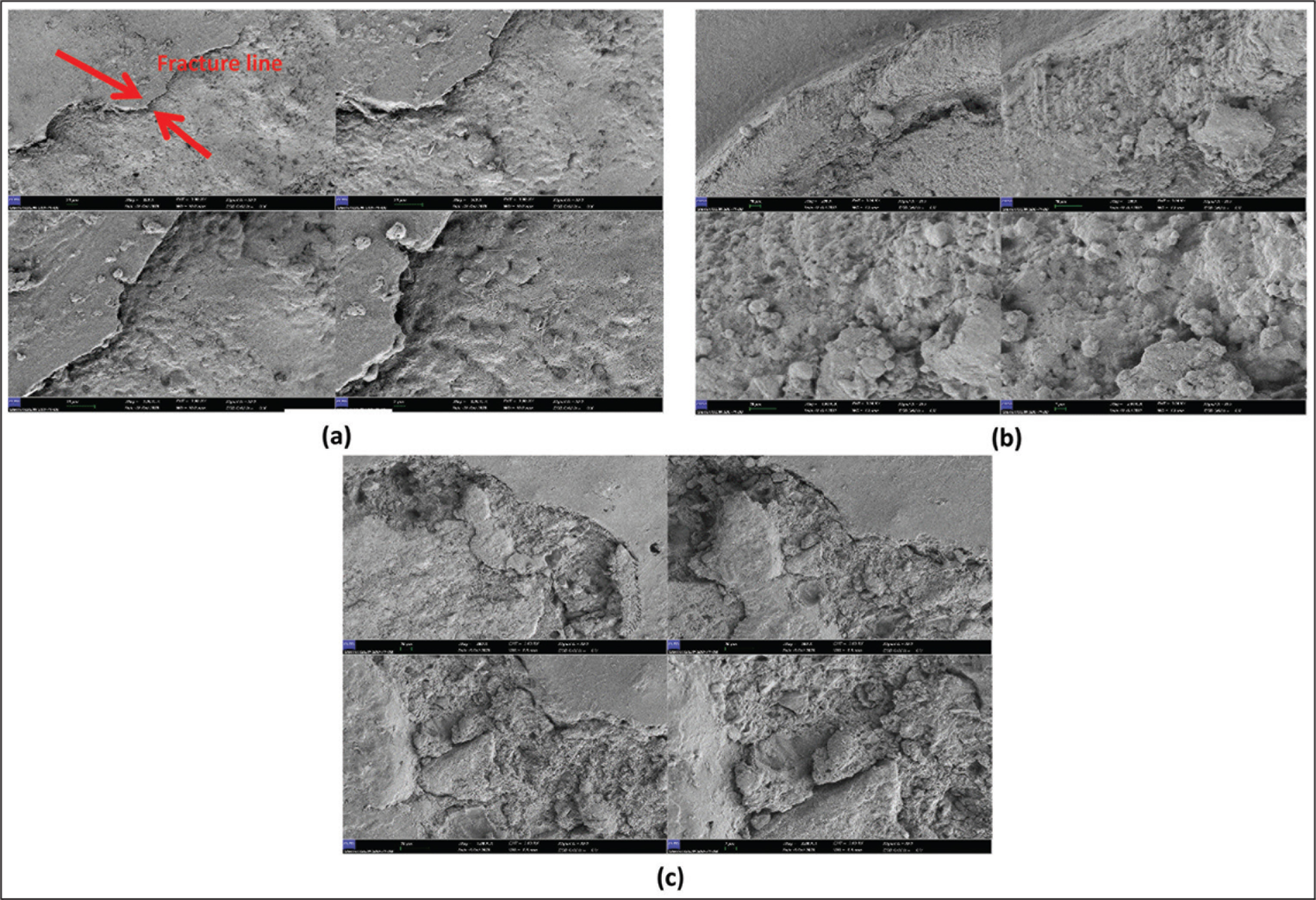
All specimens were examined under a stereomicroscope (Leica MZ 12, Leica Microsystems GmbH, Wetzlar, Germany) at ×10 magnification to determine the fracture type. The fracture at the interface of the composite/pulp capping material is adhesive fracture, and the fracture occurring in the composite layer or capping material is cohesive fracture. The adhesive and cohesive fractures together were classified as mixed fracture. In addition, images of the specimens were taken in SEM (Zeiss Gemini 500, USA) under vacuum at ×200, ×500, ×1000, ×2000 magnifications.
Statistical Analysis
The data obtained in the study were analyzed with IBM SPSS version 21 package program. Shapiro–Wilk’s was used because of the unit numbers while investigating the status of variables coming from the normal distribution. A value of 0.05 was used as the significance level. It was stated that there is a significant difference if P < .05, and if P > .05 there is no significant difference. Two-way analysis of variance was used to evaluate the effects of independent variables on the dependent variable. While examining the differences between groups, the Kruskal–Wallis H test was used because the variables did not come from the normal distribution.
Results
There is a statistically significant difference between pulp capping materials in terms of SBS (P < .05; Table 3). The SBS value of Dycal material is significantly lower than all other materials; the bond strength value of ProRoot MTA (PRMTA) is significantly lower than Calcimol LC (CLC), A.ART, and TLC materials; the SBS value of Biodentine material is significantly lower than CLC and TLC.
Pulp Capping Materials/Analysis Result Regarding the Evaluation of Adhesive Agent SBS
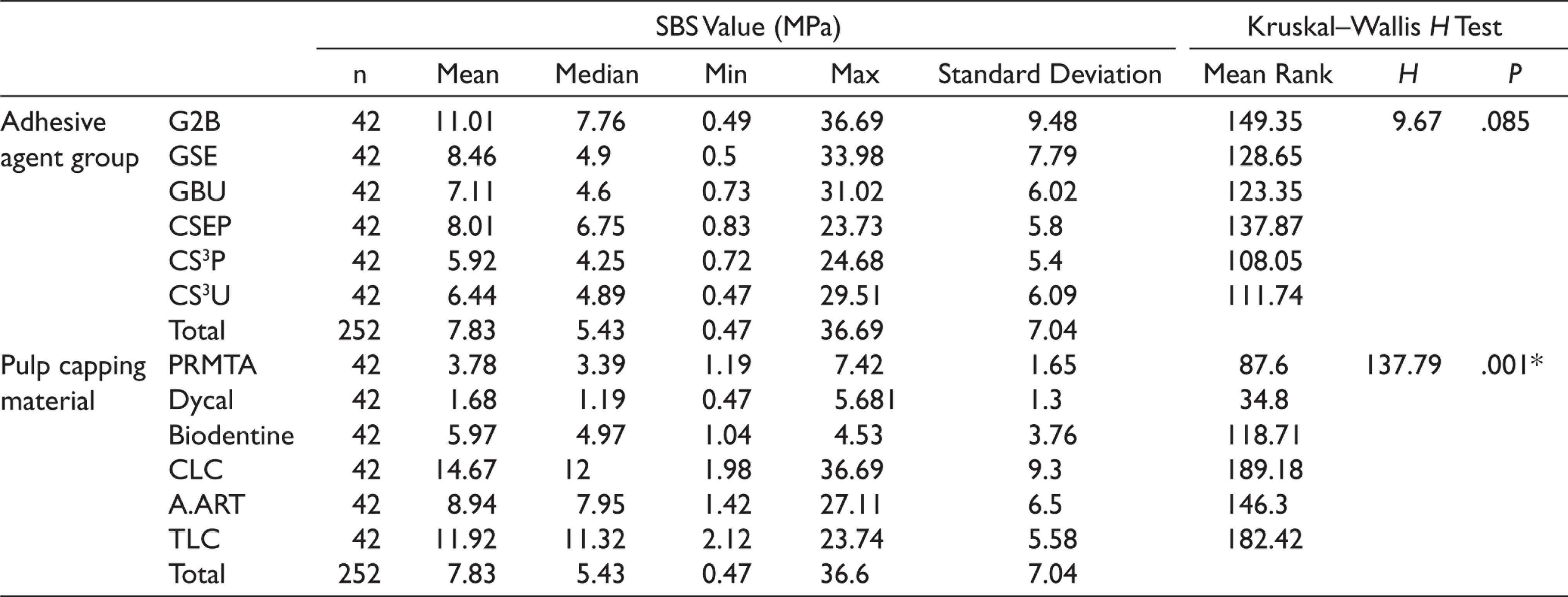
There is a statistically significant difference between the adhesive agent groups in terms of SBS in A.ART pulp capping material (P < .05). In A.ART material, the bond strength value of CS 3 P group is significantly lower than that of G2B and GBU groups. There is no statistically significant difference between the adhesive agent groups in terms of SBS of other capping materials (P > .05; Table 4).
Analysis of the Differences Between Adhesive Agent Groups in Terms of Bond Strength Values of Pulp Capping Materials
In Dycal groups, cohesive fracture within the pulp capping material was mostly observed. Although all types of fractures were seen in PRMTA, cohesive fractures were mostly detected in PRMTA. The Biodentine specimens mostly showed cohesive fractures in the composite. In TLC, adhesive and mixed fractures were seen equally and more than cohesive fractures. In A.ART groups, it can be said that the cohesive fracture in the capping material and the mixed fracture were mostly observed compared to other fracture types. Mixed fracture and cohesive fracture in a composite resin were equally observed in CLC groups.
Stereomicroscope and SEM images of the fracture types are given in Figures 2 and 3.
(a) Dycal-GSE Cohesive Fracture (in Capping Material), (b) PRMTA-CSEP Cohesive Fracture (in Capping Material), (c) Biodentine-GBU Cohesive Fracture (in Composite Resin), (d) TLC-G2B Adhesive Fracture, (e) A.ART-CS3 P Mixed Fracture, and (f) CLC-CS3U Mixed Fracture
SEM Images Taken at ×200, ×500, ×1000, ×2000 Magnifications. (a) PRMTA Adhesive Fracture (b) Biodentine Cohesive Fracture, and (c) TLC Mixed Fracture
Discussion
The most important factor affecting the success of teeth with pulp capping treatment is the use of a material with a sealing property that prevents bacterial invasion at the dentin interface with the restoration. 1 In the case of using composite resins as restorative materials, ER and SE adhesive systems are generally preferred to provide the bond with the pulp capping material. In recent years, it is known that these materials, including universal adhesives, have a clinically acceptable shear bond value of 18 to 20 MPa. 12
In studies examining the bond strength of materials such as CH, MTA, calcium silicate, and resin-modified calcium silicate, which are frequently used in pulp capping treatments, it is seen that generally two or three materials are evaluated together. We evaluated by comparing the bond strengths of six current pulp capping materials (Dycal, PRMTA, Biodentine, TLC, A.ART, and CLC) to different adhesive systems.
According to the results of our study, the bond strength value of Dycal to adhesive systems (1.68 MPa) is significantly lower than all other materials. Although Dycal’s SBS value to adhesive systems was significantly lower than other materials, it exhibited better bond strength with CSEP (2.24 MPa), which is a two-step SE system. Dycal’s low SBS value can be explained by the fact that adhesive systems with acidic pH cause softening and dissolution in this chemically setting material. Raina et al. 13 examined the bond strength of Dycal, MTA Plus, Biodentine, and TLC to fluid and bulk-fill composite resins, using a one-step SE adhesive system, and determined that Dycal has the lowest bond strength value (2.68 ± 1.66 MPa).
Alzraikat et al. 14 and Karadas et al. 15 evaluated the bond strength of MTA and TLC to composite resins using different adhesive systems in their studies, and have emphasized that the bonding capacity of MTA is lower than TLC. In our study, it was observed that the SBS value of PRMTA was significantly lower than CLC, A.ART, and TLC materials. The highest bond strength value of MTA was observed in two-step SE CSEP, and the lowest value was observed in the one-step SE CS 3 P group. This result may be because of the fact that CSEP contains a functional monomer (10-MDP) that has the property of ionically bonding to Ca+ ions in hydroxyapatite. We are of the opinion that CS 3 P, which has a pH value of 2.3 even though it contains 10-MDP, cannot exert enough etching effect for mechanical interlocking on the MTA surface because of its low acidity.
Nowadays, ER systems are still considered the gold standard for bonding to enamel and dentin. 16 In many studies, when using ER adhesive systems, the bond strength of MTA to restorative materials has been found to be higher than SE systems.14,17–19 This result was explained by the lower acidity of SE adhesives and the reduction of polymerization of SE adhesives by the presence of moisture in the MTA. 14 In addition, the bond strength of MTA to ER adhesives reaches the highest value after final setting (96 h). 20
Shin et al. 21 and Cantekin and Avci 22 reported that the bond strength of Biodentine to adhesive systems is higher than MTA. Deepa et al. 23 in their research explained that the bond strength of Biodentine to composite resins is weaker compared to TLC. In our study, Biodentine showed higher bond strength than MTA, and lower bond strength than TLC and CLC. Higher bond strength values were observed between Biodentine and 10-MDP-containing adhesive groups. While the highest SBS value was seen in two-step SE CSEP, the lowest value was found in a one-step GSE adhesive system.
Methacryloyloxidecyl dihydrogen phosphate (10-MDP) monomer is a functional monomer that is found in SE and universal adhesive systems and has the ability to bind ionically to calcium in hydroxyapatite. Studies have shown that 10-MDP can improve micromechanical and chemical adhesion by chemically bonding Ca+ ions in Biodentine.24,25 Keleş et al. reported that the highest SBS value of Biodentine was seen in the two-step SE system. 24 Because one-step SE adhesive systems form a thinner adhesive layer structure that is prone to oxygen polymerization inhibition and is less permeable, they exhibit lower bond strength values than two-step SE. 15 One-step SE adhesive systems with low acidity demineralize dentin superficially and often smear plugs cannot be completely removed from the dentinal tubule. 20
According to the results of our study, the highest bond strength was seen in CLC and the ER system exhibited the highest bonding values. CLC was followed by TLC and A.ART, respectively. We believe that the significantly higher bond strength values of CLC and TLC compared to Biodentine are related to the bonding of these resin-containing materials to the adhesive resins by chemical adhesion as well as micromechanical interlocking. However, the buffering reaction that occurs as a result of the combination of alkaline MTA and Biodentine with acidic adhesive agents may have reduced the bonding effect.
Boby et al. 26 examined the bond strength of TLC and light-cured CH to nanocomposite resins in their study, and have found that the bond strength of light-cured CH was higher than TLC.
Comparing TLC and A.ART, Baltacıoğlu 17 found the bond strength value of A.ART to be higher. On the other hand, Karadas and Atıcı 27 have reported the bond strength values of resin-based pulp capping materials to dentin from high to low, respectively: A.ART, TLC, and CLC.
According to our study in the TLC groups, ER G2B showed the highest bond strength, whereas CS 3 U as a universal system showed the lowest bond strength value. This result can be explained by the fact that the phosphoric acid used in the ER system causes morphological changes on the substrate surface that allow monomer diffusion and micromechanical bonding to occur. Karadaş et al. 15 reported that TLC’s bond strength to the composite resin was higher when using the ER adhesive system and lower when using a single-stage SE system.
In the A.ART groups containing nanohydroxyapatite, the two-step ER system and the universal system exhibited the highest bond strength in terms of bond strength to adhesive systems, respectively. In our study, the bond strength value of CLC with a two-step ER adhesive system was found to be 21.77 MPa. Micro-tension and micro-shear bond tests are frequently used in determining the bond strength of adhesive systems. These tests allow the measurement of high bond values in dentin without causing a cohesive fracture. While adhesive fractures occur at the bonding interface, it suggests that there is no strong bond strength. 13 Cohesive fractures that occur within the material make the bonding activity acceptable. 14 Compared to other test methods, macro-shear test was used in our study because of its advantages such as easy setup and sample preparation in shear tests and low level of deviations in force application.
In our study, cohesive fractures were generally observed in the pulp capping material in Dycal groups. We are of the opinion that these fractures are not because of the high adhesive bond but because of the low internal resistance of the material. Although all types of fractures are encountered in MTA groups, cohesive fractures in the material are the most common. Biodentine specimens showed cohesive fractures more than other fracture types. The reason for this can be attributed to the poor structure of the material in the early setting stage. In TLC, adhesive and mixed fractures were seen in equal numbers and more than other fracture types. Although all types of fractures were seen in A.ART, mixed and cohesive fractures were mostly seen. Cohesive fracture in the composite resin and mixed fracture were equally observed in CLC groups.
In this in vitro study, early bond strength was investigated without aging the specimens. Different results can be obtained after aging process. This was the limitation of the study.
Both in vitro and in vivo studies are needed to evaluate the bond strength of pulp capping materials to adhesive systems.
Conclusion
In traditional pulp capping materials such as Dycal, MTA, and Biodentine, using a two-step SE adhesive system can result in higher bond strength values. In resin-based TLC, A.ART, and CLC materials, which exhibit higher bonding strength compared to conventional CH and calcium silicate-based pulp capping materials, it may be recommended to use a two-step ER adhesive system. Although adding a resin monomer to these materials increases the physical resistance of the material, we believe that it should be subjected to cytotoxicity studies because it poses a risk in terms of biocompatibility.
Footnotes
Acknowledgements
The authors acknowledge Faculty of Dentistry Research Laboratory, Erciyes University and Faculty of Dentistry, Dicle University for the support for this work.
Declaration of Conflicting Interests
The authors declared no potential conflicts of interest with respect to the research, authorship, and/or publication of this article.
Funding
This work was supported by the Dicle University Scientific Research Projects Commission Presidency with the project number DİŞ 19.004.