
Abstract
Aim:
To analyze the remineralization efficacy of casein phosphopeptide–amorphous calcium phosphate (CPP–ACP) and 8% arginine on artificial early enamel lesions on primary teeth enamel samples.
Materials and Methods:
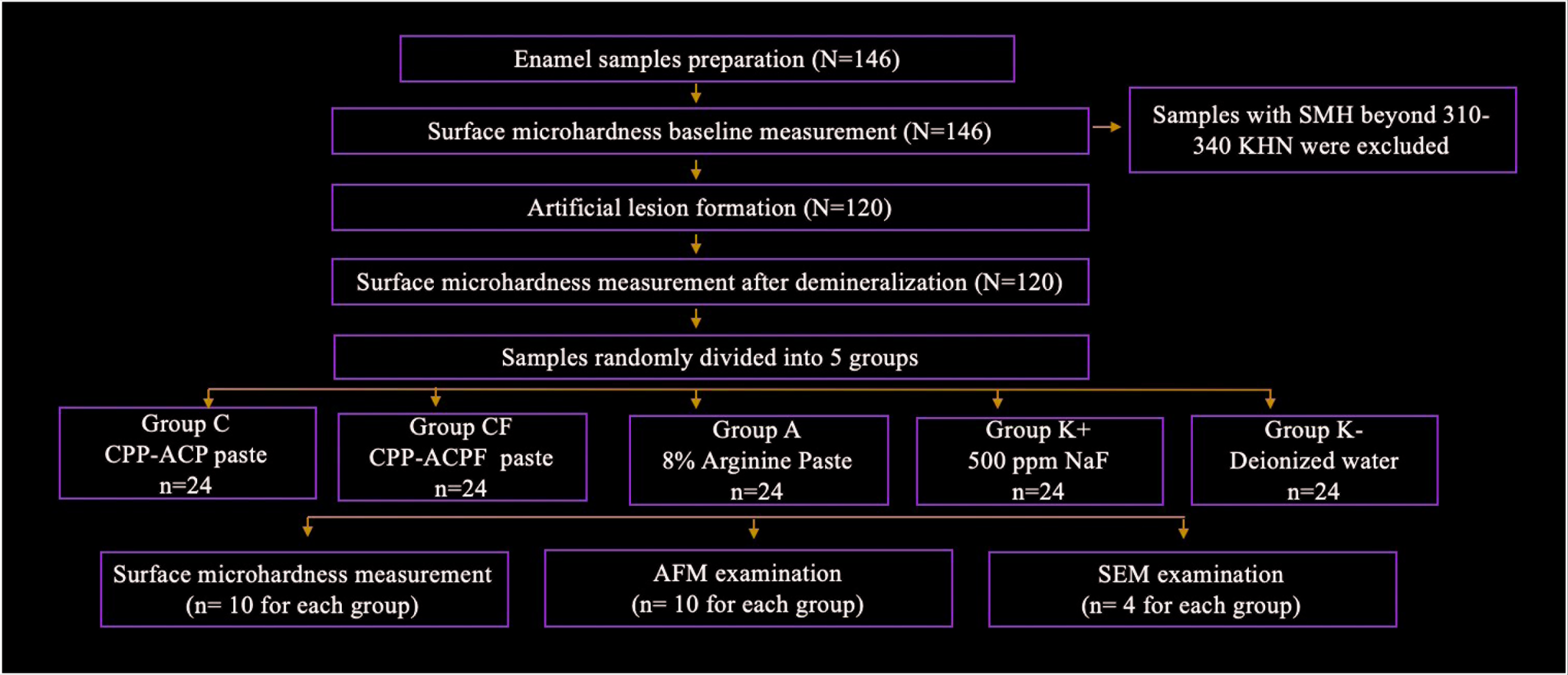
In this in vitro study, artificial enamel lesions were created in 120 primary teeth. Teeth were randomly divided into 5 groups: group C: CPP–ACP paste; group CF: CPP–ACPF paste with 900 ppm flour; group A: paste with 8% arginine; group K+: 500 ppm NaF as a positive control; and group K–: deionized water as a negative control. After 4 weeks of the remineralization process, the effect of remineralization on samples of agents used the microhardness, atomic force microscope, and scanning electron microscope analyses. Statistical analysis was done using Statistical Package for the Social Sciences (SPSS version 20.0, SPSS Inc., Chicago, IL, USA). ANOVA and Tukey Post-Hoc multiple comparison test were applied (p < .05).
Results:
Increasing percentage values of the microhardness at groups C, CF, and A were significantly better than group K– (p < .05), whereas there is no statistically significant difference with group K+ (p > .05). The average surface roughness values of groups C and CF were similar with group K+ in AFM analysis, whereas average surface roughness values of group A were significantly higher than K+ group.
Conclusion:
The CPP–ACP, CPP–ACPF, and arginine are effective agents for remineralization of early childhood caries lesions.
Keywords
Introduction
Early childhood caries (ECC) is a special form of severe dental caries affecting babies and small children, and it is a multifactorial disease caused by cariogenic microorganisms (Streptococcus mutans), fermentable carbohydrates, and inappropriate nutritional habits, impairing the quality of life of children and the parents. 1 Preventing the development of ECC or treatment of early carious lesions using appropriate methods is vitally important in improving the affected quality of life of both the children and the family. 2 ECC is an aggressive progressive disease, so it is very important to prevent ECC in the early period as endodontic treatment or tooth extraction are stressful treatments in children. 3
Although fluorides are known to be the most effective agents in the remineralization of early enamel caries lesions, the risk of fluorosis has been considered as a consequence of its widespread use.4,5 Therefore, an appropriate nonfluoride anticaries agent is required. One such agent involves the application of a casein phosphopeptide– amorphous calcium phosphate (CPP–ACP) agent. Casein is a phosphoprotein, which is present in bovine milk in the form of 30 to 300 nm diameter particles and in high amounts, constituting about 80% of the total protein, and its most important feature is stabilizing calcium and phosphate in protein complexes. 6 Studies have demonstrated that the CPP–ACP complex is dissociated in an acidic media, and that a saturated plaque can be formed, providing for the liberation of calcium and phosphate ions. This reaction supports remineralization through the blockage of the demineralization of the enamel surface of the tooth.7,8
Another agent, arginine, has been closely associated with the bacterial ammonium metabolism, although it is a natural compound of human saliva. Various nonpathogenic organisms, including oral streptococci, lactobacilli, and spirochetes, degrade arginine to produce ammonium through the arginine deiminase pathway. Plaque acids are neutralized after the release of ammonium, resulting in intraoral pH becoming less acidic, thus supporting remineralization and decreasing demineralization. In addition, free calcium ions are produced from insoluble calcium compounds, providing further support to remineralization. 9
The aim in this in vitro study is to analyze the remineralization efficacy of CPP–ACP, fluoride-added CPP–ACP, and paste of 8% arginine containing on artificial carious lesions in enamel formed in primary teeth, based on the findings of a microhardness analysis, atomic force microscope (AFM), and scanning electron microscope (SEM) analyses.
The null hypothesis tested was that CPP–ACP, fluoride-added CPP–ACP, and paste of 8% arginine contains have no contribution to the remineralization of artificial enamel caries lesion formed on primary teeth.
Materials and Methods
Preparation of the Samples
The necessary ethics committee approval for the study was obtained from the Clinical Research Ethics Committee Directorate of Erciyes University (no. 2014–192). A total of 120 teeth without caries were collected from 6- to 7-year-old patients who had undergone a tooth extraction due to indications of physiological root resorption in the lower anterior primary teeth. Any remains of plaque or soft tissue on the teeth were removed by brushing under running water. The roots of the teeth were removed under water cooling at the junction of crown and root using a diamond separator. The teeth were stored in deionized water, including a 0.2% thymol crystal at 4 ºC until the time of experiment. Teeth with carious lesions in the enamel surface, hypocalcification, and defects caused during extraction were excluded from the study. Each tooth was embedded in acrylic resin in cylinder-shaped plastic molds with their labial surfaces parallel to the base. The surfaces of the enamel samples were polished with 2400 grit silicon carbide discs (Struers, Copenhagen, Denmark) under water cooling in order to obtain clear images, and smooth, even, and shiny enamel surfaces were obtained. The samples were washed with deionized water (Generic Standard Elix 35 60L Tank, Millipore, USA) for 10 s to clear any remnants from their surfaces.
Production of Lesion
The contents of the demineralization solution that was used to form the early enamel lesions included 2.2 mM Ca(NO3)2 (AR, Beibei Chemical Factory, Chongqing, China), 2.2 mM KH2PO4 (AR, Kelong Chemical Factory, Chengdu, China), 0.1 ppm NaF (AR, Kelong Chemical Factory), and 50 mM acetic acid (pH 4.5; AR, Kelong Chemical Factory). The samples were stored in a demineralization solution at 37 °C for 48 hours and were dried at room temperature following washing with deionized water subsequent to the demineralization procedure. 10
Remineralization
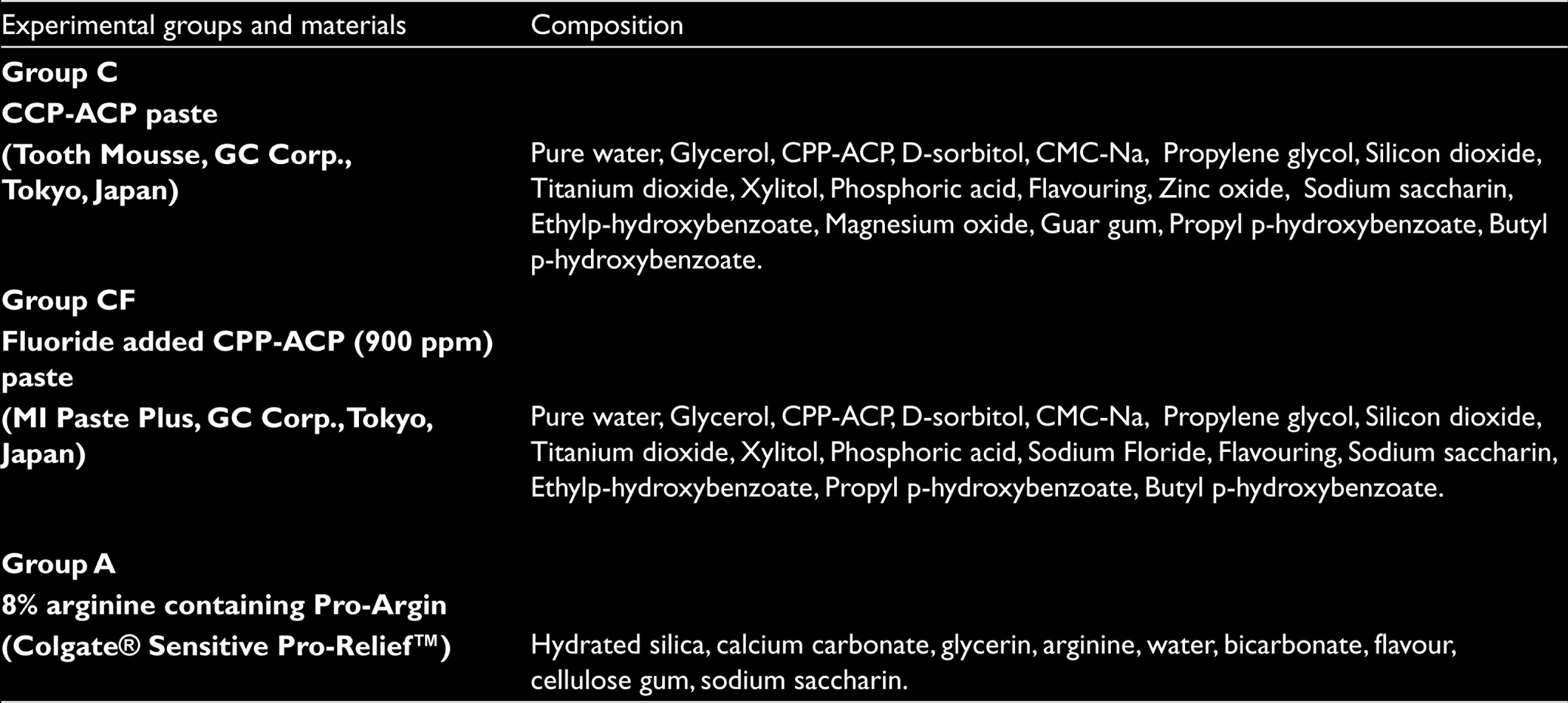
The samples were divided randomly into five groups: CPP–ACP (Tooth Mousse [TM]; GC Corp., Tokyo, Japan) paste formed group C; CPP–ACP paste (CPP–ACPF) (MI Paste Plus, TM; GC Corp., Tokyo, Japan), including 900 ppm fluorine, formed group CF; 8% arginine-containing paste (Colgate Sensitive Pro-Relief) formed group A; 500 ppm NaF solution (positive control group) formed group K+, and deionized water (negative control group) formed group K– (Table 1 and Figure 1). Artificial saliva was used for the remineralization solution, as per the recommendation of ten Cate and Duijsters. 11 In groups C and CF, a thin layer of CPP–ACP and CPP–ACPF pastes were applied to the surface of the enamel samples using a microbrush, left undisturbed for 3 minutes, and then stirred at about 100 rpm in the artificial saliva for 30 min twice daily for the 4-week remineralization process. In group A, a thin layer of 8% arginine paste was applied to the surface of the enamel samples once a week throughout the 4-week process with a low-speed dental hand piece for 3 to 5 s and was kept on the surface for 3 min. In Group K+, the samples were stirred at about 100 rpm in the 500 ppm NaF solution for 3 min twice daily for the 4-week remineralization process. In group K–, the surface of the samples was given no treatment, and the samples were kept in the deionized water for 3 min twice daily for the 4-week remineralization process. All samples were washed in deionized water following the application of the test materials and were kept in artificial saliva solution until the time of the experiment. The enamel samples were stored in a cold and dry environment after the remineralization process until the tests were applied.
Schematic of Study Design
Composition of Remineralizing Agents
Measurement of Surface Microhardness
The microhardness of the enamel samples was measured using an enamel surface microhardness tester (Struers Corp., Japan), based on the Knoop hardness (KHN) measurement principle. Surface microhardness measurements (SMH) were made by applying a 10 g load to the enamel samples for 15 s, with measurements taken at baseline, after an artificial carious lesion was formed and after the experimental materials had been applied, with three measurements obtained for each sample at each time point. The data obtained for each enamel sample were recorded, and their mean values were calculated. To ensure standardization in the sample group, samples with an initial SMH value of 310 to 344 KHN were included in the study. 10
For the determination of the percentage increase or decrease of hardness, the following formula was used:
where SMH 1 is the average of the baseline, SMH 2 is the average of the after an artificial carious lesion was formed, and SMH 3 is the average of the after the experimental materials had been applied. 10
Evaluation With an Atomic Force Microscope
The roughness of the enamel surfaces was evaluated using an AFM device (Veeco Multimode 8, Mannheim, Germany) following the application of the test materials. A gold-plated pyramidal silicon tip was used for the surface roughness measurements, which were performed at a 1.6 Hz screening rate. Vibration frequency was determined as 10 kHz and areas of 10 µm × 10 µm were screened, with 256 × 256 pixel three-dimensional views of the enamel samples and mean surface roughness (Ra) values determined and recorded. Calibration was repeated at each measurement phase. AFM measurements were performed three times for each enamel sample, and the mean value was calculated to obtain the Ra value.
Evaluation With Scanning Electron Microscope
Following the application of the test materials to the enamel samples to produce early enamel lesions, an SEM (LEO 440, K61n, Leike Zeiss; Cambridge, UK) device was used for the assessment of the structural changes to the enamel surfaces. The demineralized enamel samples, the healthy samples, the samples subjected to no treatment procedure, and the samples in the test and control groups were coated with gold–palladium mixture in 100 Å thickness, and evaluated at different magnification rates (×1000 to ×10,000).
Statistical Analysis
The statistical analysis of the obtained data was made using the “Statistical Package for the Social Sciences” software (SPSS 20 for Windows, SPSS Inc.).
A power analysis (G*Power 3.1, Franz Faul, Kiel, Germany) was carried out to determine the number of samples in the experimental groups.
The in-group evaluation of the results of the microhardness and AFM tests was made using a one-way analysis of variance with repeated measures. The intergroup findings related to microhardness and surface roughness were evaluated statistically using a one-way ANOVA, a Shapiro–Wilk normality test was used to determine the level of normality between the groups required for the ANOVA test, and a Levene’s test was used for the determination of variance homogeneity. The origin of difference and statistical significance were determined by way of a Tukey’s HSD multiple comparison test (p < .05).
Results
Findings of Microhardness Test
A quantitative evaluation was made of the effects of different remineralization agents on the artificially developed early enamel lesion and the respective percentages of remineralization (SMH %) are presented in Table 2. No difference was noted in the microhardness measurements of the healthy enamel surface between the groups (p = .476). Microhardness values were observed to be significantly decreased in all groups following the 48-hour demineralization process (p < .05), although the difference in values between the groups was not significant (p = .254). Measurements after the 4-week remineralization process revealed that microhardness values of all groups except group K– were significantly increased when compared to the measurements performed after demineralization (p < .001). While there was no statistically significant difference in the increased level of remineralization between groups C, CF, A, and K+ (p > .05), their remineralization levels were found to be statistically significantly superior to that of group K– (p < .05).
The SMHs Alterations Initial, Before and After Treatment Cycles
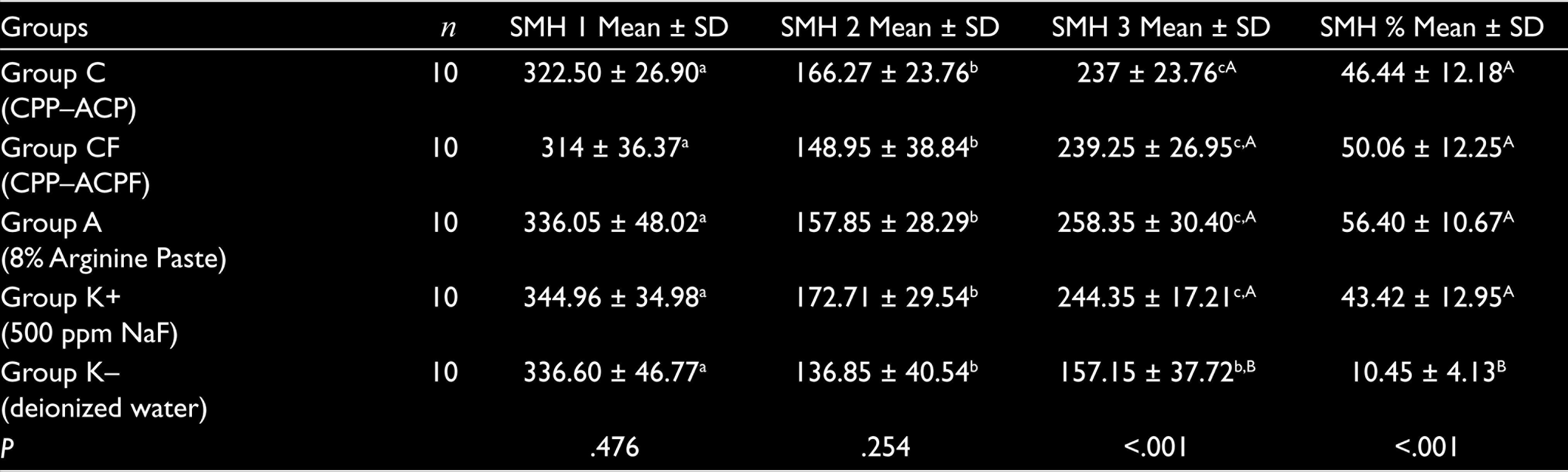
a–c Statistically different (p < .001).
A,B Statistical difference (p < .001).
Findings of the AFM Analysis
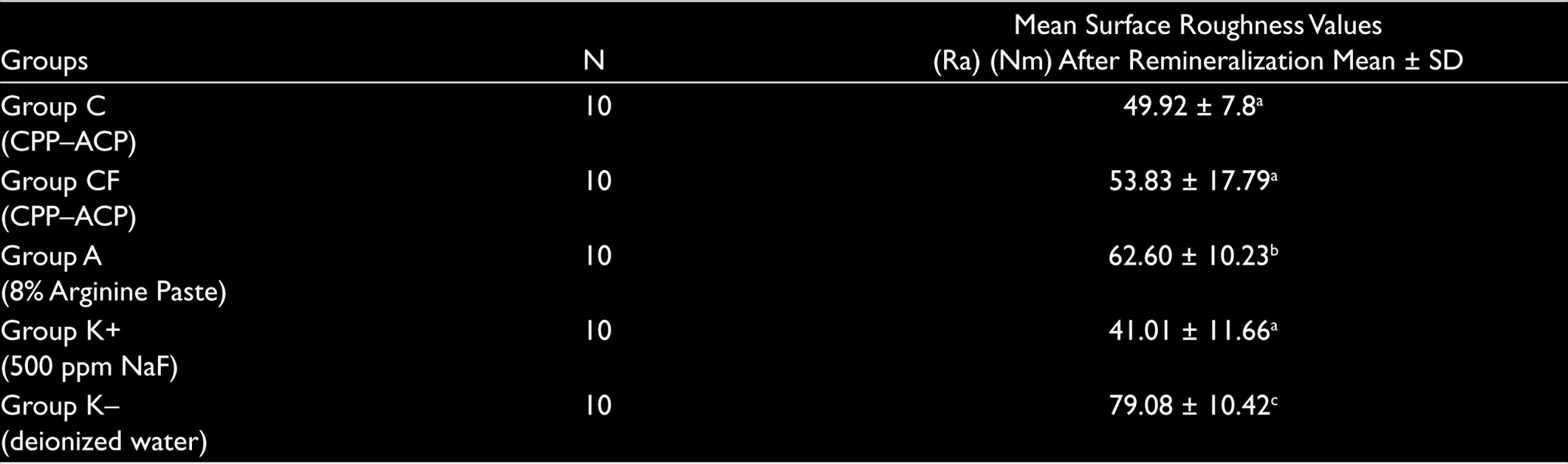
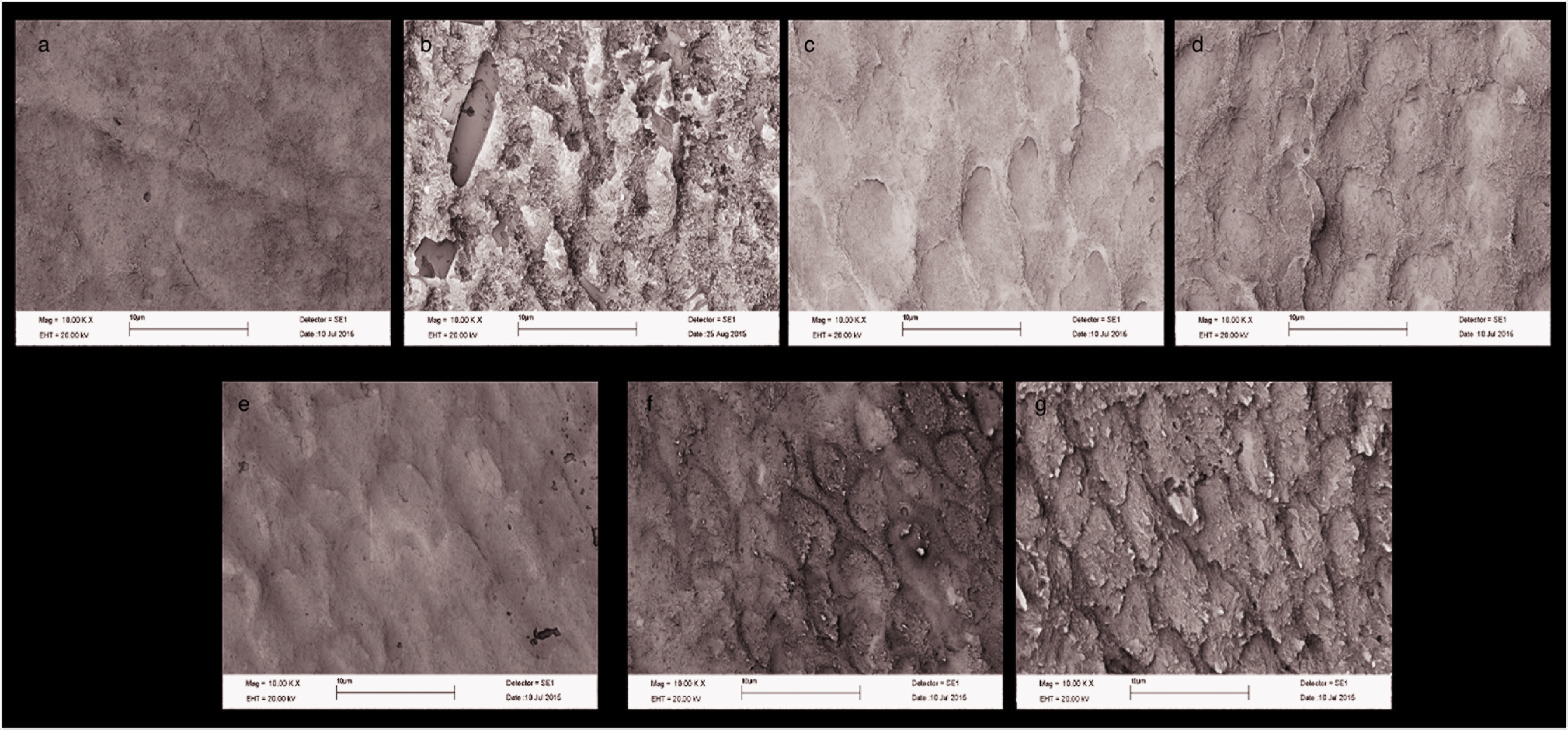
The surface topography of the enamel samples following the remineralization process is shown in Figure 2. The mean Ra values obtained after remineralization are presented in Table 3. Group K– was found to have the roughest surface according to the obtained data (p < .001). No statistically significant difference was found in the mean Ra values between groups C, CF, and A (p = .168). While the mean Ra values of groups C and CF were similar to the values of group K+ (p = .019), the mean Ra value of group A was found to be significantly higher when compared to group K+ (p < .001).
Atomic Force Micrographs in Different Groups: (a) Healthy Enamel, (b) Demineralized Enamel, (c) CPP–ACP (Group C), (d) CPP–ACPF (Group CF), (e) 8% Arginine Paste (Group A), (f) 500 ppm NaF (Group K+ ), and (g) Deionized Water (Group K–)
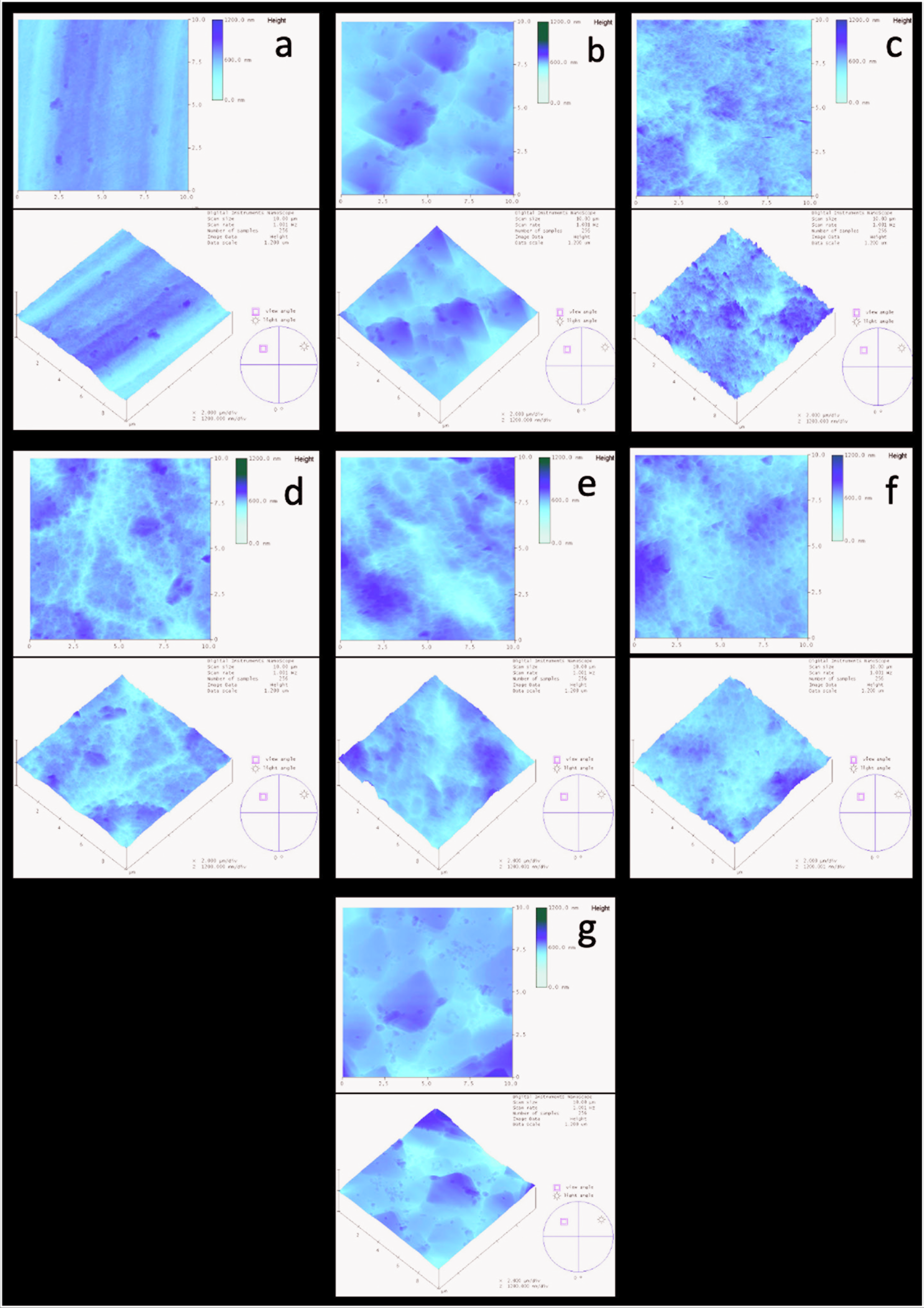
Average Surface Roughness (Ra) Values of the Experimental and Control Groups
Findings of SEM Analysis
The nondemineralized healthy enamel surfaces were found to be smooth and to have a homogeneous appearance due to the regular localization of enamel prisms. In contrast, the demineralized enamel prisms were found to be distributed irregularly and their diameters were found to be increased. In addition, due to the removal of the prism cores during the demineralization process, typically, keyhole-like patches had developed. The porous structure of the surface was noted to be lost in the group C samples, while the gaps formed after demineralization were found to have been closed through mineral precipitation. Similar to group C, a slightly smoother surface layer was seen in group CF due to the F- content. In group A, the typical appearance of demineralized enamel and keyhole patches were found to be masked, although some gaps in the interprismatic regions were found to have not been filled completely. In group K+, no homogeneous smooth layer was identified on the surface, and the layer was shaped consistent with the morphology of an artificial carious lesion. The accumulation of mineral particles was observed in the pits that were formed in the core regions of the prisms. In group K–, the deionized water was found to have caused no change to the surface morphology, although a porous layer could be observed, as was in the case with the demineralized enamel surface (Figure 3).
The SEM Images of the Enamel Surfaces in the Different Groups (×10,000): (a) Healthy Enamel, (b) Demineralized Enamel, (c) CPP–ACP (Group C), (d) CPP–ACPF (Group CF), (e) 8% Arginine Paste (Group A), (f) 500 ppm NaF (Group K+ ), and (g) Deionized Water (Group K–)
Discussion
This in vitro study evaluated the remineralization efficacy of different agents applied for the prevention of ECC and for the treatment of early carious lesion in enamel. It was noticed that all of the tested experimental materials used in this study supported remineralization on primary enamel surface, so that the null hypothesis was rejected.
The enamel on primary teeth is thinner than that of permanent enamel teeth and contains lower levels of calcium and phosphate. The diffusion coefficient of primary teeth is greater than that of permanent teeth, and while the enamel of primary teeth is significantly softer and less aesthetic, it is also less sensitive to erosion. Accordingly, the demineralization and remineralization cycles of primary teeth and permanent teeth are different. 12
The organization of the demineralization–remineralization cycle, as a dynamic process in the development of caries, plays an important role in the prevention of caries and in the treatment of early carious lesions. The ideal means of encouraging remineralization is through the reconstitution of hydroxyapatite crystals, as the inorganic component in enamel. The most frequently used agent for this purpose is fluoride, 13 although the use of toothpaste containing 1000 ppm of fluoride and higher has been reported to cause fluorosis in children younger than 6 years of age. 10 For this reason, a 500 ppm fluoride solution was used as the positive control group in the present study.
Indirect mineral losses or gains can be determined through a microhardness analysis method, the suitability of which has been demonstrated based on quantitative information on the mineral changes that occur to the enamel surface, and its status as a repeatable, fast, and easily applicable process.14,15 Many in vitro studies to date have demonstrated that CPP–ACP compounds dissolve in an acidic environment and increase the saturation and activity of calcium and phosphate ions in plaque. Accordingly, an oversaturated environment has been reported to prevent demineralization and to support remineralization.16–18 In addition, the use of CPP–ACP paste together with fluoride has been reported to encourage the passage of F- ions together with calcium and phosphate ions, and to have a synergistic effect, hence increasing remineralization. 19 Rehder Neto et al. 14 and Turssi et al. 19 evaluated the remineralization efficacy of toothpastes containing CPP–ACP and CPP–ACPF with a microhardness test and reported that surface hardness was increased following remineralization in their experimental groups, although the hardness values were similar between the groups. In the same study, similar to the findings of more recent studies, no statistically significant differences were found in the microhardness values of the CPP–ACP, CPP–ACPF, and NaF groups.
Arginine plays an important role in the ammonia metabolism, while neutralizing the acidic environment and supporting remineralization. 20 While the effects of pastes containing arginine on the remineralization of permanent tooth have been investigated in studies, there has been no study which analyzed their effects on primary teeth remineralization. Cheng et al., 21 in a study evaluating the remineralization efficacy of a 2.5% arginine-containing solution and fluoride containing agents on artificial carious lesions, demonstrated that the arginine solution had a significantly higher microhardness value than the deionized water group, and a lower microhardness value when compared to the NaF solution. In the present study, no statistically significant difference was found in the surface microhardness of the arginine group and the NaF group. In contrast, Cheng et al. 21 used a solution containing 2.5% arginine, while an 8% arginine paste was used in the present study. The differences between the findings of the two studies are considered to be based on the differences in arginine concentrations and between the enamel surface properties of primary teeth and permanent teeth.
The AFM method is frequently used in the determination of enamel surface characterizations in studies on demineralization and remineralization.22,23 For these reasons, the AFM test was used in the present study to evaluate the effects of different agents applied on the early carious lesions in enamel of the primary teeth on enamel surface roughness.
The layers forming on enamel surfaces following the application of remineralization agents have been reported in studies to occur as a result of the globular organization of minerals. In a study by Zhou et al., 24 the 4-, 8-, 12-, and 24-hour efficacy of CPP–ACP on early carious lesions in enamel was evaluated by an AFM, and surface roughness was found to decrease with the increased duration of application of the material. CPP–ACP applied to enamel surfaces demineralized with acidic beverages resulted in AFM appearances of a thick surface with a homogeneous distribution in the treatment group, while a typical honeycomb pattern was noted on the demineralized surfaces. 25 In addition, CPP–ACP was found to fill the interprismatic holes and to form a protective layer, partially covering the prisms. 26 The honeycomb appearance on the enamel surface with early carious lesions disappeared in the CPP–ACP, CPP-ACPF, and positive control groups, and a protective nonhomogeneous surface formation with a globular-type mineral accumulation was observed, while previous studies have identified different surface topographies in AFM evaluations following the CPP–ACP applications. This difference has been attributed to the varying frequencies and durations of application of the agents and the varying depth of the formed lesions.
After identifying the positive effects of arginine paste on remineralization, the investigators have turned their attention to such arginine-containing pastes. In a study in which erosion-like lesions to the enamel surface were created using acidic beverages, the remineralization effects of zinc hydroxy apatite, calcium phosphosilicate, and 8% arginine-containing pastes were evaluated using an AFM. The smoothest surface was found to be achieved by the 8% arginine-containing paste in an evaluation of roughness values and surface topographies. 22 Poggio et al. 27 compared the remineralization efficacy of pastes either containing CPP–ACP or 8% arginine on erosion-like lesions on the enamel surface, and found that both agents decreased surface roughness at similar rates, although the arginine-containing paste was found to form a more homogeneous and thick surface layer when the surface topography was analyzed. In the present study, the 8% arginine-containing paste and the CPP–ACP group paste had similar roughness values and surface topography appearances. The surface roughness findings obtained in the present study with the 8% arginine-containing paste were similar to the findings in the literature, although the surface topography findings were somewhat different. These differences were thought to originate from the different origins of the enamel samples (like bovine teeth and permanent human teeth), the different depths of the formed enamel surface lesions, and the different paste application durations.
In vitro studies evaluating demineralization and remineralization processes are considered to provide the most sensitive and detailed information about changes in the enamel surface morphology in SEM studies. 28 The effects of the agents used in the present study on the morphological structure of the enamel were evaluated with the SEM analysis.
In the present study, a SEM evaluation of the effects of pastes containing CPP–ACP and CPP–ACPF, and the positive control group on carious lesions in enamel revealed similar surface properties in all groups. The surface was observed to be covered with a new remineralization layer, and this layer filled the holes formed by pitting of the prism cores and masked the porous appearance of the surface, with circular globules observed to have formed on the newly formed surface layer. In an in vitro study evaluating the effect of 500 ppm NaF and CPP–ACP on demineralized primary teeth through a SEM analysis, numerous particles and amorphous crystals were identified on the surfaces of both the study groups, and these crystals were aligned more homogeneously in the CPP–ACP group. 10 Jayarajan et al., 29 in their study evaluating the remineralization efficacy of CPP–ACP and CPP–ACPF pastes on early carious lesions in enamel with a SEM analysis, observed a similar surface morphology in both the groups, although calcification was slightly more prominent in the CPP–ACPF group. Similar to the findings of the present study, the pastes containing CPP–ACP and CPP–ACPF were found to form a remineralization layer on the enamel surface that was found to increase resistance to demineralization.
The efficacy of 8% arginine on remineralization was evaluated with a SEM analysis in a study in which erosion-like lesions were produced on the enamel surface, while enamel prisms and the interprism regions were found to be uncovered in the control group. Furthermore, the enamel surface was observed to be covered with a thick, homogeneous, uniform, and compact layer in the arginine group. 22 The findings related to the 8% arginine paste in that study support the findings of the present study.
It is not possible to provide all conditions of the complex oral environment in the in vitro studies, so this can be considered as the limitation of the study. Also, for more precise results, further in vivo studies are needed.
Conclusion
In the present study, CPP–ACP and arginine were found to support remineralization on early carious lesions in enamel of primary teeth, and while in vitro and in vivo studies are required, this study can be considered as providing the preliminary information for future research works.
Footnotes
Declaration of Conflicting Interests
The authors declared no potential conflicts of interest with respect to the research, authorship, and/or publication of this article.
Funding
This study received support from the Scientific and Technological Research Council of Turkey (TUBITAK; project no. 114S970).