
Abstract
Background:
Having knowledge about the anatomical and pathological relationship between the maxillary posterior root tip and the maxillary sinus floor is very significant for preoperative treatment planning.
Aims and Objectives:
The purpose of the present study was to compare the accuracy of original panoramic radiographs and digital panoramic images over cone beam computed tomography (CBCT) images in evaluating the relationship between the maxillary posterior teeth and the maxillary sinus floor, and to verify the accuracy of the signs of the relationship between the roots of the maxillary posterior teeth and maxillary sinus on the panoramic radiographs over CBCT images.
Materials and Methods:
From 106 subjects (47 males and 59 females; mean age = 39 years; range = 18-67 years) referred to our university, a pair of panoramic and CBCT images was selected for further analysis. The relationship between the maxillary posterior teeth, the maxillary sinus, and panoramic radiography signs (root projection into the sinus, interruption of the maxillary sinus floor cortex, absence of lamina dura, darkening of the root apex, and curvature of the sinus floor on the root apex) associated with the protrusion of root apexes into the sinus was evaluated.
Results:
The P values of the data obtained from the original and invert enhanced panoramic images were .53 and .52, respectively, and there was no statistically significant difference in the accuracy of the 2 methods (P > .05).
Conclusion:
The root projection into the sinus is an indicative sign of root protrusion into the sinus on CBCT.
Introduction
Having knowledge about the intimate relationship between the maxillary posterior teeth and maxillary sinus is important for accurate diagnosis and treatment planning. The extraction of the maxillary posterior teeth is a routine procedure in which sometimes complications occur. 1 Considering the anatomic proximity between the maxillary posterior root tips and maxillary sinus, oroantral communications may occur after tooth extraction. 2 This has been a source of concern for clinicians. The close relationship between the root apex and the maxillary sinus floor may be an effect of bone resorption induced by chronic apical periodontitis, the process of development of the tooth, aging, maxillary sinus pneumatization, and the impaction status of the third molar. 3
The simplest way of assessing the relationship between maxillary posterior roots and maxillary sinus is imaging the relevant area appropriately. Panoramic radiography, which is the main imaging technique used in dentistry, is the first choice for diagnosis and treatment planning. It is a simple and easily accessible technique for the initial evaluation. This method, which gives a 2-dimensional image, has some disadvantages such as the superposition of surrounding tissues, magnification, distortion of images, and perspective problems. 4 Digital panoramic radiographs have been developed to provide better quality images with increased technological developments. Digital panoramic images allow the original image to be replaced with different development tools. In the literature, there are studies on the efficiency of digital panoramic images.5-8
After the discovery of X-rays, computed tomography (CT), considered the most important development in radiology, also allowed the 3-dimensional evaluation of maxillofacial structures. The cost of CT is high, and its use in dentistry is limited due to the fact that it is not present at all centers, especially because the amount of radiation exposed by patients is high, the duration of the scan and image processing is long, and resolution is insufficient to detect small apical and alveolar lesions. 9 For eliminating the problems of conventional CT, the imaging system called cone beam computed tomography (CBCT) has been developed. CBCT has many advantages over CT. Because the X-ray beam can be limited in CBCT and X-rays are directed only at the field to be imaged, the radiation dose to the patient is much less. Because of the isotropic nature of voxels in CBCT, accurate and precise measurements can be made.10,11
The purposes of our work were as follows: (a) to compare the accuracy of the original and invert enhanced panoramic images over CBCT images in evaluating the relationship between the maxillary posterior teeth and maxillary sinus, and (b) to verify the accuracy of the signs of the relationship between the roots of the maxillary posterior teeth and the maxillary sinus on the panoramic radiographs over CBCT images. Thus, a reasoning to make a decision on the need for preoperative CBCT scans will be presented to the clinicians before the dentoalveolar surgery in the posterior maxilla.
Materials and Methods
In the present retrospective study, 106 subjects, who were presented to the faculty to undergo both panoramic and CBCT examinations as part of their routine examination, determining the position of the maxillary molars in relation to the maxillary sinus and implant or orthognathic surgery treatment planning, were enrolled. Forty-seven of these patients were male and 59 were female. In the study, 1008 teeth were examined. This study has been approved by the ethical committee of our university.
CBCT images showing the relationship of the maxillary posterior teeth with the maxillary sinus (axial, coronal, and sagittal images).
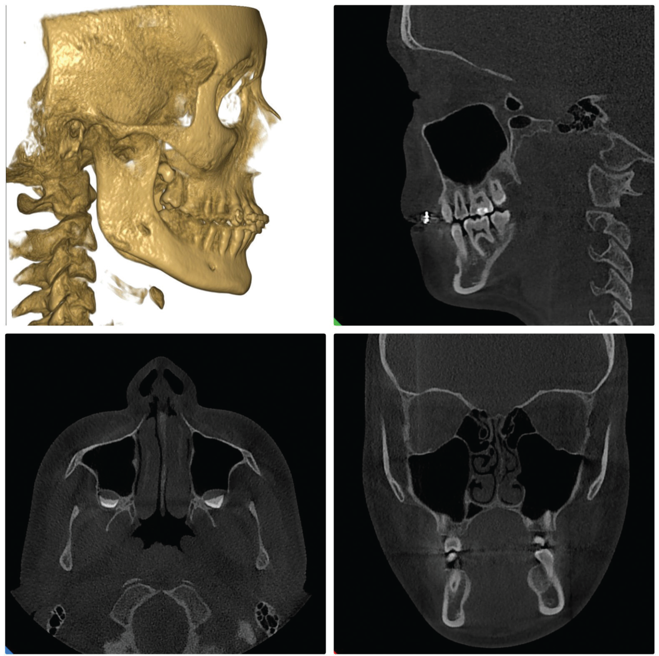
Multiplanar reformatted reconstructions in the axial planes were created. All the images were evaluated in 3 planes of axial, coronal, and sagittal (Figure 1). CBCT images were evaluated according to the relationship between the maxillary posterior teeth and maxillary sinus, and were grouped on the basis of the classification of Shahbazian et al.
12
The images were classified as follows:
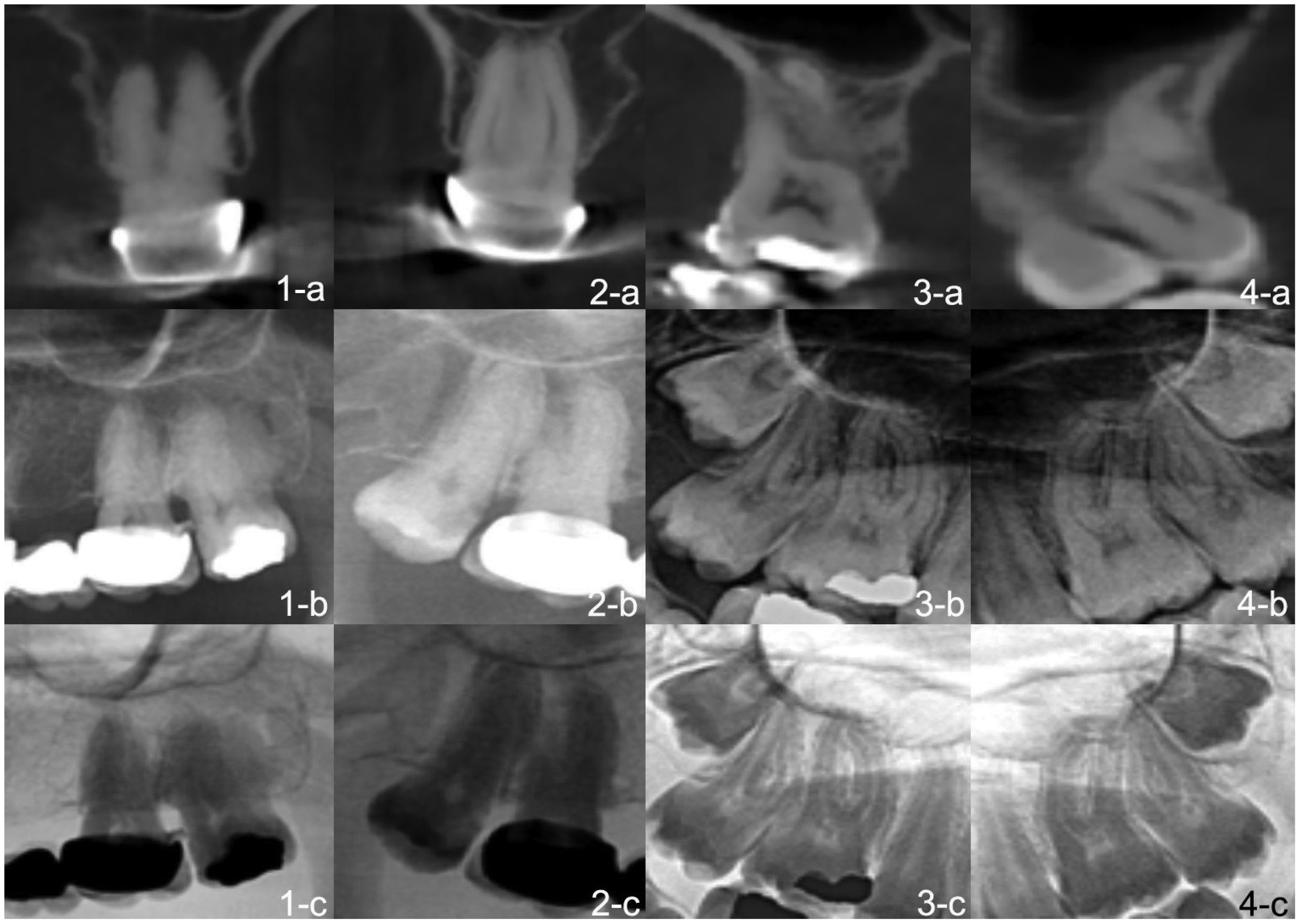
Class 1: There is a sufficient distance between the root and the maxillary sinus. Class 2: There is <0.5-mm distance between the root tip and the maxillary sinus floor. Class 3: Roots are closely related to the maxillary sinus floor and roots are outside the sinus. Class 4: Roots are closely related to the maxillary sinus floor and are inside the sinus (Figure 2).
(a) CBCT images, (b) original panoramic images, (c) invert enhanced panoramic images of the classification of maxillary posterior teeth and its relationship with the inferior wall of the sinus; the remote distance between 26 root tips and the maxillary sinus floor shown in CBCT (1-a) original, (1-b) invert enhanced panoramic image, and (1-c) close contact between root tips of tooth 17 and maxillary sinus shown in (2-a) CBCT, (2-b) original, and (2-c) invert enhanced panoramic image; lateral projection of the mesiobuccal root tip of tooth 16 on the maxillary sinus floor shown in (3-a) CBCT, (3-b) original, and (3-c) invert enhanced panoramic image; intimate relationship between the palatal root tip of tooth 27 and maxillary sinus shown in (4-a) CBCT, (4-b) original, and (4-c) invert enhanced panoramic image.
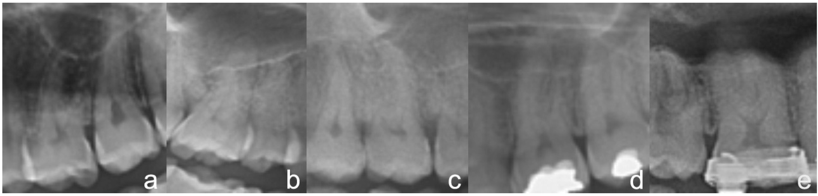
The CBCT images of teeth in Classes 3 and 4, the ones that are in a close relation with the maxillary sinus, were evaluated based on the findings of the sinus reflection of the root apexes, interruption of the maxillary sinus floor cortex, absence of lamina dura, darkening of the root apex, and curvature of the sinus floor on the root apex on panoramic radiographs 13 (Figure 3). The panoramic device original image, invert tool image, and CBCT images were evaluated and compared between the groups when the maxillary posterior teeth were closely related to the maxillary sinus.
Cropped panoramic radiographs showing a close approximation of maxillary sinus with maxillary teeth. (a) Darkening in the root apexes, (b) projection of the root apexes, (c) superiorly curving sinus floor enveloping the associated tooth root, (d) absence of lamina dura, and (e) interruption of the maxillary sinus floor.
Statistical evaluations were performed with the help of Statistical Product and Service Solutions version 21.0 (IBM Corp., New York, NY; formerly SPSS Inc., Chicago, IL). The Kolmogorov-Smirnov test was applied to all continuous variables to be analyzed to determine whether the distribution provided normality hypothesis. CBCT findings were accepted as gold standard. Contingency crosstables with scores given for panoramic radiographic images and for the reference standard, CBCT, were performed. The McNemar-Bowker test evaluated the disagreement between the CBCT and panoramic imaging modality, concerning the anatomical relationship between the upper posterior root tip and the maxillary sinus floor. All data were evaluated at a significance level of P < .05. According to the null hypothesis, there was no discrepancy between CBCT and original and inverted panoramic modality. Additionally, positive-predictive value (possibility of true-positive result occurring) and negative-predictive value (possibility of true-negative result arising) were calculated.
Results
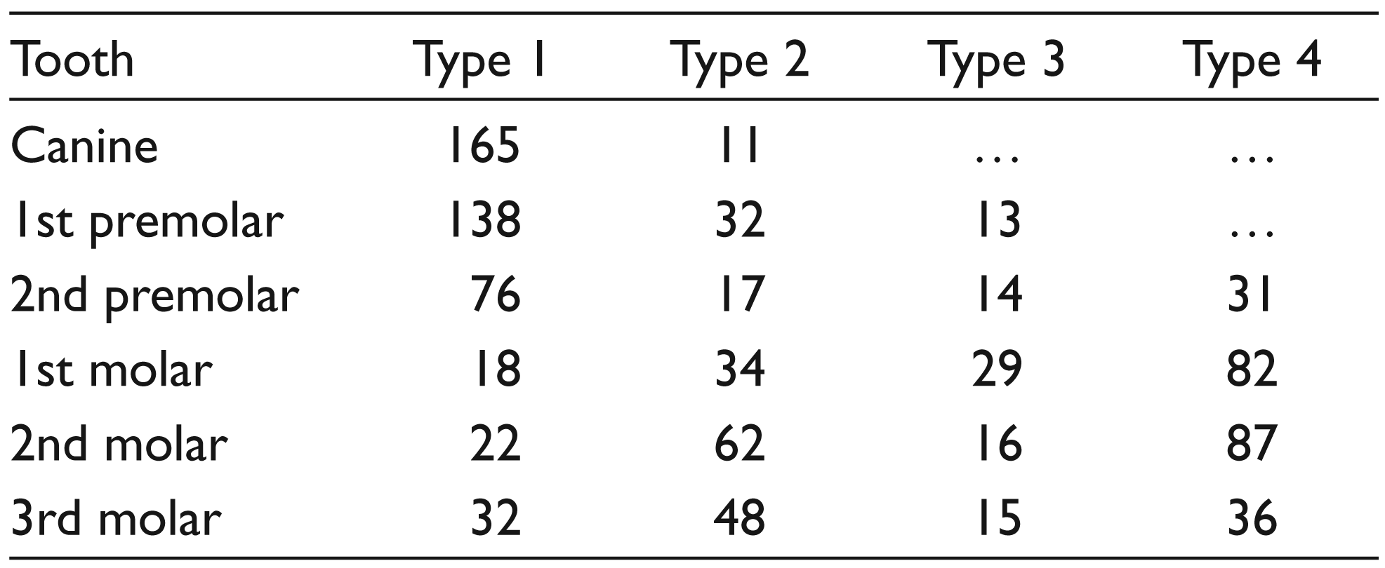
The mean age of the 106 patients was 39.16 (range 18-67) years, and 1008 teeth were grouped according to the classification of Shahbazian et al. 12 Out of these teeth, 481 were molars, of which 163 teeth were the first molar, 187 teeth were the second molar, and the remaining 131 teeth were the third molar. Among the 1008 teeth that were grouped, 351 teeth were premolars (183 teeth of the first premolar and 168 teeth were of the second premolar), and 176 teeth were canine. CBCT examinations revealed that there were 353 teeth included in Classes 3 and 4 (Table 1).
Distributions of the Maxillary Teeth According to Shahbazian’s Classification 12
Original panoramic radiographs of Classes 3 and 4 with a close relation in CBCT images showed the same results in 296 cases (83.8%; Table 2). In 57 cases, false-positive response was seen. In invert enhanced panoramic radiographs, in 309 (87.5%) cases, maxillary posterior teeth were closely associated with maxillary sinus and were found to be consistent with the results found in CBCT images (Table 2).
Equivalence Relationship Between Original and Invert Enhanced Panoramic Images and CBCT (Gold Standard)

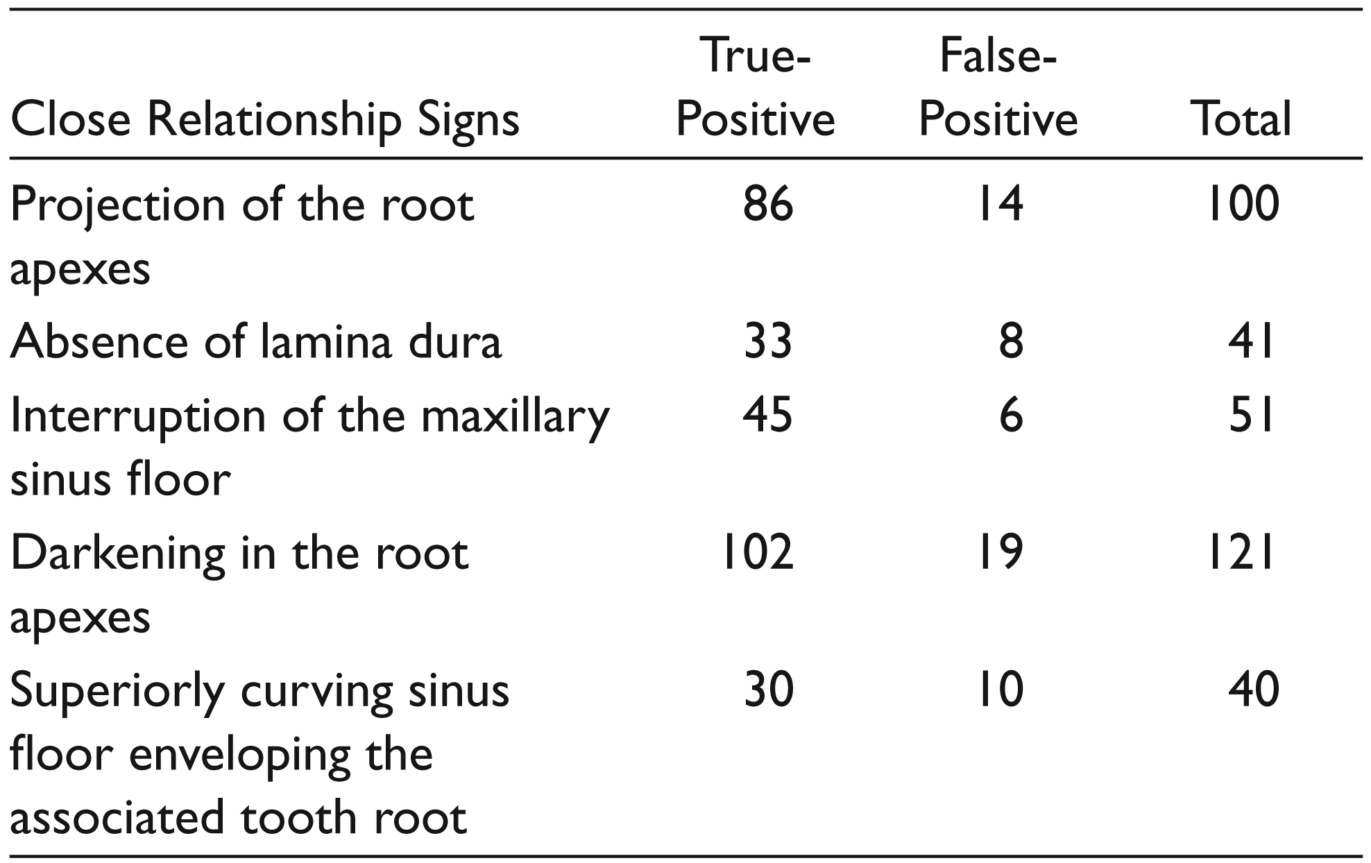
Original panoramic and inverted enhanced panoramic radiographs showing positive and negative responses were compared with CBCT images to assess which indications of the close relationship between maxillary sinus and teeth roots corresponded to 3-dimensional results (Tables 3 and 4).
Comparison of the Signs of Close Relationship Found in the Original Panoramic Image Modality and CBCT Image Results (Gold Standard)
Comparison of the Signs of Close Relationship Found in the Invert Enhanced Panoramic Image Modality and CBCT Image Results (Gold Standard)
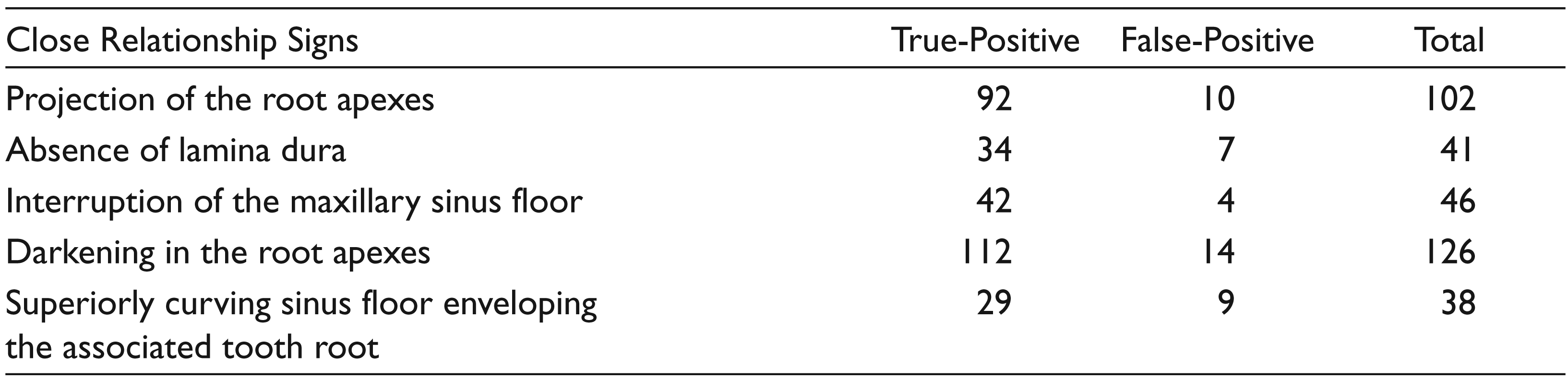
When the data obtained from the panoramic and invert enhanced panoramic images were evaluated, it was found that their P values were .53 and .52, respectively, and there was no statistically significant difference in the accuracy of the 2 methods (P > .05). In both imaging techniques, only root apexes reflected in the sinus cavity showed that the root of the tooth was within the maxillary sinus (P < .05; Tables 5 and 6).
Multiple Logistic Regressions for Possible Radiographic Signs Indicating Root Protrusion into the Maxillary Sinus (MS) for Panoramic Images
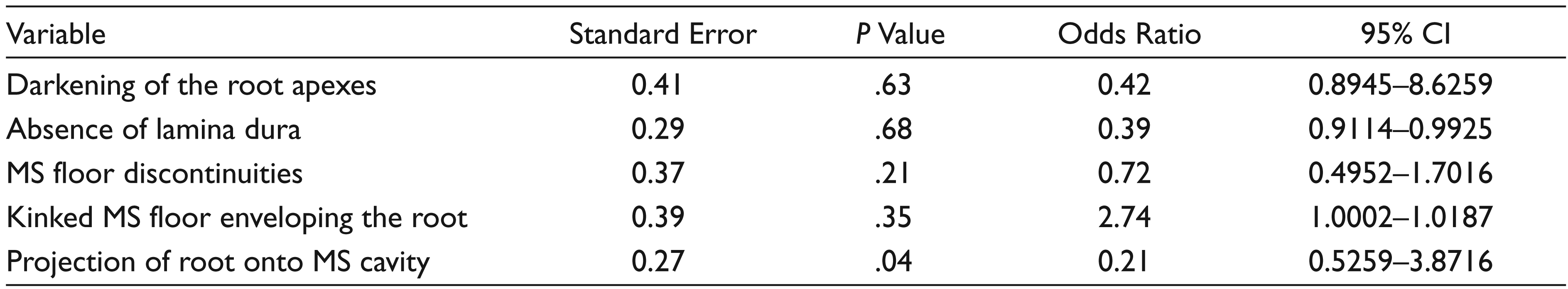
*P < .05; CI, confidence interval; MS, maxillary sinus.
Multiple Logistic Regressions for Possible Radiographic Signs Indicating Root Protrusion into the Maxillary Sinus (MS) for Invert Enhanced Panoramic Images
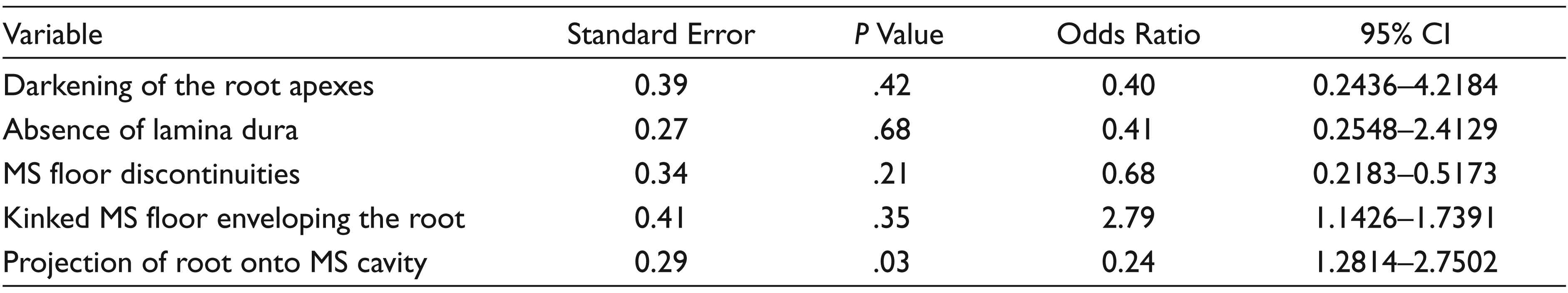
*P < .05, CI, confidence interval; MS, maxillary sinus.
Discussion
The anatomy of the maxillary sinus floor is excessively variable. This condition can be affected by factors such as aging, maxillary sinus size, and maxillary sinus pneumatization. Maxillary posterior teeth root and maxillary sinus floor are sometimes separated only by sinus mucosa. 14 In such cases, important complications such as endodontic treatment, orthodontic tooth movement, oroantral fistula, and the displacement of the root to the sinus space may be encountered.15-17 The relationship between the maxillary sinus floor and maxillary posterior tooth root is important for the preoperative treatment planning of the maxillary sinus area. Considering the proximity of the maxillary sinus floor and the maxillary posterior teeth, clinicians should be more careful when performing dental procedures in this region.
Panoramic radiography is the main imaging technique used to evaluate the close relationship between the maxillary posterior roots and maxillary sinus; however, the assessment of the relationship between the upper teeth roots and maxillary sinus has shown that panoramic radiography and CBCT are significantly different techniques.18,19 Despite image-distortion problems and lack of acutance, panoramic radiography has been reported to be useful in most routine clinical situations and an important tool in determining the need for preoperative CBCT scanning.20,21
In regard to maxillary sinus root emergence, our study is compatible with other 35% of the sample studies.12,22 When the roots are close to the sinus, the difference between imaging methods is obvious. In the study of Lopes et al, 13 the close relationship between the roots and maxillary sinus was evaluated as symptoms of the “projection of the root apexes to the sinus cavity, interruption of the maxillary sinus floor cortex, the absence of lamina dura, the darkening of the root apexes, and the curvature of the sinus floor on the root apex.” In our study, we used these symptoms to determine the accuracy of panoramic radiographs and invert enhanced panoramic images with CBCT. To our knowledge, this present work is the first study to use invert enhanced panoramic images in evaluating the relationship between the maxillary posterior teeth and maxillary sinus.
When assessing the effect of panoramic images and enhancement tool on the digital panoramic image detection of the close relationship between the maxillary posterior roots and the maxillary sinus, there was no statistically significant difference (P > .05) However, when comparing the results found for each modality with CBCT, panoramic images—both original and invert enhanced—presented a high number of false-positive results. Nevertheless, panoramic radiography provides a quick assessment of the clinically diagnosed dental diseases. Clinicians chose to use panoramic radiographs instead of its alternatives because of its easy accessibility, cost-effectiveness, and low radiation dosage when evaluating signs of the close relationship between the upper tooth roots and the maxillary sinus (False positive; invert enhanced panoramic: 44, panoramic: 57).
Lopes et al 13 in 51% of the cases where the roots were reflected toward the maxillary sinus found that the maxillary sinus base was interrupted. In our study, this rate was 63%. According to our study, this sign should be accepted as a sign of the actual root outreaching to the maxillary sinus. In our study, radiological signs such as the reflection of the root apex to the sinus cavity indicates that the root apexes were within the maxillary sinus (P < .05).
Conclusion
Based on the results of the present study, there was no significant difference between the original panoramic radiographs and the invert enhanced panoramic radiographs in terms of the signs indicating that the root of the tooth was within the maxillary sinus. However, less false-positive results were found in inverted enhanced panoramic images. Although it is difficult for panoramic radiography to analyze the maxillary sinus anatomy, radiographic signs, such as root apexes reflected in the sinus cavity, suggest that the roots may be within the maxillary sinus. If the panoramic radiographic examinations of the roots have a reflection of the maxillary sinus, in such cases, important complications such as endodontic treatment, orthodontic tooth movement, oroantral communication, and the displacement of the root to the maxillary sinus may be encountered.
Recommendations
In the presence of these radiographic signs, CBCT requested by clinicians is ideal before surgical operations, and endodontic and orthodontic treatment.
Invert enhanced panoramic modality can be useful for the assessment of the relationship between the maxillary posterior roots and maxillary sinus.
Declaration of Conflicting Interest
The authors declared no potential conflicts of interest with respect to the research, authorship, and/or publication of this article.
Funding
The authors received no financial support for the research, authorship, and/or publication of this article.