
Abstract
Purpose:
To compare the biomechanical properties of a high-tensile strength suture and high-tensile strength tape in tendon graft fixation using two needleless suture wrapping techniques, the modified Prusik knot and modified rolling hitch.
Methods:
Two needleless suture wrapping techniques, the modified rolling hitch (MR) and modified Prusik knot (MP), were utilized. Meanwhile, two kinds of suture materials, a No. 2 braided nonabsorbable high-strength suture (S) and a 1.3 mm high-tensile strength tape (T), were used. A total of 40 porcine tendons were used, which were randomly divided into four groups. Each group was assigned to one of the following groups: MRS, MRT, MPS, and MPT. Each specimen was pretensioned to 100 N for three cycles, cyclically loaded from 50 to 200 N for 200 cycles, and finally loaded to failure.
Results:
The MRT group (34.1 ± 3.5%) had a significantly higher value compared with the MRS (29.7 ± 2.3%), MPS (27.1 ± 3.6%) and MPT (29.5 ± 4.0%) groups in term of elongation after cyclic loadings (p = 0.002). In terms of ultimate failure load, there were no significant differences in the MRS (401 ± 27 N), MRT (380 ± 27 N), MPS (398 ± 44 N) and MPT (406 ± 49 N) values (p = 0.539). All specimens failed due to suture breakage at the knots.
Conclusion:
Compared with the high-tensile strength suture, using the high-tensile strength tape lead to greater elongation after cyclic loading when the modified rolling hitch was used. No differences in terms of elongation after cyclic loading and load to failure were found between the high-tensile strength suture and tape using the modified Prusik knot.
Keywords
Introduction
Acquiring a sturdy suture-tendon construct is not only important in ligament reconstruction surgery, such as anterior cruciate ligament (ACL) reconstruction and tendon transfer in ankle stabilization. 1 To achieve strong fixation, different techniques have been described. 2 –5 Among these techniques, the Krackow suture technique 3 is especially popular due to its superior biomechanical properties. 6 –8 However, it is worth noting that passing sutures through tendons is time-consuming 9 and will possibly cause tendon damage; additionally, potential risk of needlestick injury is also a concern.
To overcome the disadvantages of the previous suture techniques with needles, several needleless suture wrapping techniques have been proposed. 10 –12 Some studies have evaluated the biomechanical properties of these suture wrapping techniques in tendon graft fixation, and several of them, such as the modified Prusik knot 11 and modified rolling hitch, 10 have been reported to be attractive options due to their comparable or superior biomechanical properties. 13 –16 Additionally, these techniques have also been proven to be less time-consuming. 9,13
From the biomechanical perspective, tape-type sutures can prevent sutures from pulling through the tendons. 17,18 Gnandt et al. conducted a biomechanical study of tendon graft fixation with different suture techniques and found that high-tensile strength tape withstood a significantly higher load than a high-tensile strength suture during a single load to failure test when whipstitch and Krackow suture techniques were used. 17 Similarly, Ono et al. reported that tape-type sutures had significantly greater stiffness values than standard sutures sourced from the same company in their sheep infraspinatus tendon model. 18
The friction between the suture and tendon was the major fixation mechanism for tendon graft fixation with suture wrapping techniques, which was different from the traditional suture technique. The previous studies have suggested several suture wrapping techniques to be attractive options in tendon graft fixation with braided nonabsorbable high-strength sutures. Although using tape-type sutures in tendon graft fixation with specific suture techniques may lead to greater tendon graft fixation strength, 17 –19 it remains unclear whether tape-type sutures have superior biomechanical properties to round sutures in tendon graft fixation using needleless suture wrapping techniques.
The purpose of this study was to compare the biomechanical properties of a high-tensile strength suture and high-tensile strength tape in tendon graft fixation using two needleless suture wrapping techniques, the modified Prusik knot and modified rolling hitch. Our hypothesis was that the suture-tendon construct with the high-tensile strength tape would have comparable elongation after cyclic loading and a greater maximal failure load compared with the high-tensile strength suture.
Methods
Study design
This study was an in vitro biomechanical study using porcine tendons. The study was granted an exemption from the Institutional Review Board at a national medical center. The independent variables were type of sutures and suture wrapping techniques, whereas the dependent variables were biomechanical properties, including elongation after the cyclic loadings and ultimate failure loads.
Tendon specimens
Porcine hindleg flexor profundus tendons were selected for this study since a previous study suggested them to be surrogates for human semitendinosus tendons. 20 In accordance with previous studies. 14 –16,21 –25 the entire tendons were excised from adult male fresh porcine (mean age, 2 years) hindleg trotters and were cut into equal lengths (18 cm). Next, the tendons were evaluated carefully for possible degenerative or pathologic lesions. After confirming the quality of the tendons, the attached soft tissue, including the synovial sheath, was removed. A 5 mm thick transverse section was acquired from the end of the distal tendon. With the assistance of a calibration scale, a digital camera (EOS 60D; Canon, Tokyo, Japan), and image processing software (ImageJ, version 1.52p; National Institutes of Health, USA), a cross-sectional area of each tendon was measured. During tendon preparation and biomechanical testing, 0.9% saline solution was sprayed on the tendons to keep them moist.
Suture materials and suture wrapping techniques
Both braided nonabsorbable high-strength suture and tape were used, including No. 2 FiberWire (Arthrex, Naples, FL) and 1.3 mm SutureTape (Arthrex, Naples, FL). Meanwhile, two kinds of suture wrapping techniques for tendon graft fixation were selected, the modified Prusik knot and modified rolling hitch. These two techniques were selected because they exhibited less suture-tendon elongation in a previous biomechanical study as compared to other alternatives. 16 The wrapping suture techniques were conducted 1 cm from the end of the distal tendon. The modified rolling hitch begins by wrapping the suture around the tendon, followed by making a second and a third wrap. The working limb of the suture is then crossed over the other limb, and the procedure is completed with a half hitch by making a turn around the tendon and passing the working limb through it (Figure 1A–B). The modified Prusik knot starts with passing the two free suture ends through its own loop when wrapping the tendon. This step is repeated to wrap the tendon again and pass the two free suture ends through the same suture loop. Finally, the knot is tightened (Figure 1C–D).

Illustration of the (A) MRS, (B) MRT, (C) MPS and (D) MPT groups. Porcine hindleg flexor profundus tendons were used, and the wrapping suture techniques were performed 1 cm from the end of the distal tendon.
Grouping
There were four subgroups in our study: A FiberWire suture with a modified Prusik knot (MPS group), a FiberWire suture with a modified rolling hitch (MRS group), SutureTape with a modified Prusik knot (MPT group), and SutureTape with a modified rolling hitch (MRT group). A total of 40 porcine tendons were used and were randomly divided into four subgroups of 10 specimens each.
Biomechanical testing
A material-testing system (AG-X; Shimadzu, Tokyo, Japan) was utilized for biomechanical testing. When mounting the prepared specimens, the proximal ends of the tendons were fixed with a sinusoid clamp, making the length of the free tendons equal in length (9 cm). The ends of the suture limbs were knotted together and looped over a post on the adapter connected to the load cell (Figure 2). The biomechanical testing parameters and testing protocol were in accordance with those used in previous studies. 2,6,12,15,16,19,21,23,26,27 Initially, three cycles of pretensioning force ranging from 0 to 100 N at a rate of 100 mm/min were conducted. After preloading to 50 N for 1 minute, each specimen was cyclically loaded between 50 and 200 N with a cross-head speed of 200 mm/min for 200 cycles. Finally, the specimens were loaded to failure at a constant rate of 20 mm/min. Failure was defined as when the maximum tensile force suddenly dropped or discontinued in the load-displacement curve. The ultimate failure load and the mode of failure in each specimen was recorded.

The biomechanical testing setup. A sinusoid clamp was used to fix the tendon. The ends of the sutures were knotted and looped over a post on the load cell connected to the materials-testing machine. A digital camera was used for recording the images.
A dark-blue line drawn 5 cm from the distal end of the tendon and two dark-blue dots marked on both suture limbs where they extend from the tendon were used as indicators for measuring the elongation of each suture-tendon construct. The elongation value after cyclic loading was acquired by calculating the distance difference in the cyclic loading test between the dark-blue line and two dark-blue dots. A digital camera (HDR-XR 269; Sony, Tokyo, Japan) was used for recording images. Image processing software (ImageJ, version 1.52p; National Institutes of Health, USA) was used to calculate the distance between the indicators.
Statistical analysis
A pilot study was designed to calculate the required sample size. A total of 16 specimens were divided equally and randomly assigned to the MRS, MRT, MPS, and MPT groups. G*Power, version 3.1.9.2 (Heinrich Heine-University of Dusseldorf, Dusseldorf, Germany) software was used for the priori power analysis. An α equal to 0.05 and a power (1 − β) of 0.80 were given for this a priori power analysis model, and an effect size of 0.56 was obtained. Finally, the required sample size was determined to be 40 specimens, with 10 specimens in each group. A statistical analysis was conducted using IBM SPSS Statistics (version 20; IBM SPSS Inc., Chicago, IL, USA). Descriptive statistics, including means and standard deviations, were obtained for all of the subgroups. The Kruskal-Wallis H test, a non-parametric method, was used to compare the parameters among the four groups. The statistical significance was set as p < .05. The Mann-Whitney U test with a Bonferroni correction was used for the post-hoc analysis, resulting in a significance level set at p < 0.0125.
Results
The cross-sectional areas of the porcine tendons among four groups were not significantly different. The cross-sectional areas of tendon in the MRS, MRT, MPS, and MPT groups were 43.3 ± 4.4 mm2, 43.8 ± 4.4 mm2, 42.5 ± 4.5 mm2, 42.4 ± 4.5 mm2, respectively (p = 0.879).
In terms of elongation after the cyclic loadings, the MRT group had the largest value compared with the other groups. There were significant differences in elongation after cyclic loading among the MRS (29.7 ± 2.3%), MRT (34.1 ± 3.5%), MPS (27.1 ± 3.6%) and MPT (29.5 ± 4.0%) groups (p = 0.002). The post-hoc analysis showed that elongation after cyclic loading in the MRT group was significantly greater than that in the MRS, MPS, and MPT groups (p = 0.009, 0.001, and 0.009, respectively); otherwise, there were no differences between the MRS and MPS groups (p = 0.089), the MRS and MPT groups (p = 0.739) or the MPS and MPT groups (p = 0.123) (Figure 3).

Elongation after cyclic loading for the MRS, MRT, MPS and MPT groups. The MRT group had the highest value compared with the MRS, MPS and MPT groups. The data of gray bars and error-bars were mean and standard deviation, respectively. A p value <0.0125 was considered statistically different.
In terms of ultimate failure load, all four groups had similar values without any significant differences. The ultimate failure loads in the MRS, MRT, MPS and MPT groups were 401 ± 27 N, 380 ± 27 N, 398 ± 44 N, 406 ± 49 N, respectively (p = 0.539). Each specimen in the four groups failed during the load to failure test, where breakage of suture material at the knot was the failure mode (Table 1).
Biomechanical properties of the MRS, MRT, MPS and MPT groups.
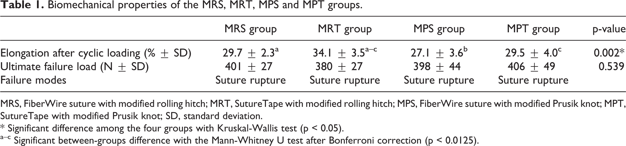
MRS, FiberWire suture with modified rolling hitch; MRT, SutureTape with modified rolling hitch; MPS, FiberWire suture with modified Prusik knot; MPT, SutureTape with modified Prusik knot; SD, standard deviation.
* Significant difference among the four groups with Kruskal-Wallis test (p < 0.05).
a–c Significant between-groups difference with the Mann-Whitney U test after Bonferroni correction (p < 0.0125).
Discussion
The major findings in the present study indicated that the high-tensile strength tape did not offer superior biomechanical properties in tendon graft fixation compared to the high-tensile strength suture when needleless suture wrapping techniques were applied. Obtaining a reliable suture-tendon construct is clinically important, especially when a post-tie fixation technique is used. In recent decades, several needleless suture wrapping techniques were reported to be attractive surgical options. 10 –12 On the other hand, tape-type sutures were developed to prevent sutures from pulling through the tendons and were believed to provide greater tendon graft fixation strength compared to round-shaped sutures. 17 –19 However, it remains unclear whether tape-type sutures can provide superior biomechanical properties in tendon graft fixation using needleless suture wrapping techniques. The present study then further investigated the use of tape-type suture in tendon graft fixation using needleless suture wrapping techniques.
The tape-shaped suture has become popular in recent years. With the ongoing development of sutures, a newly developed high-tensile strength tape, SutureTape (Arthrex), has been proposed to avoid the solid cores of sutures (FiberWire (Arthrex)) cutting through soft tissue. 28 The tape-shaped design was aimed to spread the force across a broader tissue area, potentially resulting in greater resistance to pull-out forces. 17,28 The use of tape-shaped sutures in tendon graft fixation with whipstitches or Krackow stitches was supported by a previous biomechanical study conducted to evaluate the maximum loads at failure during a single load to failure test. 17 The present study further investigated the use of tape-shaped sutures in tendon graft fixation with needleless suture wrapping techniques. However, our study did not support the use of SutureTape for tendon graft fixation with needleless suture wrapping techniques compared to FiberWire because only similar or worse biomechanical properties were found when SutureTape was used. The possible reason for the aforementioned findings could be the poorer constriction effect of the tape-shaped suture since the constriction force was spread across a broader tissue area, resulting in poor resistance to the pulling force.
Both the modified rolling hitch and modified Prusik knot are attractive needleless suture wrapping technique for tendon graft fixation. 9,15,16 Previous biomechanical studies indicated that both the modified rolling hitch and modified Prusik knot can achieve equal elongation after cyclic loading and ultimate failure loads, 9,16 and both of them can achieve equal or superior biomechanical properties as compared to the Krackow suture. 9,15 Similar to previous studies, the present study showed that the MRS and MPS groups had equal elongation after cyclic loading and ultimate failure loads. Despite the fact that both the modified rolling hitch and modified Prusik knot are considered attractive alternatives for tendon graft fixation, our study suggested that tape-shaped sutures should not be used for these needleless suture wrapping techniques, especially the modified rolling hitch, due to the poor biomechanical properties found in the present study.
Most of the experimental results in our study were consistent with those in previous studies although some of them were not. Some previous studies 9,14,15 reported ultimate failure loads of 330–360 N in both the modified rolling hitch and modified Prusik knot groups; meanwhile, the rupture of sutures at the knot was the failure mode in all specimens in both groups. Similar to previous studies, 9,14,15 the present study found 401 ± 27 N and 398 ± 44 N ultimate failure loads in the MRS and MPS groups, respectively, and all specimens failed due to sutures broken at the suture knot. Regarding elongation after cyclic loading, however, the experimental results in our study seemed to be higher than those in the previous study. 9 The present study found 29.7 ± 2.3% and 27.1 ± 3.6% elongation after cyclic loading in the MRS and MPS groups, respectively, which were greater than the values ranging from 20–22% in a previous study. 9 Different brands of high strength-tensile sutures may have contributed to the aforementioned finding. In the present study, FiberWire (Arthrex, Naples, FL) sutures were used, whereas ULTRABRAID sutures (Smith & Nephew, MA, USA) were used in the previous study. 9
This study had a number of limitations. First, porcine flexor tendons, instead of human tendons, were used in this biomechanical study. Although some structural differences exist between porcine and human tendons, porcine flexor tendons are regarded as reasonable surrogates for human semitendinosus tendons. 20 Second, a cyclic loading test was designed to imitate repetitive movements during rehabilitation, and greater elongation after cyclic loading implied graft loosening, potentially affecting graft healing. Nevertheless, the actual physiologic loading on tendons could not be entirely simulated. Finally, the needleless suture wrapping techniques were performed by a surgeon, and the individual surgeon’s technique could affect the elongation of the suture-tendon construct. To reduce surgical errors, in the present study, all specimens were completed by a single surgeon.
Conclusions
Using high-tensile strength tape led to greater elongation after cyclic loading when the modified rolling hitch was used in tendon graft fixation compared with the high-tensile strength suture. No differences in terms of elongation after cyclic loading and load to failure were found between the high-tensile strength suture and the tape in tendon graft fixation using the modified Prusik knot. The biomechanical findings from this model could be a reference for clinical practice especially when a post-tie fixation technique is used.
Footnotes
Authors’ note
This research was conducted in Skeleton Materials and Bio-compatibility Core Lab, Research Center of Clinical Medicine, National Cheng Kung University Hospital, Tainan, Taiwan.
Author contributions
C-KH, F-CK and C-HC contributed to the concept. C-KH and K-LH contributed to the methodology. C-KH and H-CC conducted the biomechanical testing. C-KH and drafted the manuscript. YC and W-RS contributed to critical review of the manuscript. W-RS supervised the study.
Acknowledgments
We thank Skeleton Materials and Bio-compatibility Core Lab, Research Center of Clinical Medicine, National Cheng Kung University Hospital, Tainan, Taiwan for assistance with this project.
Declaration of conflicting interests
The author(s) declared no potential conflicts of interest with respect to the research, authorship, and/or publication of this article.
Funding
The author(s) disclosed receipt of the following financial support for the research, authorship, and/or publication of this article: This study was funded by Ministry of Science and Technology, Taiwan.